
Development of right atrium
Reading File
Now let me search the medical library for content on right atrium development.
Finding Sources
Finding Sources
Finding Sources
Finding Sources
Finding Sources
Finding Sources
Excellent - found the key sources. Let me read the relevant sections from "The Developing Human" and "Langman's Medical Embryology" simultaneously.
Reading File
Reading File
Excellent content obtained. Let me read more from both sources to get a complete picture, and also fetch the key diagrams.
Reading File
Reading File
I now have comprehensive material from both major embryology textbooks. Let me fetch the key diagrams to include in the answer.
Loading Image
I have all the information needed for a comprehensive answer. Here is the complete, well-structured response:
Development of the Right Atrium
The adult right atrium is derived from two embryological components:
- The primordial (primitive) right atrium - forms the rough trabeculated part and right auricle
- The right horn of the sinus venosus - forms the smooth-walled part (sinus venarum)
Step 1: Formation of the Primordial Heart Tube and Sinus Venosus
In the middle of the 4th week, the sinus venosus receives venous blood from both right and left sinus horns. Each horn receives blood from three veins:
- The vitelline (omphalomesenteric) vein
- The umbilical vein
- The common cardinal vein
Initially, communication between the sinus venosus and the primordial atrium is wide, and both horns are roughly equal in size.
Step 2: Left-to-Right Shunts and Enlargement of the Right Horn
Progressive enlargement of the right sinus horn occurs due to two left-to-right shunts:
- First shunt: Transformation of vitelline and umbilical veins directs blood to the right
- Second shunt: The left and right anterior cardinal veins are connected by an anastomosis, which becomes the left brachiocephalic vein, channeling blood to the right side
As a result:
- The right umbilical vein and left vitelline vein obliterate by the 5th week
- The left sinus horn loses its importance and eventually becomes the oblique vein of the left atrium and the coronary sinus
- The right anterior and right common cardinal veins become the SVC
By the end of the 4th week, the right horn is noticeably larger, and the sinoatrial (SA) orifice shifts to the right, opening into the portion of the primordial atrium that will become the adult right atrium.
Step 3: Incorporation of the Right Sinus Horn
As the right sinus horn enlarges, it is progressively incorporated into the right atrium. This gives rise to:
- The smooth-walled posterior portion of the right atrium = sinus venarum (also called sinus venarum cordis)
- This area receives the SVC, IVC, and coronary sinus
The primordial right atrium (original trabeculated portion) forms:
- The anterior rough trabeculated part of the right atrium
- The right auricle (a conical muscular pouch)
Step 4: Crista Terminalis and Related Structures
The boundary between the two portions is marked by:
| Structure | Nature |
|---|---|
| Crista terminalis | Internal vertical muscular ridge separating smooth from rough atrial wall |
| Sulcus terminalis | Corresponding external shallow groove on the outer surface |
The crista terminalis represents the cranial part of the right sinuatrial valve (right valve of the SA orifice). The caudal part of this right SA valve forms the:
- Eustachian valve (valve of the IVC)
- Thebesian valve (valve of the coronary sinus)
The left SA valve fuses with and is incorporated into the septum secundum as part of the interatrial septum.
Step 5: Atrial Septation
The atrial septum develops in two stages:
Septum Primum
- At the end of the 4th week, a sickle-shaped crest - the septum primum - grows from the roof of the common atrium downward toward the endocardial cushions
- The gap between its lower edge and the cushions = ostium primum (foramen primum)
- As ostium primum closes (by fusion with endocardial cushion extensions), apoptotic perforations appear in the upper septum primum, coalescing to form the ostium secundum - ensuring continued right-to-left blood flow
Septum Secundum
- When the sinus horn is incorporated and the right atrium expands, a new crescent-shaped fold - the septum secundum - grows from the roof, to the right of the septum primum
- It never forms a complete partition; its free concave edge overlaps the ostium secundum, leaving an oblique opening = the foramen ovale
- The remnant of the septum primum becomes the valve of the foramen ovale
After Birth
- As lung circulation begins, left atrial pressure exceeds right atrial pressure
- The valve of the foramen ovale is pressed against the septum secundum
- Permanent fusion occurs at ~3 months, forming the fossa ovalis
- In ~20% of people, incomplete fusion results in probe patency of the foramen ovale (no hemodynamic shunt)
Summary: Components of the Adult Right Atrium
| Adult Structure | Embryological Origin |
|---|---|
| Smooth posterior wall (sinus venarum) | Right horn of sinus venosus |
| Rough anterior wall + right auricle | Primordial right atrium |
| Crista terminalis | Right sinuatrial valve (cranial part) |
| Valve of IVC (Eustachian valve) | Right sinuatrial valve (caudal part) |
| Valve of coronary sinus (Thebesian valve) | Right sinuatrial valve (caudal part) |
| Fossa ovalis | Septum primum + septum secundum |
Key Diagram
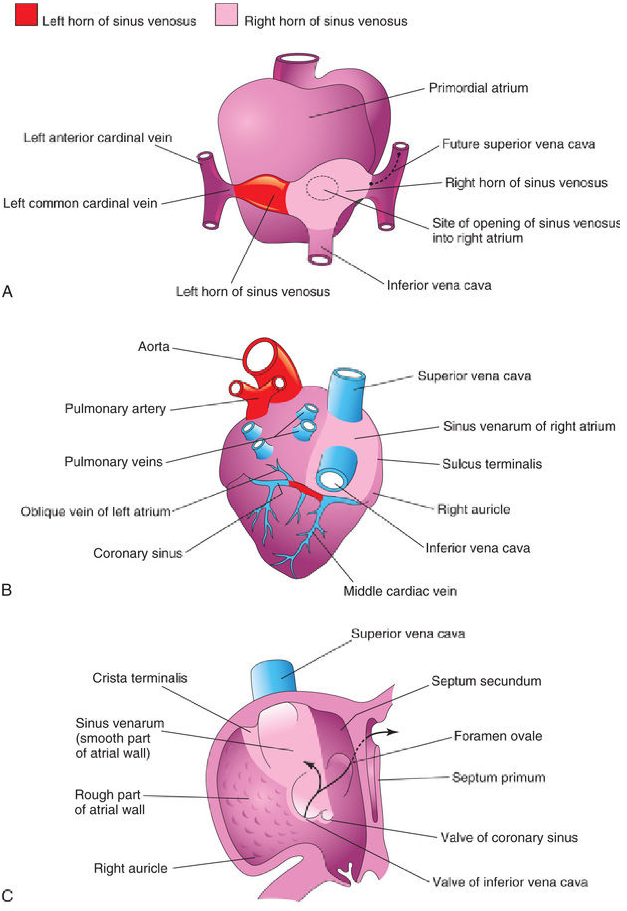
FIG 13.15 - Fate of the sinus venosus. (A) Dorsal view ~26 days showing primordial atrium and sinus venosus. (B) Dorsal view at 8 weeks after right horn incorporation showing the sinus venarum and coronary sinus. (C) Internal view of fetal right atrium showing the smooth (sinus venarum) and rough parts separated by the crista terminalis.
Sources:
- The Developing Human: Clinically Oriented Embryology (Moore & Persaud), Ch. 13
- Langman's Medical Embryology, Ch. 13 - Development of the Sinus Venosus and Septum Formation
This is a shared conversation. Sign in to Orris to start your own chat.