
COMPLEMENT SYSTEM The complement system received its name because it complements the action of antibody in killing bacteria. Early in this century Bordet discovered that if fresh serum containing antibacterial antibody was added to bacteria it would lyse the bacteria. However, if the serum was heated to 56C, it would not lyse the bacteria. Because the antibody activity was not destroyed by heating, he reasoned that there was another factor in the serum that was heat labile that complemented the activity of the antibody. We now know that this complementing activity was due to a group of proteins that forms a highly regulated enzyme cascade that can result in direct damage to infectious agents and the initiation of an inflammatory response to focus humoral and cellular defense mechanisms at the site of infection. Two related, but different, enzyme cascade pathways can initiate or “activate” the complement system. The classical pathway comprises four protein components (C1, C2, C3, and C4,) and is predominantly activated by the presence of IgM or IgG antibody bound to an antigen. When the antibody molecule binds to an antigen, a conformational change occurs in the antibody molecule exposing the complement binding site. The first component of complement classical pathway C1 (C1 consists of three separate proteins C1q, C1r, C1s bound together by calcium-dependent bonds) can then bind to the changed antibody molecule. The binding of C1 to the antibody molecule causes a conformational change in C1, which converts it to an enzyme. This new C1 enzyme can then cleave multiple molecules of C2 and C4. The split pieces of C2 and C4 can combine on a cell surface and form a second enzyme (C4b, 2b, 3b) called a C5 convertase, which can start the terminal pathway of complement by splitting C5 into C5a and C5b. The alternative pathway (so named because it was described after the classical pathway and therefore was considered to be an alternative to the classical pathway) involve four proteins (factors B and D, properdin, and C3, which is also a component of the classical pathway). The alternative pathway can be activated on some microbial surfaces in the absence of antibody and therefore can play an important role in natural immunity. The key to understanding the alternative pathway is to understand that C3 in the plasma spontaneously breaks down at a slow rate into C3a and C3b. If the C3b attaches to a foreign membrane, it will be stabilized by interacting with other components of the alternative complement pathway and lead to activation of the alternative pathway and eventually the terminal complement pathway. If the spontaneously formed C3b attaches to the surface of a normal host cell, molecules on the surface of the host cell can inactivate the C3b so that it cannot start the alternative complement pathway and damage the cell. If a small amount of C3b becomes stabilized by the other components of the alternative pathway on a bacterial cell surface, then it rapidly splits more C3 and amplifies the complement reaction. Each activation pathway involves the sequential activation of three different enzymes. Each active enzyme molecule can cleave many molecules of its substrate, thereby creating multiple active molecules of the next enzyme in the cascade. This sequential activation of enzymes gives the complement system one of its important properties: biological amplification. A small initiating stimulus (e.g. an antigen-antibody complex for the classical pathway or a suitable microbial surface for the alternative pathway) can lead to a vigorous response because it is amplified by the enzyme cascade. All of the proteins that participate in both the classical and alternative pathways are preformed but are circulating in an inactive state, which enables the complement system to be activated very rapidly after it is initiated. 75 The terminal pathway (also called the membrane attack pathway) involves five proteins (C5, C6, C7, C8, and C9) and can be activated by either the classical or alternative pathway. It is initiated when C5 is split into C5a and C5b by the third enzyme (C5 convertase) formed by either the classical or alternative pathway. C5a is a small peptide that diffuses away and has many important biological activities; C5b is a larger peptide that can bind to cell surfaces immediately adjacent to where it is formed. If the C5b binds to a cell membrane, the rest of the membrane attack complex self assembles without the need for further enzyme activity. One molecule each of C6, C7, and C8 binds to the C5b molecule, then up to 15 molecules of C9 bind to this complex and form a hollow cylinder of protein inserted into the cell membrane. This effectively produces a hole in the membrane, which allows water and ions to diffuse freely into and out of the cell. If there are enough of these membrane attack complex holes in the cell, the cell will eventually rupture as a result of increased pressure from water, which is osmotically attracted into the cell. The enzymes in the complement pathway typically cleave the substrate protein molecules into a small piece that diffuses away and a large piece that binds to surfaces very near the site of cleavage. The large pieces contribute to forming the next enzyme in the cascade (C4b, C2b, Bb, and C3b), act as an opsonin to facilitate phagocytosis of the particle (C3b), or are the starting point for the terminal pathway (C5b). The small cleavage products that diffuse away are important initiators of the inflammatory response. C3a, C4a, and C5a are called anaphylatoxins because they cause mast cell degranulation. This results in the release of histamine and other mediators, leading to vasodilation and increased vascular permeability. C5a is a strong chemoattractant for neutrophils. The end result is the diffusion of serum components (including antibody and more complement) and the egress of neutrophils into the tissues to help control the infectious agents that initiated the complement cascade. Because the complement system can be initiated rapidly and is self-amplifying, it is very important that its activation be carefully regulated. Uncontrolled, massive activation of the complement system can cause vascular damage and initiation of the coagulation system, leading to disseminated intravascular coagulation, which is rapidly fatal. Complement system activation is regulated by the fact that the activated enzymes have a short half-life, and the cleavage products that bind to membranes have only a very brief time in which to bind or they will lose their ability to do so. There is also a group of serum proteins that help to slow complement activation by inhibiting various complement components that bind to their surface or by accelerating their decay. This helps to prevent complement damage to normal cells. However, these protective molecules can be overwhelmed by massive activation of the complement system. C3a, C4a, and C5a are anaphylatoxins causing mast cell degranulation, vasodilatation and greater vascular permeability. C5a is chemotactic for neutrophils. In fact, it is the presence of the complement inhibitory molecules in normal host cell membranes that protect the host from the alternative complement pathway. The alternative pathway is continuously activated by spontaneous, low-level cleavage of C3 into C3a and C3b. Host cells have molecules that rapidly inactivate C3b before it can lead to amplification of the alternative pathway. Bacterial surfaces lack the inhibitory molecules, thus allowing the alternative pathway to amplify and attack the bacteria. The presence or absence of inhibitors is a crude method for the complement system to distinguish self from non-self. 76 Complement pathways The complement system is especially important for control of bacterial infections. People and animals that are deficient in key complement components (C3) are more susceptible to recurrent bacterial infections Sunmarize into flowcharted essay
The Complement System: A Flowcharted Essay
🔬 ORIGIN & NAMING
Bordet's Observation → Fresh serum lysed bacteria | Heated serum (56°C) did not ↓ Antibody survived heating, but a heat-labile factor did not ↓ This factor was named complement - because it complemented antibody activity
⚙️ TWO ACTIVATION PATHWAYS
PATH 1 - CLASSICAL PATHWAY
Antigen binds IgM or IgG antibody
        ↓
Conformational change in antibody exposes complement-binding site
        ↓
C1 complex (C1q + C1r + C1s, held by Ca²⁺ bonds) binds to antibody
        ↓
Conformational change in C1 → C1 becomes an ENZYME
        ↓
C1 cleaves C2 and C4
        ↓
C4b + C2b assemble on cell surface → form C3 CONVERTASE
        ↓
C3 convertase cleaves C3 → C3a (diffuses away) + C3b (binds surface)
        ↓
C4b + C2b + C3b form C5 CONVERTASE
        ↓
→ TERMINAL PATHWAY (see below)
PATH 2 - ALTERNATIVE PATHWAY
C3 in plasma spontaneously hydrolyzes (slow, continuous) → C3a + C3b
        ↓
        ┌─────────────────────────────────────────┐
        │                                         │
C3b binds HOST cell surface            C3b binds FOREIGN (microbial) surface
        │                                         │
Host inhibitory molecules              No inhibitory molecules present
INACTIVATE C3b                                    │
        │                               C3b STABILIZED by Factors B & D
No cascade → HOST PROTECTED              and Properdin
                                                  │
                                    C3b amplifies → cleaves more C3
                                                  │
                                    C3b + Bb form C5 CONVERTASE
                                                  │
                                    → TERMINAL PATHWAY (see below)
THE PRINCIPLE OF AMPLIFICATION
Small initiating stimulus (1 immune complex or 1 microbial surface)
        ↓
Each active enzyme cleaves MANY substrate molecules
        ↓
Each substrate molecule becomes the NEXT enzyme
        ↓
Exponential amplification → VIGOROUS RESPONSE from tiny trigger
💥 TERMINAL PATHWAY (Membrane Attack Pathway)
C5 CONVERTASE cleaves C5
        ↓
C5a (small) → diffuses away    |    C5b (large) → binds adjacent cell membrane
        ↓                                          ↓
[Inflammatory effects]          C6 binds C5b
                                        ↓
                                C7 binds → complex inserts into membrane
                                        ↓
                                C8 binds → initiates pore formation
                                        ↓
                              Up to 15 molecules of C9 bind
                                        ↓
                         MEMBRANE ATTACK COMPLEX (MAC) formed
                         (hollow protein cylinder in cell membrane)
                                        ↓
                    Free diffusion of water & ions into/out of cell
                                        ↓
                        Osmotic influx of water → cell RUPTURE & LYSIS
🔥 INFLAMMATORY MEDIATORS (Small Cleavage Fragments)
| Fragment | Activity |
|---|---|
| C3a | Anaphylatoxin → mast cell degranulation → histamine release → vasodilation + increased vascular permeability |
| C4a | Anaphylatoxin → same as C3a (weaker) |
| C5a | Anaphylatoxin (strongest) + potent chemoattractant for neutrophils |
C3a / C4a / C5a released
        ↓
Mast cell degranulation → Histamine + other mediators
        ↓
Vasodilation + increased vascular permeability
        ↓
Serum proteins (antibody, more complement) flood into tissue
        ↓
C5a recruits NEUTROPHILS to infection site
        ↓
Amplified local immune defense at infection site
🛡️ LARGE CLEAVAGE FRAGMENTS (Surface-Bound)
| Fragment | Role |
|---|---|
| C4b, C2b, Bb | Build the next enzyme (convertases) in the cascade |
| C3b | Opsonin - coats pathogens to facilitate phagocytosis |
| C5b | Starting point for MAC assembly |
⚖️ REGULATION OF THE COMPLEMENT SYSTEM
- Vascular damage
- Coagulation cascade activation
- Disseminated Intravascular Coagulation (DIC) → rapidly fatal
Activated enzymes have SHORT HALF-LIVES → self-limiting
        +
Membrane-binding cleavage products have a BRIEF WINDOW to bind
        +
Serum inhibitor proteins slow activation and accelerate decay of components
        +
Host cell membrane INHIBITORY MOLECULES inactivate C3b on self-surfaces
🧩 SUMMARY: THE BIG PICTURE
INFECTION DETECTED
      │
      ├─── Antibody present? ──YES──► CLASSICAL PATHWAY
      │                                       │
      └─── Microbial surface? ──YES──► ALTERNATIVE PATHWAY
                                              │
                              Both converge at C3 CLEAVAGE
                                              │
                               ┌──────────────┴──────────────┐
                               │                             │
                          C3a/C4a/C5a                      C3b (opsonin)
                        (inflammation,                  Enhances phagocytosis
                        neutrophil recruitment)               │
                               │                             │
                          C5 CONVERTASE formed               │
                               │                             │
                        TERMINAL PATHWAY                      │
                               │                             │
                         MAC → CELL LYSIS                     │
                                         └───────────────────┘
                                          PATHOGEN ELIMINATED
🏥 CLINICAL RELEVANCE
- C3 deficiency → recurrent bacterial infections (C3 is the central convergence point of both pathways)
- Deficiency in terminal components (C5-C9) → susceptibility to Neisseria infections specifically
- Unregulated complement → DIC, organ damage, shock
- Complement inhibitor deficiency (e.g., CD59, DAF) → host cells attacked → as in Paroxysmal Nocturnal Hemoglobinuria (PNH)
Create a visual infographic poster of the complement cascade
A detailed scientific infographic poster titled "THE COMPLEMENT CASCADE" showing the full complement system pathway. Layout: Dark navy blue background with bright, color-coded pathways. TOP SECTION - Two Activation Pathways side by side: LEFT (Classical Pathway - blue theme): Shows antigen-antibody complex (IgG/IgM) → C1q/C1r/C1s complex binding → C1 becomes enzyme → cleaves C2 and C4 → C4b+C2b form C3 Convertase (C4b2b) → arrows pointing down RIGHT (Alternative Pathway - green theme): Shows microbial surface with no inhibitors → spontaneous C3 hydrolysis → C3b binds foreign surface → stabilized by Factor B, Factor D, Properdin → forms C3 Convertase (C3bBb) → arrows pointing down MIDDLE SECTION - Convergence Point (bright yellow/gold): Both pathways converge at a large central node labeled "C3 CLEAVAGE" Arrow left: C3a (small fragment, orange glow) labeled "ANAPHYLATOXIN - Mast cell degranulation, Vasodilation, Vascular permeability" Arrow right: C3b (large fragment, gold) labeled "OPSONIN - Coats bacteria for phagocytosis" Below: C5 Convertase forms → cleaves C5 → C5a + C5b C5a labeled "Strongest anaphylatoxin + Neutrophil chemoattractant" (red/orange) C5b labeled "Initiates MAC" (purple) BOTTOM SECTION - Terminal Pathway / MAC (purple/red theme): Linear assembly diagram: C5b → C5b+C6 → C5b+C6+C7 → C5b+C6+C7+C8 → C5b+C6+C7+C8+C9(x15) Final image: Cross-section of a cell membrane showing a hollow cylinder pore (MAC) inserted through it, with water rushing in and the cell swelling/lysing Labeled: "MEMBRANE ATTACK COMPLEX (MAC) → Cell Lysis" RIGHT SIDE PANEL - Regulatory Mechanisms (grey/silver): Box titled "REGULATION" listing: Short enzyme half-lives, Brief binding windows, Serum inhibitor proteins, Host cell CD59/DAF inhibitors Small diagram showing C3b inactivated on host cell vs amplified on bacterial cell BOTTOM BAR - Clinical Notes (red alert box): "C3 Deficiency → Recurrent bacterial infections | Terminal component deficiency → Neisseria susceptibility | Unregulated activation → DIC" LEGEND (bottom left): Color-coded key for Classical (blue), Alternative (green), Terminal (purple), Inflammatory mediators (orange) Style: Clean medical textbook illustration style, white labels with colored arrows, professional scientific poster, all text legible, high detail
400, message: Got more than 4194304 bytes when reading: b'data: {"candidates": [{"content": {"role": "model","parts": [{"inlineData": {"mimeType": "image/png"...'.
We encountered an error while processing your request. Please try again or reach out to us for help.
Retry
Scientific infographic poster of the complement cascade system on dark navy background. Three color-coded pathways: Classical pathway (blue, left side) showing antibody-antigen complex activating C1q/C1r/C1s then cleaving C2 and C4 to form C3 convertase. Alternative pathway (green, right side) showing microbial surface with C3b stabilized by Factor B, D, and Properdin forming C3 convertase. Both pathways converge at central C3 cleavage node (gold/yellow). C3a fragment (orange) branches off labeled anaphylatoxin causing mast cell degranulation and vasodilation. C3b fragment (yellow) labeled opsonin for phagocytosis. Below convergence: C5 convertase cleaves C5 into C5a (red, chemotactic for neutrophils) and C5b (purple). Terminal pathway (purple, bottom center): sequential assembly C5b plus C6 plus C7 plus C8 plus 15xC9 forming Membrane Attack Complex MAC shown as cylindrical pore inserted into cell membrane causing cell lysis. Right side box shows regulation mechanisms. Bottom clinical notes bar. Clean medical illustration style, white text labels, professional scientific poster design
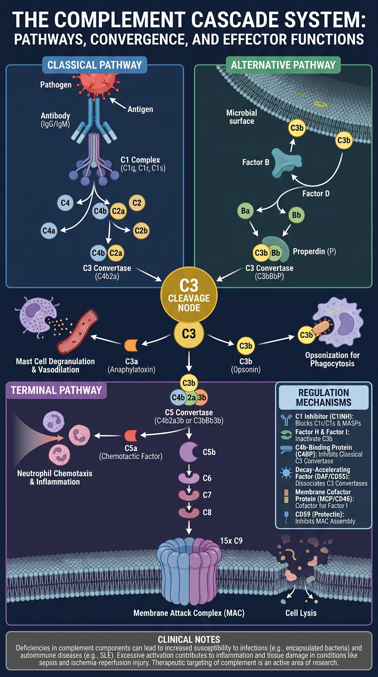
- Classical Pathway (blue, left) - antibody-antigen complex triggering C1 activation through to C3 convertase
- Alternative Pathway (green, right) - microbial surface C3b stabilization via Factors B, D, and Properdin
- Central convergence at C3 cleavage (gold), with C3a as anaphylatoxin and C3b as opsonin branching off
- C5 convertase cleaving into C5a (chemoattractant) and C5b (MAC initiator)
- Terminal Pathway / MAC (purple, bottom) - the sequential C5b-C6-C7-C8-C9 assembly forming the membrane pore and cell lysis
- Regulation mechanisms and clinical notes included
Lectin pathway not included
Scientific infographic poster titled "THE COMPLEMENT CASCADE - Three Activation Pathways" on dark navy blue background. Three clearly labeled parallel activation pathways at the top, all converging downward to a shared terminal pathway. LEFT - Classical Pathway (blue): Antigen-Antibody complex (IgG/IgM) → conformational change exposes complement site → C1q C1r C1s complex binds (calcium dependent) → C1 activated as enzyme → cleaves C4 and C2 → C4b+C2b = C3 Convertase CENTER - Lectin Pathway (teal/cyan): Mannose-binding lectin MBL binds mannose on microbial surface → MBL-associated serine proteases MASP-1 and MASP-2 activated → MASP-2 cleaves C4 and C2 → C4b+C2b = C3 Convertase (same as classical) RIGHT - Alternative Pathway (green): Spontaneous C3 hydrolysis in plasma → C3b deposits on microbial surface (no inhibitors present) → Factor B binds C3b → Factor D cleaves Factor B → C3bBb stabilized by Properdin = C3 Convertase All three pathways converge with a bold downward arrow to central gold node: C3 CLEAVAGE C3 → C3a (orange bubble, anaphylatoxin, mast cell degranulation, vasodilation) and C3b (yellow bubble, opsonin, phagocytosis) C3b joins convertase → C5 Convertase → cleaves C5 C5a (red, anaphylatoxin, neutrophil chemoattractant) and C5b (purple, initiates MAC) BOTTOM - Terminal Pathway (purple): C5b + C6 + C7 + C8 + poly-C9 → Membrane Attack Complex MAC shown as barrel pore in lipid bilayer membrane → osmotic water influx → cell lysis Side panel: Regulation box listing host inhibitory proteins CD55 DAF and CD59 protecting self cells. Clinical pearls box: C3 deficiency leads to recurrent infections, MAC deficiency leads to Neisseria susceptibility. Clean professional medical illustration, bright color-coded arrows, white bold labels, all three pathways visually distinct, high detail scientific poster style
400, message: Got more than 4194304 bytes when reading: b'data: {"candidates": [{"content": {"role": "model","parts": [{"inlineData": {"mimeType": "image/png"...'.
We encountered an error while processing your request. Please try again or reach out to us for help.
Retry
Medical infographic poster: Complete Complement Cascade with all three activation pathways on dark navy background. Top row shows three pathways side by side. Left: Classical Pathway in blue - IgG/IgM antibody bound to antigen activates C1qrs complex, cleaves C4 and C2, forms C3 convertase C4b2b. Center: Lectin Pathway in teal - MBL binds mannose on microbe surface, activates MASP1 MASP2, cleaves C4 and C2, forms same C3 convertase C4b2b. Right: Alternative Pathway in green - spontaneous C3b on microbial surface, Factor B and Factor D and Properdin stabilize, forms C3 convertase C3bBb. Three arrows converge on central C3 node. C3 splits into C3a anaphylatoxin orange and C3b opsonin yellow. Then C5 convertase forms, splits C5 into C5a red chemotaxin and C5b purple. Bottom: terminal pathway showing MAC assembly C5b C6 C7 C8 C9 forming membrane pore causing cell lysis. Clean scientific diagram white labels colored pathways
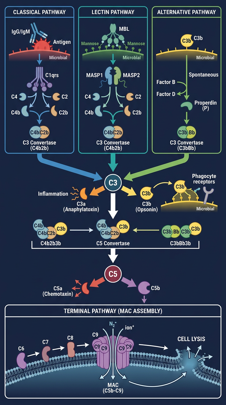
| Pathway | Trigger | Key Proteins |
|---|---|---|
| Classical (blue) | Antigen-IgG/IgM complex | C1q, C1r, C1s → C4, C2 |
| Lectin (teal) | MBL binds mannose on microbe | MBL, MASP-1, MASP-2 → C4, C2 |
| Alternative (green) | Spontaneous C3b on microbial surface | Factor B, Factor D, Properdin |
Give each patheay separately in each image
Medical infographic poster titled "CLASSICAL PATHWAY - Complement System" on dark navy blue background. Step by step vertical flowchart with blue color theme. Step 1: Antigen binds IgG or IgM antibody, showing Y-shaped antibody bound to bacterial antigen. Step 2: Conformational change in antibody exposes complement binding site. Step 3: C1 complex binds - showing C1q C1r C1s held together by calcium ions. Step 4: C1 undergoes conformational change and becomes an active enzyme. Step 5: Active C1 cleaves C4 into C4a and C4b, and cleaves C2 into C2a and C2b. C4a and C2a labeled as small fragments that diffuse away. Step 6: C4b and C2b assemble on cell surface forming C3 Convertase C4b2b shown as enzyme complex on membrane. Step 7: C3 Convertase cleaves C3 into C3a small orange fragment labeled anaphylatoxin causing mast cell degranulation vasodilation and C3b large yellow fragment labeled opsonin for phagocytosis. Step 8: C3b joins complex forming C5 Convertase C4b2b3b. Arrow pointing down labeled "feeds into Terminal Pathway". Side notes in boxes: Requires antibody so part of adaptive immunity, Activated by IgM or IgG, C1 held by calcium bonds so EDTA inhibits. Clean professional medical diagram, white bold labels, blue arrows, high detail scientific illustration style
Medical infographic poster titled "LECTIN PATHWAY - Complement System" on dark navy blue background. Step by step vertical flowchart with teal and cyan color theme. Step 1: Microbial surface showing mannose sugar residues on bacteria or fungi surface, labeled pattern recognition. Step 2: Mannose-Binding Lectin MBL circulating in blood recognizes and binds to mannose patterns on microbial surface. MBL shown as bouquet-shaped protein. Step 3: MBL-Associated Serine Proteases MASP-1 and MASP-2 are recruited and activated. Step 4: MASP-2 cleaves C4 into C4a diffuses away and C4b binds surface. MASP-2 also cleaves C2 into C2a and C2b. Step 5: C4b and C2b assemble forming C3 Convertase C4b2b on microbial membrane surface. Step 6: C3 Convertase cleaves C3 into C3a small orange fragment labeled anaphylatoxin and C3b large yellow fragment labeled opsonin. Step 7: C5 Convertase C4b2b3b forms. Arrow pointing down labeled feeds into Terminal Pathway. Side notes: Does not require antibody so part of innate immunity, Activated early in infection before antibody production, MBL is an acute phase protein. Clean professional medical diagram, white bold labels, teal cyan arrows, high detail scientific illustration
Medical infographic poster titled "ALTERNATIVE PATHWAY - Complement System" on dark navy blue background. Step by step vertical flowchart with green color theme. Step 1: Spontaneous low-level C3 hydrolysis in plasma shown as C3 molecule splitting slowly into C3a and C3b continuously, labeled tick-over mechanism. Step 2: Two branches diverging. Left branch: C3b lands on HOST cell surface, host inhibitory molecules CD55 DAF and CD59 rapidly inactivate C3b, labeled host protected. Right branch: C3b lands on MICROBIAL surface, no inhibitory molecules present, C3b survives and binds. Step 3 right branch continues: Factor B binds to surface-bound C3b forming C3bB complex. Step 4: Factor D cleaves Factor B in the complex, releasing Ba small fragment and Bb stays bound forming C3bBb. Step 5: Properdin binds and stabilizes C3bBb complex preventing decay. C3bBb is now active C3 Convertase. Step 6: C3 Convertase cleaves many more C3 molecules into C3a orange anaphylatoxin and C3b yellow opsonin, showing amplification loop with curved arrow. Step 7: C3b joins convertase forming C5 Convertase C3bBbC3b. Arrow down labeled feeds into Terminal Pathway. Side notes: No antibody required innate immunity, Amplification loop provides rapid escalation, Distinguishes self from non-self via inhibitors. Clean professional medical diagram, white bold labels, green arrows, scientific illustration style
429 Too Many Requests. {'message': '{\n "error": {\n "code": 429,\n "message": "Resource exhausted. Please try again later. Please refer to https://cloud.google.com/vertex-ai/generative-ai/docs/error-code-429 for more details.",\n "status": "RESOURCE_EXHAUSTED"\n }\n}\n', 'status': 'Too Many Requests'}
We encountered an error while processing your request. Please try again or reach out to us for help.
Retry
Medical infographic poster titled "LECTIN PATHWAY - Complement System" on dark navy blue background. Vertical flowchart with teal and cyan color theme. Step 1: Microbial surface with mannose sugar residues on bacteria. Step 2: Mannose-Binding Lectin MBL binds to mannose patterns, MBL shown as bouquet-shaped protein. Step 3: MASP-1 and MASP-2 serine proteases recruited and activated. Step 4: MASP-2 cleaves C4 into C4a and C4b, and C2 into C2a and C2b. Step 5: C4b and C2b form C3 Convertase on membrane surface. Step 6: C3 Convertase cleaves C3 into C3a anaphylatoxin orange and C3b opsonin yellow. Step 7: C5 Convertase forms, feeds into Terminal Pathway. Side notes: innate immunity, no antibody required, MBL is acute phase protein. White bold labels, teal arrows, clean scientific medical illustration
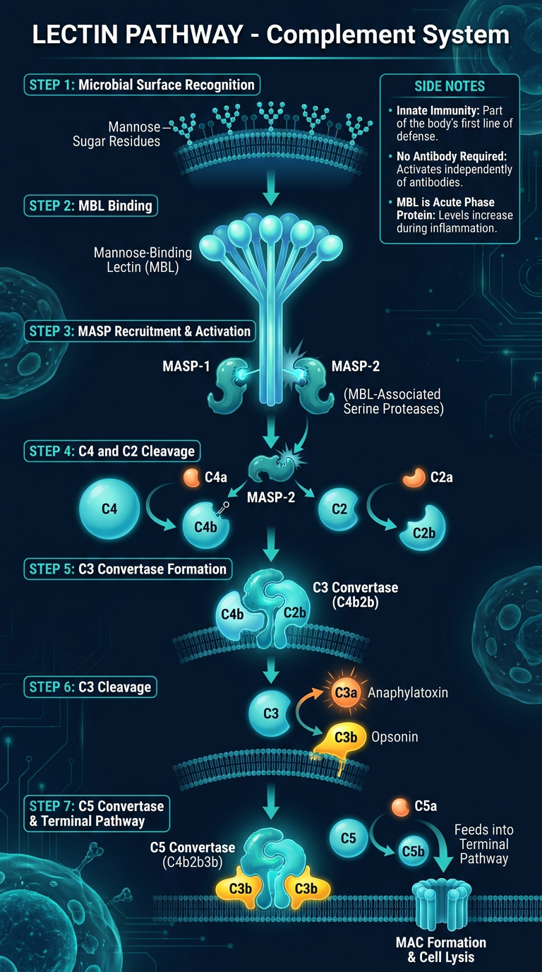
| Poster | Trigger | Immunity Type | Color |
|---|---|---|---|
| Classical | Antigen-IgG/IgM complex | Adaptive | Blue |
| Lectin | MBL binds mannose on microbe | Innate | Teal |
| Alternative | C3b on microbial surface (no inhibitors) | Innate | Green |
Retry 1 and 2
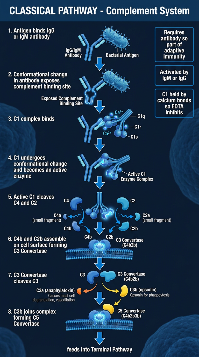
Medical infographic poster titled "CLASSICAL PATHWAY - Complement System" on dark navy blue background. Vertical flowchart blue color theme. Shows: antigen bound to IgG IgM antibody, conformational change exposes complement site, C1q C1r C1s complex binds held by calcium ions, C1 becomes active enzyme, C1 cleaves C4 into C4a diffuses away and C4b binds surface, C1 cleaves C2 into C2a and C2b, C4b plus C2b assemble as C3 Convertase on cell membrane, C3 Convertase cleaves C3 into C3a anaphylatoxin orange causing mast cell degranulation and vasodilation, and C3b opsonin yellow coating bacteria for phagocytosis, C3b joins to form C5 Convertase C4b2b3b, arrow down to Terminal Pathway. Side box: requires IgG or IgM antibody, part of adaptive immunity, calcium dependent C1 complex. Professional clean medical diagram white labels blue arrows
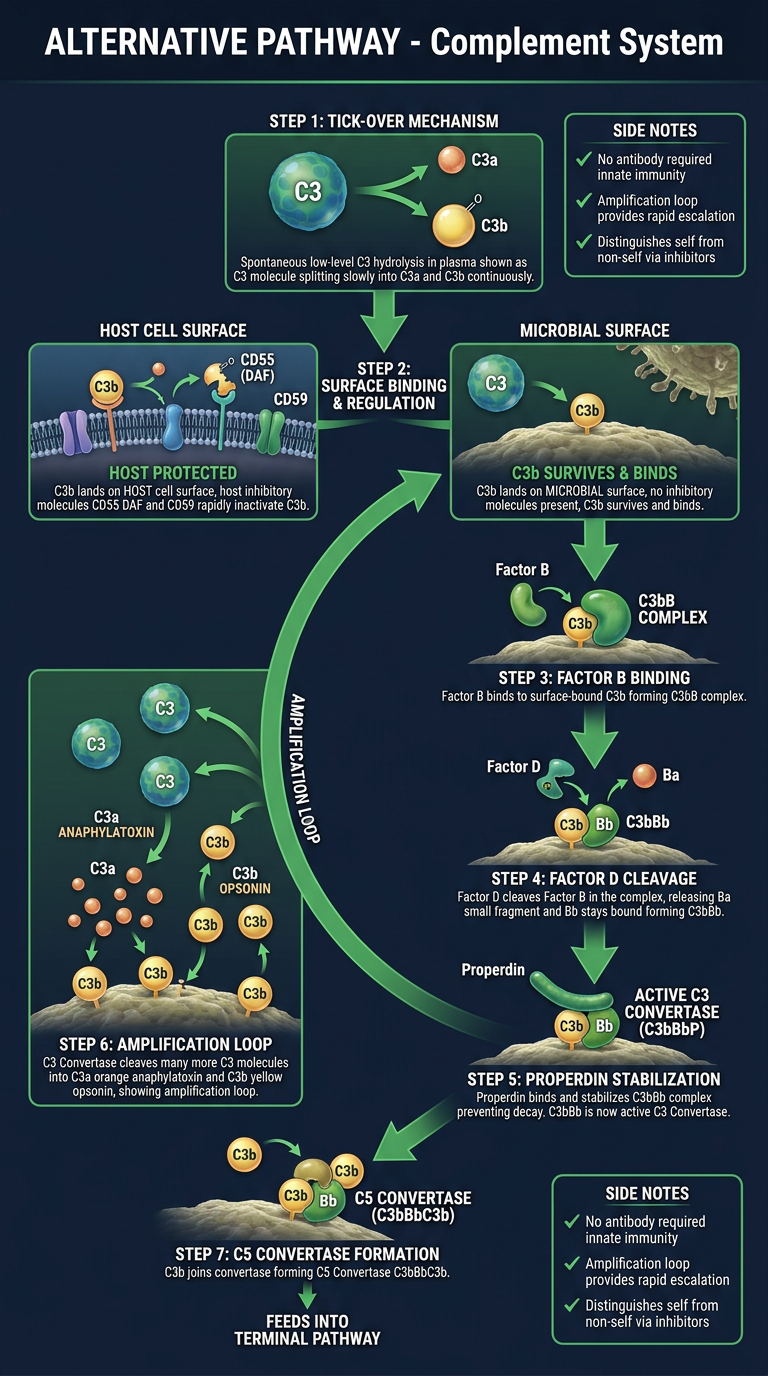
Medical infographic poster titled "ALTERNATIVE PATHWAY - Complement System" on dark navy blue background. Vertical flowchart green color theme. Shows: spontaneous slow C3 hydrolysis in plasma labeled tick-over, C3 splits into C3a and C3b continuously, two branches: left branch C3b on host cell surface inactivated by CD55 and CD59 inhibitors labeled host protected, right branch C3b on microbial surface survives because no inhibitors present, Factor B binds C3b forming C3bB complex, Factor D cleaves Factor B releasing Ba fragment, C3bBb active C3 Convertase, Properdin binds and stabilizes complex, amplification loop showing C3 Convertase cleaves many more C3 molecules, C3a orange anaphylatoxin and C3b yellow opsonin produced, C5 Convertase C3bBbC3b forms, arrow to Terminal Pathway. Side box: no antibody needed innate immunity, amplification loop key feature, self vs non-self discrimination via inhibitors. Professional clean medical diagram white labels green arrows
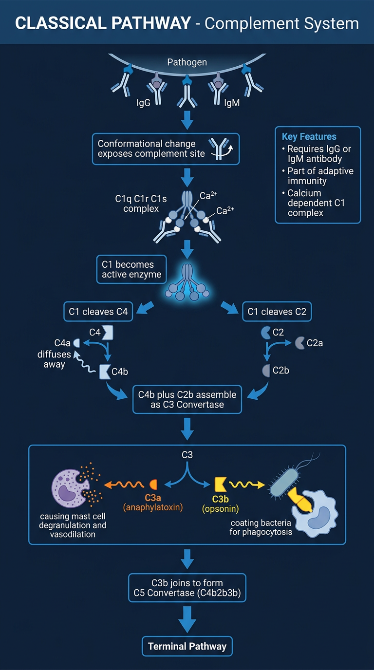
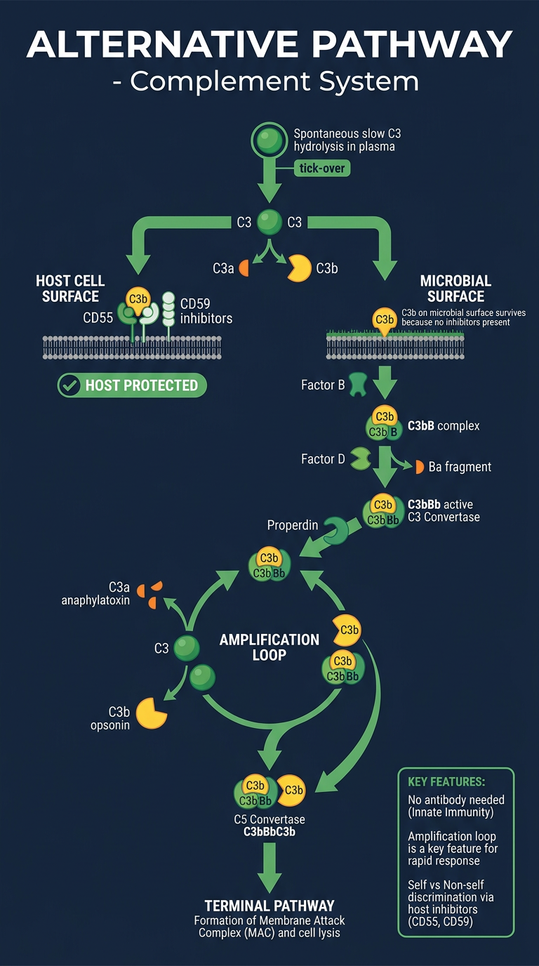
| # | Pathway | Image |
|---|---|---|
| 1 | Classical (blue) | Above |
| 2 | Alternative (green) | Above |
| 3 | Lectin (teal) | Generated previously |
Terminal pathway
Medical infographic poster titled "TERMINAL PATHWAY - Membrane Attack Complex (MAC)" on dark navy blue background. Purple and red color theme. Vertical flowchart showing: Step 1: Both classical and alternative pathways feed in, showing C5 Convertase enzyme complex on cell membrane surface. Step 2: C5 Convertase cleaves C5 into two fragments. C5a small fragment red labeled strongest anaphylatoxin, causes mast cell degranulation, vasodilation, increased vascular permeability, potent neutrophil chemoattractant, diffuses away into tissue. C5b large fragment purple labeled binds adjacent cell membrane immediately. Step 3: C5b anchored to membrane, C6 binds to C5b forming C5b6 complex. Step 4: C7 binds forming C5b67 complex, begins inserting into lipid bilayer membrane. Step 5: C8 binds forming C5b678, initiates slow membrane penetration creating small pore. Step 6: Up to 15 molecules of C9 bind sequentially, polymerizing around the complex, forming a large hollow cylindrical barrel pore through the membrane. Labeled Membrane Attack Complex MAC. No further enzyme activity needed, self assembles. Step 7: Cross section diagram of MAC pore inserted through phospholipid bilayer membrane showing hollow channel. Water and ions freely diffuse in and out shown by arrows. Osmotic water influx causes cell swelling. Step 8: Cell ruptures and lyses shown as bursting cell. Labeled cell death by osmotic lysis. Side information boxes: MAC components are C5b C6 C7 C8 C9. Effective against gram negative bacteria. Deficiency in C5-C9 increases susceptibility to Neisseria meningitidis and Neisseria gonorrhoeae. Regulation by CD59 protectin on host cells blocks C9 polymerization. Bottom note: C5a biological effects listed with icons for inflammation chemotaxis and mast cell activation. Clean professional medical diagram, white bold labels, purple arrows, detailed scientific illustration style, all steps clearly numbered
400, message: Got more than 4194304 bytes when reading: b'data: {"candidates": [{"content": {"role": "model","parts": [{"inlineData": {"mimeType": "image/png"...'.
We encountered an error while processing your request. Please try again or reach out to us for help.
Retry
Medical infographic poster "TERMINAL PATHWAY - Membrane Attack Complex" on dark navy background, purple color theme. Numbered vertical flowchart: 1) C5 Convertase cleaves C5 into C5a red fragment labeled anaphylatoxin and neutrophil chemoattractant, and C5b purple fragment binds cell membrane. 2) C6 binds C5b. 3) C7 binds forming C5b67 inserting into lipid bilayer. 4) C8 binds initiating pore. 5) Up to 15 C9 molecules polymerize forming hollow cylindrical MAC pore through membrane. 6) Cross-section diagram of MAC barrel pore in phospholipid bilayer with water ions flowing freely through it. 7) Osmotic water influx causes cell swelling and lysis. Side box: MAC deficiency causes Neisseria susceptibility. CD59 on host cells blocks C9 and protects self. White labels purple arrows scientific illustration style
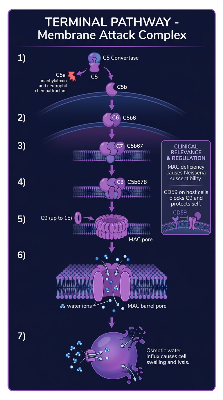
| # | Pathway | Key Feature |
|---|---|---|
| 1 | Classical (blue) | Antibody-dependent, adaptive immunity |
| 2 | Alternative (green) | Spontaneous C3b, innate, amplification loop |
| 3 | Lectin (teal) | MBL binds mannose, innate, no antibody |
| 4 | Terminal / MAC (purple) | C5b-C9 pore assembly, cell lysis |