
Hodgkin lymphoma
Reading File
Now I'll search the medical library and PubMed in parallel for Hodgkin lymphoma.
Finding Sources
Searching PubMed
"Hodgkin lymphoma"[MeSH Terms] AND treatment
Good findings. Now I'll read the most comprehensive sources - Robbins Pathology for pathology/classification, Goldman-Cecil for clinical management, and search for more content in parallel.
Reading File
Reading File
Searching PubMed
"Hodgkin lymphoma"[MeSH Terms] AND treatment
Let me read more from Robbins Pathology for morphology/RS cell details, and find the dedicated Goldman-Cecil chapter on Hodgkin lymphoma.
Reading File
Finding Sources
Excellent content from Robbins. Let me fetch the RS cell image and read more morphology + the Goldman-Cecil dedicated chapter.
Reading File
Reading File
Loading Image
Excellent content. Let me now read the Goldman-Cecil chapter continuation (staging, treatment) and fetch key histology images.
Reading File
Loading Image
Now let me read the treatment/staging section from Goldman-Cecil.
Reading File
I now have comprehensive content to write a thorough response. Let me compile everything.
Hodgkin Lymphoma
Overview
Hodgkin lymphoma (HL) is a B-cell lymphoma defined by the presence of distinctive neoplastic Reed-Sternberg (RS) cells within a background of reactive inflammatory and immune cells. It is one of the most curable cancers in medicine, even in advanced stages. The two major categories are classic Hodgkin lymphoma (cHL) (~90% of cases) and nodular lymphocyte-predominant Hodgkin lymphoma (NLPHL) (~10%).
- Goldman-Cecil Medicine, p. 1964
Epidemiology
-
~30,000 new cases/year in North America and Europe
-
Incidence: ~2.7 per 100,000; mortality: ~0.5 per 100,000
-
Bimodal age distribution: early peak at 25-30 years, second peak at >50 years
-
Slightly more common in men; higher incidence in Whites vs. Blacks; very rare in Asian populations
-
Highest incidence in the US, Canada, Switzerland, and northern Europe
-
Cumulative lifetime risk: ~1 in 250-300 in North America
-
Goldman-Cecil Medicine, p. 1964
Pathobiology and Etiology
The exact cause remains unknown. The leading etiologic suspect is Epstein-Barr virus (EBV):
- EBV DNA and proteins are detected in RS cells in up to 70% of mixed-cellularity cases and a smaller fraction of other classic subtypes
- The EBV integration site is identical in all RS cells within a given patient, indicating infection precedes clonal transformation
- A history of infectious mononucleosis raises the risk of HL 2-3x; this elevated risk persists for >20 years after the EBV illness
- EBV likely is one of several steps in transformation, particularly for mixed-cellularity subtype
HL is not associated with radiation exposure, chemicals, or health care-related occupational exposure.
- Robbins & Kumar Basic Pathology, p. 416; Goldman-Cecil Medicine, p. 1964
Cell of Origin
Microdissection studies established that every RS cell in a given case carries the same immunoglobulin gene rearrangements with somatic hypermutation, proving that HL arises from germinal center B cells. Despite this B-cell origin, RS cells paradoxically down-regulate most B-cell markers.
Reed-Sternberg Cell
The RS cell is the pathognomonic neoplastic cell of HL.
Morphology:
- Large cell, 15-45 μm in diameter
- Enormous multilobate nucleus with exceptionally prominent nucleoli
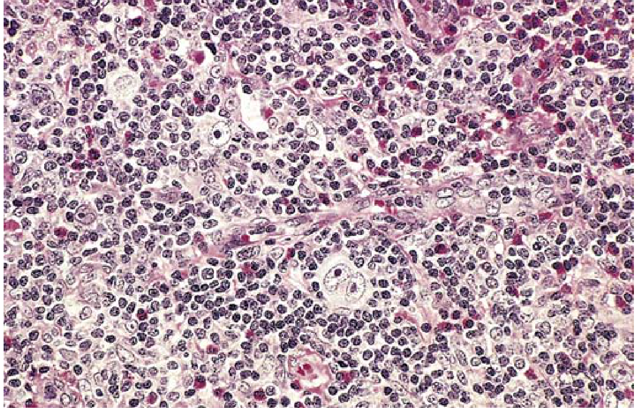
- Binucleate cells with mirror-image lobes, each with a large acidophilic nucleolus surrounded by a clear zone - the classic "owl-eye" appearance
- Abundant, slightly eosinophilic cytoplasm
Immunophenotype of classic RS cells:
| Marker | Status |
|---|---|
| CD30 | Positive (90-100%) |
| CD15 | Positive (75-85%) |
| PAX5 (BSAP) | Positive (>90%) |
| CD45 (LCA) | Negative |
| CD20 | Variable (~40% positive) |
| T-cell markers | Negative |
In NLPHL, the neoplastic cells are called lymphocytic and histiocytic (L&H) cells or "popcorn cells", which express CD20 and other germinal center B-cell markers but are CD15-negative and CD30-variable.
- Robbins & Kumar Basic Pathology, p. 417-418; Goldman-Cecil Medicine, p. 1964-1965
Histology of classic HL - mixed-cellularity type showing a Reed-Sternberg cell (large, binucleate, owl-eye nucleoli) surrounded by eosinophils, lymphocytes, and histiocytes:
Classification: Five Subtypes
1. Nodular Sclerosis (most common, ~70%)
- Equally frequent in men and women
- Typical age: adolescents and young adults
- Predilection for lower cervical, supraclavicular, and mediastinal lymph nodes
- Morphology: lacunar cells (RS variant with pale cytoplasm that retracts in formalin-fixed tissue, leaving nucleus in an empty space/lacune); broad bands of collagen subdivide tumor into nodules
- Prognosis: excellent
2. Mixed Cellularity (~20-25%)
- More common in men; median age slightly older
- Strongly associated with EBV (up to 70%)
- Classic binucleate RS cells in a mixed background of eosinophils, lymphocytes, plasma cells, and histiocytes
- Often presents at a more advanced stage
3. Lymphocyte-Rich (<5%)
- Must be distinguished from NLPHL
- Good prognosis; RS cells set in a predominantly lymphocytic background
4. Lymphocyte Depletion (rare, <1%)
- Older patients; often HIV-associated
- Paucity of lymphocytes; abundant RS cells or fibrosis
- Poorest prognosis among the classic subtypes; often presents in advanced stage
5. Nodular Lymphocyte Predominant (NLPHL, ~10%)
-
Separate entity from classic HL
-
Characteristic "popcorn" L&H cells expressing CD20, CD79a, BCL6
-
CD15-negative; indolent course; late relapses can occur
-
Robbins & Kumar Basic Pathology, p. 417-419; Goldman-Cecil Medicine, p. 1964-1965
Immune Evasion Mechanism
RS cells are a cardinal example of tumor immune escape:
-
Loss of β2-microglobulin function → failure to express class I MHC → CD8+ T cells cannot recognize tumor
-
High expression of PD-L1 and PD-L2 (encoded on chromosome 9q24, which is often amplified) → engagement of PD-1 on T cells suppresses anti-tumor immune response
-
This is the mechanistic basis for the remarkable clinical efficacy of anti-PD-1 antibodies (pembrolizumab, nivolumab) in relapsed/refractory HL
-
Robbins & Kumar Basic Pathology, p. 417-419
Clinical Features
Presentation:
- Painless lymphadenopathy is the cardinal feature (often cervical, supraclavicular)
- Mediastinal involvement is common in the nodular sclerosis subtype and may cause cough, dyspnea, or SVC syndrome
- Alcohol-induced pain at sites of disease (classic but uncommon)
B Symptoms (systemic):
- Unexplained fever >38°C (recurrent)
- Drenching night sweats
- Unexplained weight loss >10% body weight over 6 months
Other features:
- Pruritus (can be severe)
- Anemia
- Pel-Ebstein fever (cyclical high fevers, characteristic but rare)
Spread pattern: HL spreads in a stepwise, contiguous fashion through anatomically adjacent lymph node regions - unlike NHL, which spreads hematogenously. This pattern guided the historical use of involved-field radiation.
Staging: Modified Ann Arbor System
| Stage | Involvement |
|---|---|
| I | Single lymph node region (I) or one extralymphatic site (IE) |
| II | Two or more lymph node regions on the same side of the diaphragm (II), or local extralymphatic extension plus one or more lymph node regions, same side (IIE) |
| III | Lymph node regions on both sides of the diaphragm (III); may include local extralymphatic extension (IIIE) |
| IV | Diffuse involvement of one or more extralymphatic organs |
Suffix A: No B symptoms
Suffix B: B symptoms present (fever, night sweats, or weight loss)
- Goldman-Cecil Medicine, p. 1965
Workup and Staging Investigations
-
Full history and physical examination (lymphadenopathy, organomegaly)
-
Complete blood count, ESR
-
LFTs, renal function, LDH, alkaline phosphatase, serum albumin, protein electrophoresis
-
HIV, hepatitis B and C serology
-
Chest X-ray (PA and lateral)
-
CT scan of neck, thorax, abdomen, and pelvis with contrast
-
FDG-PET/CT - now mandatory; more sensitive and specific than CT alone for staging and response assessment; has replaced bone marrow biopsy for staging purposes
-
Goldman-Cecil Medicine, p. 1965-1966
Treatment
Limited Stage (Stages I and II, favorable)
- Brief combination chemotherapy (typically 2-4 cycles) is the cornerstone
- Standard regimen: ABVD (doxorubicin, bleomycin, vinblastine, dacarbazine) for 3-4 months
- Involved-site radiation therapy (ISRT) may be added in selected patients (bulky disease or inadequate PET response after chemotherapy)
- PET/CT after 2 cycles guides further therapy (response-adapted approach)
- 5-year OS: >90% for stage IA/IIA
Advanced Stage (Stages III and IV)
- 6 months of ABVD or escalated BEACOPP (bleomycin, etoposide, doxorubicin, cyclophosphamide, vincristine, procarbazine, prednisone)
- Escalated BEACOPP gives higher complete remission rate but more toxicity; ABVD remains standard in most centers
- Brentuximab vedotin (anti-CD30 antibody-drug conjugate) + AVD is an alternative first-line regimen for advanced disease
- Interim PET/CT after 2-4 cycles stratifies subsequent treatment intensity
- 5-year disease-free survival for stage IVA/IVB: ~50%
Relapsed/Refractory Disease
-
High-dose chemotherapy + autologous hematopoietic stem cell transplantation (auto-HSCT) can cure ~50% of relapsed patients
-
Brentuximab vedotin (anti-CD30): effective in relapsed/refractory cHL post-transplant; approved as maintenance post-auto-HSCT
-
Anti-PD-1 checkpoint inhibitors - nivolumab and pembrolizumab: highly effective in relapsed/refractory HL (response rates ~65-87%); even tumors resistant to all other therapies often respond; now being integrated into earlier lines of therapy
-
Goldman-Cecil Medicine, p. 1966-1968; Robbins & Kumar Basic Pathology, p. 419
Prognosis and Long-Term Complications
| Stage | 5-year Survival |
|---|---|
| Stage I-A / II-A | >90% |
| Stage IV-A / IV-B | ~50% (disease-free) |
HL is among the most curable cancers. However, long-term survivors face significant late effects from treatment:
From radiotherapy:
- Secondary malignancies: lung cancer, breast cancer (especially after mantle field radiation), and other solid tumors
- Cardiovascular disease: accelerated coronary artery disease, valvular disease, pericarditis
From chemotherapy (ABVD/BEACOPP):
- Pulmonary toxicity (bleomycin)
- Cardiotoxicity (doxorubicin)
- Secondary leukemia and myelodysplasia (especially alkylating agents in BEACOPP)
- Infertility
The major challenge in contemporary HL management is maintaining high cure rates while reducing late toxicity - driving the shift toward response-adapted, de-escalated therapy guided by interim PET/CT.
- Goldman-Cecil Medicine, p. 1966-1968; Robbins & Kumar Basic Pathology, p. 419
Key Summary Table
| Feature | Classic HL | NLPHL |
|---|---|---|
| RS cell marker | CD15+, CD30+, CD45- | CD20+, CD45+, CD15- |
| Most common subtype | Nodular sclerosis | - |
| EBV association | Yes (esp. mixed cellularity) | No |
| Behavior | Stepwise nodal spread | Indolent, late relapses |
| Treatment | ABVD ± radiation | Watch-and-wait or rituximab ± chemo |
Sources: Robbins & Kumar Basic Pathology (Robbins Pathology), pp. 416-419; Goldman-Cecil Medicine International Edition, Chapter 172 (Connors & Savage), pp. 1964-1968
This is a shared conversation. Sign in to Orris to start your own chat.