
Arches of foot 10 marks knruhs theory ms ortho paper 1
Arches of the Foot (10 Marks - MS Ortho Theory)
Introduction
The foot has three arches, not a flat surface, so that body weight is transmitted to the ground through a resilient, spring-like framework. This arrangement absorbs and distributes downward forces from the body during standing, walking, running, and jumping on uneven surfaces (Gray's Anatomy for Students, p. 754). The arches also protect plantar vessels and nerves and provide leverage for propulsion during gait.
There are two longitudinal arches (medial and lateral) and one transverse arch.
1. Medial Longitudinal Arch
- Bones (posterior to anterior): Calcaneus - Talus - Navicular - 3 Cuneiforms - Medial 3 metatarsals
- Highest point (keystone): Head of the talus
- Pillars:
- Posterior pillar: Calcaneal tuberosity
- Anterior pillar: Heads of the medial 3 metatarsals
- This arch is high and resilient - it is the main shock-absorbing and weight-transmitting arch of the foot.
2. Lateral Longitudinal Arch
- Bones: Calcaneus - Cuboid - Lateral 2 metatarsals
- Keystone: Cuboid
- Much lower and flatter than the medial arch, and rests almost directly on the ground, so it functions more for weight-bearing than shock absorption.
3. Transverse Arch
- Formed across the tarsometatarsal region by the cuboid, 3 cuneiforms, and bases of the metatarsals.
- Highest in the coronal plane passing through the head of the talus (region of the cuneiforms) and flattens out near the metatarsal heads, where the bones are held together by the deep transverse metatarsal ligaments (Gray's Anatomy for Students, p. 754).
- The medial and lateral longitudinal arches act as pillars supporting this transverse arch (like a Roman arch/dome), formed together with the anterior metatarsal arch.
Weight Distribution (Tripod concept)
Body weight is transmitted from the talus to three points of ground contact forming a tripod:
- Calcaneal tuberosity (posteriorly)
- Head of 1st metatarsal (anteromedially)
- Head of 5th metatarsal (anterolaterally)
Factors Maintaining the Arches
A. Bony factors
- Shape and interlocking of tarsal/metatarsal bones (wedge-shaped bones act as keystones, e.g., talar head, cuboid)
B. Ligamentous factors (passive/static support - most important at rest)
- Plantar calcaneonavicular ligament (spring ligament) - supports the head of the talus, most important for the medial longitudinal arch
- Plantar calcaneocuboid ligament (short plantar ligament)
- Long plantar ligament
- Plantar aponeurosis (plantar fascia) - acts as a tie-beam, most important passive support, especially during weight-bearing (windlass mechanism)
- Deep transverse metatarsal ligaments (support transverse arch anteriorly)
- Interosseous ligaments between tarsal bones
C. Muscular factors (dynamic/active support - important during activity)
- Tibialis posterior and tibialis anterior - support medial longitudinal arch
- Fibularis (peroneus) longus - forms a "tendon stirrup" with tibialis anterior/posterior across the sole, deepening the transverse arch and lifting the medial and lateral longitudinal arches
- Flexor hallucis longus and flexor digitorum longus
- Intrinsic muscles of the sole (abductor hallucis, flexor digitorum brevis, quadratus plantae)
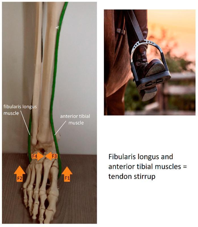
The "tendon stirrup" formed by fibularis longus and tibialis anterior/posterior - F1/F2 lift the medial and lateral longitudinal arches; F3/F4 deepen the transverse arch.
Applied Anatomy (High-yield for Ortho exam)
- Pes planus (flat foot): Collapse of medial longitudinal arch, most commonly due to laxity/failure of the spring ligament and tibialis posterior dysfunction (tibialis posterior tendon dysfunction, TPTD, is the classic acquired cause in adults).
- Pes cavus (high-arched foot): Exaggerated medial longitudinal arch, seen in neuromuscular conditions like Charcot-Marie-Tooth disease, spina bifida, and clubfoot residual deformity.
- March fracture / stress fracture: Repeated stress on the anterior pillar (2nd metatarsal neck) due to arch overload.
- Plantar fasciitis: Overuse/microtears of the plantar aponeurosis, a key passive support of the medial longitudinal arch, presenting as heel pain, classically worse with the first steps in the morning.
- Tarsal tunnel syndrome: Related to the medial longitudinal arch region and flexor retinaculum.
- Talar head as keystone: Fracture-dislocation here (e.g., in Lisfranc or subtalar injuries) can collapse the medial arch.
Summary Table
| Arch | Bones | Keystone | Main passive support |
|---|---|---|---|
| Medial longitudinal | Calcaneus, talus, navicular, 3 cuneiforms, medial 3 metatarsals | Head of talus | Spring ligament |
| Lateral longitudinal | Calcaneus, cuboid, lateral 2 metatarsals | Cuboid | Long plantar ligament |
| Transverse | Cuboid, cuneiforms, metatarsal bases | Intermediate cuneiform | Deep transverse metatarsal ligament |
Recent evidence note: A quick check for recent systematic reviews/meta-analyses on foot arch anatomy did not surface material that revises this classical anatomical description - the anatomy above is stable, well-established teaching (consistent across Gray's Anatomy for Students and standard ortho texts). If you want, I can pull specific PubMed evidence on clinical topics like tibialis posterior dysfunction or plantar fasciitis management for a viva follow-up.
This is a shared conversation. Sign in to Orris to start your own chat.