
Temporal bone , from basic anatomy to clinical aspects
Reading File
The temporal bone is one of the most anatomically complex bones in the skull, housing the middle and inner ear, the facial nerve canal, and critical vascular structures. Let me do a thorough search across the library.
Searching sources...
Finding Sources
Finding Sources
Excellent finds. Now let me read the most comprehensive sources in parallel - the dedicated anatomy chapter from Cummings, Gray's Anatomy, Scott-Brown's, and Shambaugh's.
Reading File
Reading File
Reading File
Excellent. Now let me get the detailed anatomy from Cummings and the clinical aspects (fractures, surgical landmarks) from Scott-Brown's and Cummings.
Reading File
Reading File
Finding Sources
Reading File
Reading File
Reading File
Now let me get the facial nerve anatomy and inner ear details plus the key images from Cummings:
Reading File
Reading File
I now have very rich content. Let me fetch the key images and get the remaining clinical details on the facial nerve course and surgical landmarks:
Loading Image
Loading Image
Reading File
I now have comprehensive, rich material from multiple authoritative sources. Let me compile the complete answer.
The Temporal Bone: From Basic Anatomy to Clinical Aspects
Sources: Cummings Otolaryngology Head and Neck Surgery (Ch. 126), Gray's Anatomy for Students, Scott-Brown's Otorhinolaryngology Head & Neck Surgery (Vol 2)
1. Overview and Osteology
The temporal bone is a paired, pyramidal bone that forms part of the lateral skull base. It articulates with the sphenoid, parietal, occipital, and zygomatic bones, and contributes to the cranial vault, skull base, and facial skeleton. Its four sides form:
- The middle fossa floor (superior face)
- The anterior limit of the posterior fossa (posterior face)
- Muscle attachments of the neck and infratemporal fossa (anterior-inferior face)
- The lateral surface of the head (base of the pyramid)
2. The Four Parts
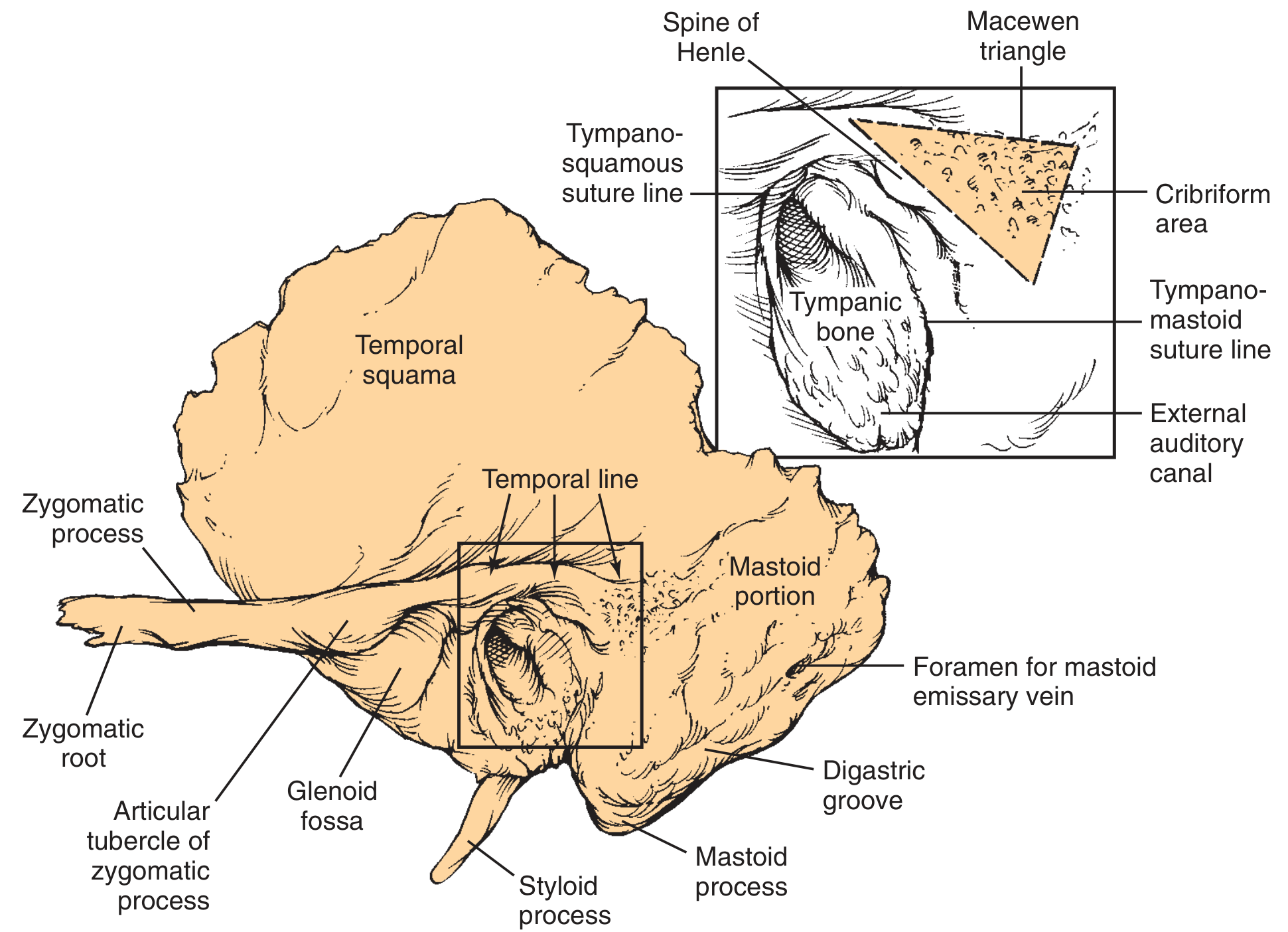
2a. Squamous Part
The largest part. It forms the convex lateral aspect of the temporal fossa and the lateral wall of the middle cranial fossa. Key features:
- Anteriorly projects the zygomatic process, which forms the bony roof of the glenoid fossa and articulates with the temporal process of the zygomatic bone to form the zygomatic arch
- The outer cortex receives the insertion of the temporalis muscle; the zygomatic process provides the masseter muscle insertion
- The temporal line (supramastoid crest) is a surface landmark aligned with the zygomatic process and runs approximately 4.7 mm above the middle fossa floor - critical for estimating the tegmen level during surgery
- The inferior surface forms the posterolateral aspect of the infratemporal fossa
- Articulates anteriorly with the greater wing of the sphenoid (sphenosquamous suture) and superiorly with the parietal bone (squamous suture)
Clinical pearl: Where the squamous temporal bone meets the frontal, parietal, and greater wing of sphenoid is the pterion - the thinnest area of the skull. A fracture here risks laceration of the middle meningeal artery and extradural haematoma. - Scott-Brown's Vol 2
2b. Mastoid Part
A bulbous structure shaped by pneumatization. Key features:
- Contains the mastoid air cells, with the central largest cell being the mastoid antrum, communicating with the middle ear via the aditus ad antrum
- Inferiorly, the pull of the sternocleidomastoid (SCM) and posterior belly of digastric elongates the bone to form the mastoid process (tip)
- The digastric ridge (sulcus) runs along the deep surface; the facial nerve exits through the stylomastoid foramen at its anterior limit
- The occipital groove, medial to the digastric notch, contains the occipital artery
- The mastoid cortex is perforated by emissary vessels that form the depressed cribriform area (key surgical landmark)
- The sigmoid sinus grooves the inner mastoid surface, itself receiving the superior petrosal sinus superiorly
Surgical landmark: The MacEwen (suprameatal) triangle is formed by the posterior-superior margin of the external auditory meatus, just posterior to the spine of Henle; the mastoid antrum lies deep to it. This is the surface landmark for cortical mastoidectomy. - Cummings Ch. 126
Developmental note: In neonates, the mastoid process is absent; the stylomastoid foramen lies near the lateral skull surface. The extracranial facial nerve is therefore very superficial and vulnerable to forceps delivery injury. The mastoid tip becomes palpable in the second year of life. - Scott-Brown's Vol 2
2c. Petrous Part
The densest bone in the body. Oriented at 45 degrees anteromedially, its apex lies between the occipital and sphenoid bones. Three surfaces deserve attention:
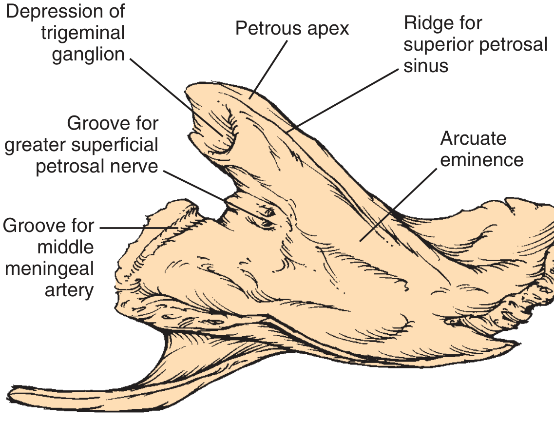
Superior surface (middle fossa floor):
- Arcuate eminence: prominence caused by the underlying superior semicircular canal - the key landmark on the middle fossa floor
- Tegmen tympani: roof of the middle ear/mastoid, lateral to the arcuate eminence
- Hiatus for the greater superficial petrosal nerve (GSPN): just anteromedial to the arcuate eminence, leading to foramen lacerum
- Trigeminal impression: smooth depression at the apex housing the trigeminal ganglion
- Superior petrosal sinus groove along the posterior ridge
Posterior surface (anterior boundary of the posterior fossa):
- Porus acusticus (Internal Acoustic Meatus, IAM): transmits CN VII and VIII (with superior and inferior vestibular divisions), and the labyrinthine artery (branch of basilar)
- At the fundus of the IAM: the falciform (horizontal) crest, Bill's bar (vertical crest), and foramina of CN VII and VIII
- Endolymphatic sac/duct occupies the operculum (depression inferolateral to the IAM)
- Subarcuate artery depression superior and lateral to the IAM
Inferior surface:
- Carotid canal: internal carotid artery (ICA) enters just anterior to the jugular fossa, traversing the petrous apex obliquely
- Jugular fossa/foramen: formed at the junction with the occipital bone; divided by the jugular spine into:
- Pars nervosa (anterior): glossopharyngeal nerve (IX) and inferior petrosal sinus
- Pars venosa (posterior): internal jugular vein; vagus (X) and accessory (XI) nerves in between
- Cochlear aqueduct opens into the pars nervosa just medial to the jugular spine; marks the superior limit of the jugular foramen
Clinical relevance: Erosion of the bony "keel" between the carotid canal and jugular fossa is an early CT finding in glomus jugulare tumour. - Scott-Brown's Vol 2
2d. Tympanic Part
A C-shaped trough that forms the majority of the external auditory canal (EAC) floor and anterior wall. Key features:
- The bony EAC is approximately 16 mm long (outer third is cartilaginous)
- Contains Santorini's fissures (between tympanic and mastoid parts) and the foramen of Huschke (developmental defect in the floor of the bony EAC, present in ~5% of adults) - potential pathways for tumour spread to and from the parotid/infratemporal fossa
2e. Styloid Part
The styloid process projects inferiorly from the undersurface, just anteromedial to the stylomastoid foramen. It anchors the stylohyoid ligament, stylohyoid muscle, styloglossus, stylopharyngeus, and digastric.
3. External Auditory Canal (EAC)
- Outer one-third: cartilaginous, continuous with the auricle
- Inner two-thirds: bony, formed by the tympanic (floor/anterior wall) and squamous (roof/posterior wall superior) parts
- The EAC ends at the tympanic membrane (TM), which sits obliquely at approximately 55 degrees to the floor
- The TM is divided by the malleus handle into the pars tensa (most of TM) and pars flaccida (Shrapnell's membrane), above the lateral process of the malleus
4. Middle Ear (Tympanic Cavity)
A mucosa-lined, air-filled space divided into:
- Epitympanum (attic): superior to the level of the TM; contains the heads of malleus and incus
- Mesotympanum: at the level of the TM; contains the ossicular chain
- Hypotympanum: inferior to the TM, bordered inferiorly by the jugular bulb
Ossicular chain:
- Malleus (hammer): attached to the TM via its handle; head in epitympanum
- Incus (anvil): body and short process in the epitympanum; long process descends to articulate with the stapes
- Stapes (stirrup): footplate sits in the oval window
Most vulnerable part: The long process of the incus has a single nutrient vessel with no collateral circulation, making it prone to aseptic necrosis from otitis media. - Cummings Ch. 126
Middle ear walls:
| Wall | Contents/Relations |
|---|---|
| Lateral | Tympanic membrane, scutum (bony EAC superior wall) |
| Medial (labyrinthine) | Promontory (basal cochlear turn), oval window, round window niche, facial nerve canal (tympanic segment), lateral semicircular canal |
| Roof (tegmen tympani) | Petrous bone separating from middle fossa dura |
| Floor | Jugular bulb beneath thin bone |
| Anterior | Eustachian tube orifice, carotid wall |
| Posterior | Aditus ad antrum, facial nerve (mastoid segment), pyramidal eminence with stapedius |
Prussak's space = recess between the pars flaccida and the neck of the malleus; the most common site for acquired attic (pars flaccida) cholesteatoma formation.
Epitympanum-mesotympanum partitioning by ossicular chain and mucosal folds leaves only two narrow openings - the isthmus tympani anticus and isthmus tympani oticus - which when blocked impair aeration of the attic.
5. Eustachian (Pharyngotympanic) Tube
- Connects the middle ear to the nasopharynx; ~35 mm long in adults
- Lateral 1/3 (~12 mm): bony, within the petrous bone, originating from the anterior mesotympanum
- Medial 2/3 (~24 mm): cartilaginous
- The isthmus (bony-cartilaginous junction) is the narrowest point
- Angled downward and medially (approximately 30-45 degrees from horizontal); in children the tube is shorter, more horizontal, and wider - predisposing to otitis media
- Opens during swallowing/yawning via tensor veli palatini (CN V3) and levator veli palatini (CN X/XI)
6. Inner Ear (Labyrinth)
Housed within the dense otic capsule of the petrous bone. Consists of an osseous labyrinth containing perilymph, within which sits the membranous labyrinth containing endolymph (the two fluids do not communicate).
Cochlea
- Coils 2¾ turns around a bony core, the modiolus
- Three scalae: scala vestibuli (perilymph, superior), scala media/cochlear duct (endolymph, middle), scala tympani (perilymph, inferior)
- Scala vestibuli opens to the oval window; scala tympani opens to the round window
- Apex directed anterolaterally toward the middle ear
- Houses the organ of Corti (hair cells, CN VIII cochlear fibers)
Vestibule
- Contains the saccule (vertical/linear acceleration) and utricle (horizontal linear acceleration)
- Communicates with the oval window laterally
Semicircular Canals (SCCs)
Three SCCs arranged in perpendicular planes:
- Superior (anterior) SCC: creates the arcuate eminence on the middle fossa floor
- Posterior SCC: oriented in the same plane as the posterior surface of the petrous bone
- Lateral (horizontal) SCC: visible as a bulge on the medial wall of the mastoid antrum; the most important surgical landmark during mastoidectomy
Endolymphatic duct and sac
- Drains from the utricle and saccule through the vestibular aqueduct
- The endolymphatic sac lies in a dural pouch on the posterior surface of the petrous bone
- Dysfunction leads to Meniere's disease (endolymphatic hydrops)
7. Facial Nerve (CN VII) in the Temporal Bone
The facial nerve travels the longest intraosseous course of any cranial nerve (~3 cm), making it uniquely vulnerable within the temporal bone. It traverses the Fallopian canal in four segments:
| Segment | Course | Length | Key Features |
|---|---|---|---|
| Meatal | From brainstem → fundus of IAM | ~8-10 mm | Anterosuperior in IAM; separated from cochlear nerve by Bill's bar |
| Labyrinthine | From fundus → geniculate ganglion | ~4 mm | Narrowest segment; no epineurium; watershed vascular supply; most vulnerable to herpes (Bell's palsy) and transverse fractures |
| Tympanic (horizontal) | From geniculate → pyramidal eminence (2nd genu) | ~11 mm | Runs over oval window, below lateral SCC; dehiscent in up to 55% of individuals |
| Mastoid (vertical) | From 2nd genu → stylomastoid foramen | ~13 mm | Posterior to the stapes, anterior to the posterior SCC |
Branches within the temporal bone:
- Greater superficial petrosal nerve (GSPN): from geniculate ganglion; preganglionic parasympathetics to lacrimal gland
- Nerve to stapedius: from mastoid segment
- Chorda tympani: from mastoid segment, crossing the middle ear (between TM and incus handle); carries taste fibers (anterior 2/3 tongue) and submandibular/sublingual secretomotor fibers
Landmark for surgery: The cochleariform process, oval window, and pyramidal eminence are key landmarks for identifying the facial nerve. The geniculate ganglion may be dehiscent at the supratubal recess (anterior attic), making it vulnerable during surgery. - Cummings Ch. 126
8. Vascular Relations
- Internal carotid artery (ICA): enters the carotid canal just anterior to the jugular foramen; traverses the petrous apex before entering the cavernous sinus. The thin bony plate separating the carotid canal from the middle ear is surgically critical
- Jugular bulb: the domed junction of the sigmoid sinus and internal jugular vein, in the jugular fossa; a high-riding jugular bulb can project into the hypotympanum and be mistaken for a middle ear mass
- Sigmoid sinus: grooves the inner mastoid cortex; the mastoid emissary vein communicates with it through the outer cortex
- Middle meningeal artery: runs in a groove on the squamous part, at risk with pterion fractures
- Labyrinthine (internal auditory) artery: branch of the basilar artery, travels with CN VII/VIII in the IAM; end-artery (no collaterals) - occlusion causes irreversible hearing loss
9. Pneumatization
The temporal bone contains an extensive network of mucosa-lined, air-filled cells:
- Mastoid: largest pneumatized region; communicates with the attic via the antrum
- Perilabyrinthine: cells surrounding the otic capsule (supralabyrinthine, infralabyrinthine, retrofacial)
- Petrous apex: when pneumatized (~30% of individuals), this can become infected (petrous apicitis - Gradenigo syndrome: sixth nerve palsy, ipsilateral facial pain, otorrhea)
- Extent of pneumatization is influenced by early otitis media history, genetics, and middle ear ventilation; poor pneumatization correlates with chronic ear disease
10. Clinical Aspects
10a. Temporal Bone Fractures
Classic classification:
| Longitudinal | Transverse | |
|---|---|---|
| Frequency | ~70-80% | ~20-30% |
| Mechanism | Lateral blow (temporal/parietal) | Frontal/occipital blow |
| Fracture line | Parallel to petrous axis, through EAC/middle ear | Perpendicular to petrous axis, through inner ear |
| Hearing loss | Conductive (haemotympanum, ossicular disruption) | Sensorineural (cochlear/labyrinthine injury) - usually permanent |
| Facial palsy | ~20% - usually delayed, incomplete | ~50% - usually immediate, complete |
| Battle's sign | May be present | Less common |
Modern classification (preferred):
- Otic capsule-sparing (OCS): 90% of fractures; predicts absence of SNHL but does not predict facial palsy or conductive loss
- Otic capsule-violating (OCV): 10%; strongly predicts SNHL and labyrinthine fistula
Investigation: HRCT temporal bone is the investigation of choice.
Site of facial nerve injury:
- Longitudinal fractures: just distal to the geniculate ganglion (1st genu)
- Transverse fractures: just proximal to the geniculate ganglion (labyrinthine segment)
Management: Immediate facial palsy warrants surgical exploration (nerve decompression); delayed or incomplete palsy is managed conservatively with steroids. CSF otorrhea usually resolves spontaneously. - Scott-Brown's Vol 2
10b. Bell's Palsy (Idiopathic Facial Palsy)
- The labyrinthine segment of the facial nerve is the most vulnerable segment due to: (1) watershed vascular supply (vertebrobasilar/external carotid junction), (2) narrow bony canal with no epineurium, (3) proximity to the geniculate ganglion where herpes simplex/zoster reactivates
- Swelling from viral neuritis causes entrapment in this narrow canal
10c. Cholesteatoma
- Arises from keratinizing squamous epithelium in the middle ear/mastoid
- Pars flaccida type: retraction of the TM into the Prussak space; erodes the scutum and lateral attic wall; spreads posteriorly through the aditus into the antrum
- Bone erosion can involve the incus (long process most vulnerable), tegmen (intracranial spread), lateral SCC (fistula, vertigo), and facial nerve canal (facial palsy)
- Surgery requires removal of incus and malleus head to address anterior attic extension fully
10d. Otitis Media Complications
The anatomy of the temporal bone defines the routes of spread:
- Subperiosteal abscess: through mastoid cortex (Bezold abscess: tracking below SCM along digastric groove)
- Petrous apicitis (Gradenigo syndrome): via air cell tracts to petrous apex → CN VI palsy, facial pain (CN V), otorrhea
- Meningitis/brain abscess: through tegmen tympani or sigmoid sinus plate erosion
- Lateral sinus (sigmoid) thrombophlebitis: erosion of the sigmoid sinus plate
- Facial paralysis: erosion of the dehiscent tympanic segment
10e. Vestibular Schwannoma (Acoustic Neuroma)
- Arises from CN VIII (usually superior vestibular division) at the glial-Schwann cell junction in the IAM
- Grows medially to fill the cerebellopontine angle (CPA)
- Presents with: unilateral SNHL, tinnitus, dysequilibrium; large tumors compress CN VII and V
- Investigation: MRI with gadolinium (gold standard); HRCT for bony changes
- Surgical approaches to the IAM utilize temporal bone landmarks (translabyrinthine, retrosigmoid, middle fossa)
10f. Glomus Tumors (Paragangliomas)
- Glomus tympanicum: on the promontory (tympanic branch of CN IX); presents as a pulsatile red mass behind TM, pulsatile tinnitus
- Glomus jugulare: in the jugular fossa; early sign on CT is erosion of the bony keel between the carotid canal and jugular fossa; can involve CN IX, X, XI (jugular foramen syndrome)
10g. Surgical Landmarks Summary
| Landmark | Significance |
|---|---|
| Temporal line | ~5 mm above middle fossa floor; guides mastoidectomy depth |
| MacEwen triangle | Surface landmark for mastoid antrum |
| Spine of Henle | Posterior-superior EAC; antrum is 15 mm deep from here |
| Digastric ridge | Inferior limit of mastoidectomy; facial nerve lies just medial to its anterior end |
| Arcuate eminence | Superior SCC on middle fossa floor |
| Lateral SCC bulge | Key intraoperative landmark in mastoidectomy |
| Short process of incus | Reliable landmark pointing to facial nerve during tympanotomy |
| Cochleariform process | Identifies the first genu of the facial nerve |
| Pyramidal eminence | Identifies the second genu of the facial nerve |
References:
- Cummings Otolaryngology Head and Neck Surgery, Ch. 126 (Francis HW) - Anatomy of the Temporal Bone, External Ear, and Middle Ear
- Gray's Anatomy for Students - Temporal bone, Skull base
- Scott-Brown's Otorhinolaryngology Head & Neck Surgery (Vol 2) - Temporal Bone Anatomy and Fractures
This is a shared conversation. Sign in to Orris to start your own chat.