
Filters used in cpb detailed notes
Reading File
CPB (cardiopulmonary bypass) uses several types of filters. Searching the medical library now.
Searching sources...
Finding Sources
Finding Sources
Finding Sources
Finding Sources
Reading File
Finding Sources
Reading File
Reading File
Reading File
Finding Sources
Reading File
Reading File
Now I have comprehensive data from multiple authoritative textbooks. Let me compile the detailed notes.
Loading Image
Here are detailed notes on Filters Used in Cardiopulmonary Bypass (CPB):
Filters Used in Cardiopulmonary Bypass (CPB)
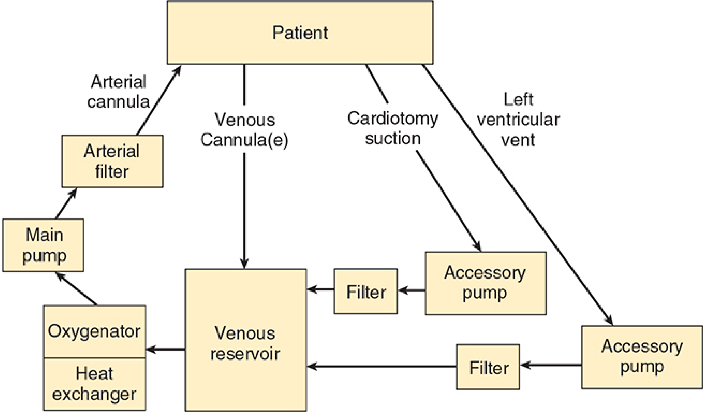
Overview
The CPB circuit contains multiple filters at different points. The six basic components of the CPB machine are: venous reservoir, oxygenator, heat exchanger, main pump, arterial filter, and connecting tubing. In addition, accessory pumps use separate inline filters before blood returns to the reservoir. A number of other filters, alarms, and in-line monitors are also typically incorporated.
1. Arterial Line Filter (Main Filter)
Location: Final inline filter, placed AFTER the main pump and BEFORE the arterial cannula (aortic return)
Pore size: Passes only particles smaller than 27-40 μm - traps everything larger
Purpose:
- Removes particulate matter - thrombi, fat globules, tissue debris that may enter via cardiotomy suction
- Traps gas bubbles (air microemboli) - has a built-in stopcock to vent these out
- Provides the last line of defense against systemic embolism before blood returns to the patient
Design features:
- Always arranged in parallel with a normally clamped bypass limb - if the filter clogs, the bypass limb can be opened to maintain perfusion
- Has a built-in stopcock to bleed off trapped gas bubbles
- Arterial inflow pressure is measured proximal to the filter (before it) so that clogging can be detected; the pressure difference across the filter reflects filter resistance
Clinical significance:
- The pressure drop across the arterial filter + arterial tubing + aortic cannula = the measured inflow pressure. Monitoring this is important for detecting problems with the arterial inflow line.
- Inflow pressures should be watched continuously for sudden rises indicating filter obstruction
(Source: Morgan & Mikhail's Clinical Anesthesiology, 7e; Barash Clinical Anesthesia, 9e)
2. Cardiotomy/Accessory Pump Filters
Location: Inline on the return lines from accessory pumps (cardiotomy suction pump and LV vent pump), positioned BEFORE blood re-enters the venous reservoir
Purpose:
- The cardiotomy suction pump aspirates blood from the surgical field and returns it to the main reservoir. This line is a portal for fat, tissue debris, thrombi, and bone fragments entering the circuit
- The filter catches this debris before it reaches the reservoir and potentially the patient
- The LV vent filter similarly screens blood drained from the left ventricle
Clinical note: This is the main reason cardiotomy suction is considered a major source of embolic and inflammatory burden - even with filtration, fat microemboli and activated inflammatory mediators can pass through.
(Source: Morgan & Mikhail's Clinical Anesthesiology, 7e - see circuit diagram above)
3. Heat Exchanger Bubble Trap / Filter
Location: Built into the heat exchanger unit (which is downstream of the oxygenator)
Purpose:
- Gas solubility decreases as blood temperature rises during rewarming
- Dissolved gas comes out of solution and forms bubbles during the rewarming phase
- A filter/trap built into the heat exchanger unit catches these bubbles before they enter the main circuit
Key point: This is especially relevant during the rewarming phase at the end of CPB when bubble formation risk is highest.
(Source: Morgan & Mikhail's Clinical Anesthesiology, 7e)
4. Leukocyte Depletion Filter (LDF)
Location: Inline in the CPB circuit (position varies - can be on the arterial line or on cardioplegia delivery lines)
Purpose:
- Selectively removes activated leukocytes from circulating blood
- Aims to reduce the systemic inflammatory response triggered by CPB (contact of blood with foreign surfaces activates complement, leukocytes, platelets, coagulation cascade)
- Leukocyte-depleted blood cardioplegia has been shown to improve myocardial preservation in some studies
- Hemofiltration (ultrafiltration) during CPB, which removes inflammatory cytokines, appears beneficial particularly in pediatric patients
Evidence:
- A review of 63 studies (Warren et al.) concluded leukocyte filtration may have some modest benefits, but definitive evidence of improvement in inflammatory-mediated complications is insufficient
- Leukocyte depletion reduces inflammation and may reduce complications, but the evidence for routine use remains weak
(Source: Morgan & Mikhail's Clinical Anesthesiology, 7e; Miller's Anesthesia, 10e)
5. Micropore / Screen Filter (Blood Transfusion Context)
Pore size: Most investigators recommend 40 μm pore size - this minimizes microembolization without causing excessive elevation in filtration pressure
Purpose:
- Used when salvaged blood or bank blood is returned during or after CPB
- Reduces risk of microembolization from microaggregates that form in stored blood (platelets, fibrin, leukocyte aggregates)
Evidence: A pore size of 40 μm is most widely supported - pore sizes from 20-170 μm have been studied; 40 μm strikes the best balance between filtration efficiency and maintaining flow without excess pressure drop.
(Source: Roberts & Hedges' Clinical Procedures in Emergency Medicine)
Summary Table
| Filter | Location | Pore Size | Primary Function |
|---|---|---|---|
| Arterial line filter | Post-pump, pre-aortic cannula | 27-40 μm | Remove particulates + trap gas emboli |
| Cardiotomy/vent filter | Post-accessory pump, pre-reservoir | Variable | Remove debris from surgical field blood |
| Heat exchanger bubble trap | Within heat exchanger unit | Gas trap | Catch bubbles formed during rewarming |
| Leukocyte depletion filter | Inline arterial or cardioplegia line | Selective | Remove activated leukocytes, reduce inflammation |
| Micropore filter | On blood salvage/transfusion return | 40 μm (recommended) | Remove microaggregates from stored/salvaged blood |
Key Clinical Points
- Arterial filter = most important filter in the standard CPB circuit - mandatory component, protects against both particulate and gaseous emboli
- Bypass limb is always present in parallel with the arterial filter - can be opened if filter clogs
- Pressure monitoring proximal to the arterial filter detects clogging early
- Cardiotomy suction is the biggest source of debris - its filter is important but not perfect (fat and activated cells still pass)
- Leukocyte filters address the inflammatory response to CPB - benefit is real but modest; not universally used
- Bubble traps in the heat exchanger are passive safety features for the rewarming phase
- In pediatric CPB, hemofiltration (ultrafiltration) as a filtering strategy to remove cytokines and excess fluid has stronger evidence than in adults
Perturbation
I need a bit more context to give you the most useful answer. "Perturbation" spans multiple fields.
Asking for Preferences
What context are you asking about perturbation in?
Reading File
Searching sources...
Finding Sources
Finding Sources
Finding Sources
Finding Sources
Finding Sources
Finding Sources
Reading File
Finding Sources
Reading File
Reading File
Reading File
I now have extensive material. Here are the detailed notes:
Hemodynamic Perturbations During Cardiopulmonary Bypass (CPB)
Hemodynamic perturbations in CPB refer to the cardiovascular disturbances and instability that occur at three critical phases: the pre-bypass (cannulation) period, the bypass period itself, and weaning/separation from CPB.
Phase 1 - Pre-Bypass Period (Before CPB)
Variable Surgical Stimulation = Variable Hemodynamics
| Surgical Step | Stimulation | Hemodynamic Effect |
|---|---|---|
| Positioning, monitor insertion, skin prep, vein harvest | Minimal | Hypotension (esp. in hypovolemic/poor LV patients) |
| Chest incision, sternal split, IMA harvest | Intense | Hypertension, tachycardia, dysrhythmias |
| Cannulation of great vessels | Minimal + mechanical | Precipitous BP fall from reduced venous return |
The anesthesiologist must be ready to treat all hemodynamic perturbations with vasopressors, inotropes, vasodilators, antiarrhythmics, and anticholinergics.
Cannulation Perturbations
Arterial cannulation (aorta) complications:
- Arterial dissection
- Hemorrhage and resultant hypotension
- Inadvertent cannulation of aortic arch vessels
- Embolic phenomena from dislodged atherosclerotic plaque or air introduction
Venous cannulation (RA/IVC/SVC) complications:
- Hypotension from blood loss
- Dysrhythmias (premature atrial contractions, SVT, AF) - manipulating the heart/great vessels triggers arrhythmias
- Surgical mechanical compression of the heart or great vessels
- Malpositioning - venous return impaired or SVC syndrome (head/neck engorgement)
- Sustained AF/SVT leads to hemodynamic deterioration - treated pharmacologically, electrically, or by emergency CPB initiation
(Morgan & Mikhail's Clinical Anesthesiology, 7e; Miller's Anesthesia, 10e)
Phase 2 - Bypass Period (On CPB)
2A. Initiation of CPB - The First Major Perturbation
What happens:
- The main CPB pump is started and venous cannula(e) unclamped
- At onset of CPB, systemic arterial pressure abruptly decreases
- Initial mean arterial pressure (radial) of 30-40 mmHg is not unusual
Mechanism of initial hypotension:
- Sudden hemodilution from priming solution (hematocrit drops to ~22-27%)
- Hemodilution reduces blood viscosity
- Reduced viscosity lowers SVR (systemic vascular resistance)
- Lower SVR → lower MAP despite normal pump flow
Treatment: Increased pump flow + vasopressors (phenylephrine, norepinephrine)
Relationship (the key equation):
MAP = Pump Flow × SVR
So MAP can be manipulated by adjusting pump flow or SVR independently.
2B. Ongoing Hemodynamic Control During CPB
Target parameters:
- Pump flow: 2.0-2.5 L/min/m² (50-60 mL/kg/min)
- MAP: 65-80 mmHg in adults
- Temperature: variable (hypothermia reduces metabolic demands, allowing lower flows)
- During deep hypothermia (20-25°C): MAP as low as 30 mmHg may still give adequate cerebral oxygen delivery
Causes of hypotension during CPB:
| Cause | Action |
|---|---|
| Hemodilution → low SVR | Vasopressors, increase pump flow |
| Inadequate pump flow (poor venous return) | Check cannulae for kinks, air locks, malposition |
| Pump malfunction | Troubleshoot pump |
| Aortic dissection | Stop CPB, repositioning cannula |
| Pressure transducer error | Check calibration |
| Excessive hypotension (<30 mmHg) | Urgent search for aortic dissection |
Causes of hypertension during CPB (MAP >100-110 mmHg):
- Dangerous - may cause aortic dissection or cerebral hemorrhage
- Treatment: decrease pump flow, increase volatile agent via oxygenator gas inlet, or infuse vasodilator (clevidipine, nicardipine, nitroprusside)
2C. Indicators of Inadequate Perfusion
(In absence of hypoxemia, all of these indicate flow is too low):
- Venous O₂ saturation <70%
- Progressive metabolic acidosis
- Reduced urinary output
2D. Electrolyte & Metabolic Perturbations
- Hyperkalemia: Cardioplegia contains high K⁺; marked rises treated with furosemide-induced diuresis
- Hyperglycemia: CPB causes stress hyperglycemia even in non-diabetics; monitor and treat
- Hematocrit: Allowed to fall to ~20%, but not below; red cell transfusion into pump reservoir if needed
- Hypothermia effects: Increases heparin half-life, prolongs ACT; metabolism slows; drug concentrations accumulate due to reduced hepatic/renal perfusion
- Pressure gradient: Arterial inflow line pressure always > systemic arterial pressure (due to pressure drop across filter, tubing, and aortic cannula); values >300 mmHg = suspect clogged filter, obstruction, or aortic dissection
2E. Aortic Cross-Clamp Perturbations
Application of aortic cross-clamp (aortic surgery):
- Sudden increase in LV afterload
- May precipitate acute LV failure and myocardial ischemia, especially with underlying LV dysfunction or coronary disease
Release of aortic cross-clamp:
- The period of greatest hemodynamic instability
- Abrupt decrease in afterload
- Plus bleeding from ischemic lower body
- Plus release of vasodilating acid metabolites from ischemic lower body
- Combined effect = severe systemic hypotension
(Morgan & Mikhail's Clinical Anesthesiology, 7e)
Phase 3 - Weaning/Separation from CPB
Pre-Conditions for Successful Weaning (CVP Mnemonic)
Before attempting separation, all of the following must be addressed:
C - Cold (Temperature):
- Rewarm to 36-37°C before weaning
- Venous blood returning to CPB circuit and nasopharyngeal temperature must NOT exceed 37°C (hyperthermia increases neurologic complication risk)
C - Conduction (Rhythm & Rate):
- Target heart rate: 80-100 bpm
- Bradycardia: treat with epicardial pacing wires ± beta-agonist inotropes
- Tachycardia (>120 bpm): undesirable; sinus tachycardia may result from anemia, hypovolemia, light anesthesia
V - Ventilation:
- Resume mechanical ventilation before separating
P - Pressure and Pump support:
- Ensure adequate BP; prepare inotropes/vasopressors
- Assess contractility (TEE is key)
- Address preload (volume)
Other issues to resolve:
- Electrolytes - specifically potassium
- Air in LV (de-airing before weaning)
- Cardiac rhythm
- Systemic BP
- Contractility
Weaning Process
- Perfusionist gradually reduces pump flow, allowing the native heart to take over
- Anesthesiologist provides inotropic support and volume as needed
- Communication between surgeon, anesthesiologist, and perfusionist is critical
- TEE used to assess LV function, filling, wall motion, and to detect residual air
Post-Bypass Perturbations
- LV distention may occur if aortic regurgitation is present - detected by TEE
- Myocardial stunning (reperfusion injury) reduces contractility
- Vasoplegic syndrome (low SVR state) may require vasopressors
- Arrhythmias (VF, heart block) may recur
Summary of Perturbations by Cause
| Perturbation | Mechanism | Treatment |
|---|---|---|
| Hypotension at CPB onset | Hemodilution → low SVR | Vasopressors, increase flow |
| Hypotension during CPB | Poor venous return, pump issue, dissection | Identify cause, fix mechanically or pharmacologically |
| Hypertension during CPB | High SVR, light anesthesia | Reduce flow, vasodilators, deepen anesthesia |
| Post-cross-clamp release hypotension | Sudden afterload drop + acidosis + vasodilation | Vasopressors, volume, anticipate |
| Post-cross-clamp application LV failure | Sudden afterload increase | Inotropes, consider urgent cardioplegia |
| Arrhythmias (cannulation) | Mechanical cardiac manipulation | Pharmacologic/electrical cardioversion or emergency CPB |
| Metabolic acidosis | Inadequate flow/perfusion | Increase pump flow, correct cause |
| Hyperkalemia | Cardioplegia | Furosemide diuresis |
| Post-CPB weaning failure | Low contractility, arrhythmia, volume issues | Inotropes, pacing, volume, TEE guidance |
Sources: Morgan & Mikhail's Clinical Anesthesiology, 7e; Miller's Anesthesia, 10e
Explain in detail about mics with proper subheading and Content
Asking for Preferences
What does 'MICS' refer to in your question?
Reading File
Searching sources...
Finding Sources
Finding Sources
Reading File
Finding Sources
Reading File
Finding Sources
Reading File
Finding Sources
Reading File
Reading File
Reading File
Reading File
I now have comprehensive material from multiple authoritative sources. Here are the full detailed notes:
Minimally Invasive Cardiac Surgery (MICS) - Detailed Notes
1. Definition and Core Concept
Minimally Invasive Cardiac Surgery (MICS) is a surgical philosophy that prioritizes avoidance of a complete median sternotomy. Surgical repair is accomplished via one or more smaller chest incisions - such as:
- Thoracotomy (anterolateral mini-thoracotomy)
- Mini-sternotomy
- Transverse sternotomy
MICS can be performed:
- With or without CPB
- With or without robotic assistance
- With or without endoscopic techniques
The overall goal is to reduce surgical trauma while achieving the same therapeutic outcomes as conventional open-heart surgery.
2. Procedures Achievable via MICS
| Procedure | MICS Approach |
|---|---|
| Myocardial revascularization (CABG) | MIDCAB, OPCAB, TECAB |
| Mitral valve repair/replacement | Right anterolateral mini-thoracotomy, robotic |
| Aortic valve replacement | Right anterior mini-thoracotomy (4th ICS) |
| Tricuspid valve surgery | Mini-thoracotomy |
| Atrial septal defect closure | Mini-thoracotomy |
| Surgical ablation of atrial fibrillation | MICS approach |
| Percutaneous valve replacement/repair | TAVR, MitraClip (selected high-risk patients) |
3. Suggested Benefits of MICS
- Less postoperative pain
- Decreased scarring
- Shorter recovery times and earlier discharge
- Decreased blood product transfusions (especially OPCAB)
- Reduced renal injury (off-pump approaches)
- Fewer wound complications
- Shorter ICU and hospital length of stay (especially robotic MIMVS)
4. MICS for Myocardial Revascularization
4A. MIDCAB - Minimally Invasive Direct Coronary Artery Bypass
Definition: Off-pump CABG performed through a small chest incision (not full sternotomy), without CPB.
Approach:
- Limited left anterolateral thoracotomy (4th or 5th left intercostal space)
- Patient positioned supine with 30-degree elevation of the left hemithorax, left arm tucked
- Chest and groins are prepped in case of emergency conversion to sternotomy/femoral-femoral bypass
Surgical steps:
- Pericardium opened to visualize the LAD through the thoracotomy
- LIMA (Left Internal Mammary Artery) dissected and harvested
- Heparin administered - target ACT >300 seconds
- LIMA-to-LAD anastomosis performed on the beating heart
- Myocardial stabilizer devices (pure pressure or vacuum-assisted) used to stabilize the anastomotic site
- Protamine administered, hemostasis achieved
- Left pleural chest tube placed, thoracotomy closed
Key features:
- Primarily for single or double-vessel disease (usually isolated LAD)
- No CPB required
- No aortic cross-clamping
Anesthetic considerations:
- Left lung isolation mandatory (double-lumen endotracheal tube or bronchial blocker) to allow surgical exposure of LIMA
- Standard ASA monitors + arterial line (radial artery preferred) + central line + ± PA catheter
- Induction same as standard CABG
- Maintenance must account for off-pump physiology - hemodynamic control via fluids and vasopressors
- Norepinephrine and phenylephrine both used effectively
- External defibrillator/pacing pads placed before draping (surgical access to heart is limited)
- No CPB-related hemodilution or inflammatory response
Outcomes:
- MIDCAB associated with less morbidity and mortality vs. on-pump and off-pump CABG
- Operative mortality ~1% in large series; 3-year mortality 3.1% (single-vessel disease)
- Meta-analysis of 12 RCTs vs. PCI: comparable mortality and MI rates, but lower revascularization requirement with MIDCAB
4B. OPCAB - Off-Pump Coronary Artery Bypass
Definition: CABG performed through a conventional median sternotomy, but on the beating heart without CPB.
Approach:
- Full median sternotomy
- LIMA and other conduits harvested for multiarterial grafting
- Heparin to target ACT ~300 seconds
Surgical steps:
- Generous pericardiotomy to allow cardiac mobilization
- LAD grafting (anterior) is performed first (most accessible)
- Inferior and lateral wall vessels require "verticalization" - lifting the heart apex to the zenith
- In verticalized position, forward ejection and stroke volume are compromised
- Suction devices achieve and maintain verticalization
- Stabilization devices (Octopus/Medtronic, Ultima II/Guidant, Acrobat-i/Macquet) compress the ventricles locally to create a still anastomotic field
- Epicardial pacing wires placed before cardiac manipulation - pace to 80 bpm to increase stroke volume
- Intracoronary shunts used to mitigate ischemia during coronary occlusion
- Grafting order: anterior → inferior → lateral wall vessels
Emergency conversion:
- Technical difficulty or refractory hemodynamic instability → abandon OPCAB → conventional CPB
- Must have CPB and mechanical circulatory support (MCS) immediately available
Anesthetic considerations (Table 39-21 summary):
| Issue | Management |
|---|---|
| Monitoring | Invasive arterial BP, CVP, TEE (essential), consider PAC if poor LV function or severe MR |
| Temperature | Normothermia - warming devices, heated circuits, optimize OR temperature |
| Hemodynamic compromise (verticalization, stabilizer application) | Trendelenburg, volume, vasopressors; anticipate and treat proactively |
| RV dysfunction | Watch closely - leads to refractory instability; may need MCS |
| Emergency preparedness | CPB and MCS must be immediately available |
| ECG/TEE limitations | May not detect ischemia during cardiac mobilization/verticalization |
Outcomes:
- Trend toward decreased perioperative blood transfusions vs. on-pump CABG
- Higher incomplete revascularization rates
- Technically challenging - outcomes vary widely by surgeon skill
- Stroke rates similar to on-pump CABG despite absence of CPB emboli
- The ROOBY trial showed increased adverse cardiac events with OPCAB vs. conventional CABG
- Cochrane review (Moller et al): no significant benefit in mortality, stroke, or MI; better long-term survival seen in on-pump CABG group
- OPCAB remains <20% of all CABG procedures in the United States
4C. TECAB - Total Endoscopic Coronary Artery Bypass
Definition: The most minimally invasive CABG approach - performed entirely through a few port sites, using a robotic system (surgeon remotely controls the ports).
Three variations:
- Arrested-heart TECAB - heart arrested; uses endoaortic occlusion balloon clamp (EAOBC)
- Beating-heart TECAB with CPB - heart beating on CPB support
- Beating-heart TECAB without CPB - fully off-pump, fully robotic
EAOBC (Endoaortic Occlusion Balloon Clamp):
- Placed via femoral vessels (or axillary artery if femoral/descending aorta is contraindicated)
- Acts as an endovascular cross-clamp - replaces the need for external aortic cross-clamping
- TEE guidance is essential for accurate EAOBC placement
Anesthetic note:
- Defibrillation pads placed preoperatively (no direct access to heart during surgery)
- Due to CO2 pneumothorax used for exposure: transthoracic electrical impedance increases - higher defibrillation energy needed
- Chest cavity may need to be deflated to improve defibrillation threshold
- Communication between surgical and anesthesia teams is critical
4D. Hybrid Coronary Revascularization (HCR)
Definition: Combines MICS surgical technique (MIDCAB or TECAB) with PCI (percutaneous coronary intervention) in the same or staged procedure.
Rationale: Uses the best of both techniques:
- Durable LIMA-to-LAD bypass graft (surgically, via MICS)
- Treat other stenoses with drug-eluting stent PCI
- Avoids large incision or CPB
Advantages:
- No full sternotomy or CPB
- Durable LAD revascularization
- Shorter hospital stay vs. OPCAB (small studies)
Disadvantages:
- Requires aggressive antiplatelet therapy (for PCI) which may increase surgical hemorrhagic complications
- Higher rates of reintervention reported in some studies
- Not widely accepted; clinical value remains debated
5. MICS for Valvular Surgery
5A. Minimally Invasive Mitral Valve Surgery
Approach: Right-sided anterolateral mini-thoracotomy
Evolution: Initially via mini-thoracotomy; now predominantly robotic using 5 small port incisions (da Vinci System)
Robotic MIMVS (R-MIMVS) - da Vinci System:
Patient positioning:
- Right shoulder elevated 30 degrees, pelvis remains supine (to allow femoral vessel access)
Surgical steps:
- One-lung ventilation (OLV) established for right lung isolation
- CO2 insufflation of right hemithorax
- Trocars introduced into 4th or 5th intercostal space
- Robot docked after exposure achieved
- CPB via femoral cannulae (peripheral CPB - femoral artery + femoral vein)
- Additional venous drainage via right internal jugular vein (superior vena cava cannula) for optimal right heart drainage
- Cardioplegia delivered into coronary vasculature
- Ascending aorta cross-clamped
- Mitral valve repaired or replaced
- TEE used to evaluate valve function after replacement
- Aortic cross-clamp removed, weaned from CPB
- Double-lumen ETT exchanged for single-lumen ETT if patient remains intubated
Exclusion criteria for robotic mitral valve repair:
- Severely calcified mitral annulus
- Severe pulmonary hypertension
- Ischemic heart disease
- Surgery requiring multiple valve repairs
- Previous surgery to the right hemithorax
- Severe aortic and peripheral atherosclerosis
Anesthetic considerations:
- OLV mandatory - double-lumen ETT or bronchial blocker
- CO2 pneumothorax causes increased pulmonary vascular resistance - poorly tolerated in patients with pre-existing mitral valve-induced pulmonary hypertension
- Optimize volume; prevent tachyarrhythmias and hypoxia during OLV
- TEE essential throughout - guides CPB cannula placement, valve assessment, de-airing, weaning
Outcomes:
- MIMVS is effective and safe
- Associated with fewer long-term and short-term complications
- Shorter ICU and hospital length of stay vs. conventional open mitral surgery
5B. Minimally Invasive Aortic Valve Replacement
Approach: Right anterior mini-thoracotomy, just lateral to the sternum in the 4th intercostal space
Key features:
- Anterior position of AV in thorax allows access through a 3-cm incision
- Right lung isolation required (same as mitral MICS)
- Peripheral CPB required (femoral cannulation)
- Superior vena cava (SVC) cannula via right IJV NOT required (unlike mitral MICS) - because the AV is positioned anteriorly, complete right heart drainage is not needed for visualization
6. Peripheral CPB in MICS
Because MICS uses small incisions without full sternal opening, central cannulation (direct aortic/RA) is not possible in most cases. Therefore:
- Femoral artery - arterial return cannula
- Femoral vein - primary venous drainage
- Right internal jugular vein - SVC cannula for additional drainage (mandatory in mitral MICS, not needed in AV MICS)
- TEE guides cannula positioning and checks for adequate drainage
7. Lung Isolation in MICS
MICS via thoracotomy requires access to the right hemithorax - the right lung must be collapsed for surgical exposure. Options:
- Double-lumen endotracheal tube (DLT) - preferred by most cardiac anesthesiologists for MIDCAB and MICS
- Single-lumen ETT with bronchial blocker - alternative
After robotic MIMVS, if the patient is left intubated, the DLT is exchanged for a single-lumen ETT in the ICU.
8. Monitoring Requirements for MICS
| Monitor | Indication |
|---|---|
| Invasive arterial line (radial artery) | All MICS cases |
| Central venous catheter/introducer | All MICS cases |
| PA catheter | Poor LV function, severe MR |
| TEE | Essential in all MICS - guides cannulation, valve assessment, de-airing, weaning |
| External defibrillation/pacing pads | Placed pre-draping (limited access to heart) |
| ECG | Standard - note limitations during cardiac mobilization |
9. Conversion to Conventional Surgery
Always prepare for emergency conversion in any MICS case:
- Chest and groins prepped for possible sternotomy + femoral-femoral bypass
- Systemic heparin prepared and immediately available
- CPB standby team ready
- Indications for conversion: Hemodynamic collapse, technical failure, uncontrolled bleeding, inability to complete revascularization/repair
10. Summary Comparison
| Feature | MIDCAB | OPCAB | TECAB | MICS Valve |
|---|---|---|---|---|
| Incision | Left mini-thoracotomy | Median sternotomy | Port sites only | Right mini-thoracotomy or ports |
| CPB | No | No | ± (arrested/beating) | Yes (peripheral femoral) |
| Aortic cross-clamp | No | No | ± (EAOBC) | Yes |
| Vessels | Single (LAD) | Multi-vessel | Single/multi | N/A |
| Lung isolation | Yes (left) | No | Yes (bilateral) | Yes (right) |
| Robotic | Possible | No | Yes (da Vinci) | Yes (da Vinci) |
| TEE | Yes | Essential | Essential | Essential |
Sources: Barash Clinical Anesthesia, 9e; Miller's Anesthesia, 10e; Schwartz's Principles of Surgery, 11e
This is a shared conversation. Sign in to Orris to start your own chat.