
Classify the muscles based on the arrangement of fibers.
Reading File
Now let me search the medical library and look for images in parallel.
Finding Sources
Searching Images
classification of muscle fiber arrangement pennate parallel
Finding Sources
Finding Sources
Searching Images
pennate muscle bipennate unipennate multipennate diagram anatomy
Reading File
Reading File
I found a great diagram. Let me fetch it and also search for more detailed classification content.
Loading Image
Finding Sources
I now have excellent source material. Here is the full classification:
Classification of Muscles Based on Arrangement of Fibers
The fundamental distinction in fiber arrangement is whether the muscle fascicles run parallel to the line of pull or are angled (pennate) to it. This architecture directly determines whether a muscle is built for range of motion and speed or for force production.
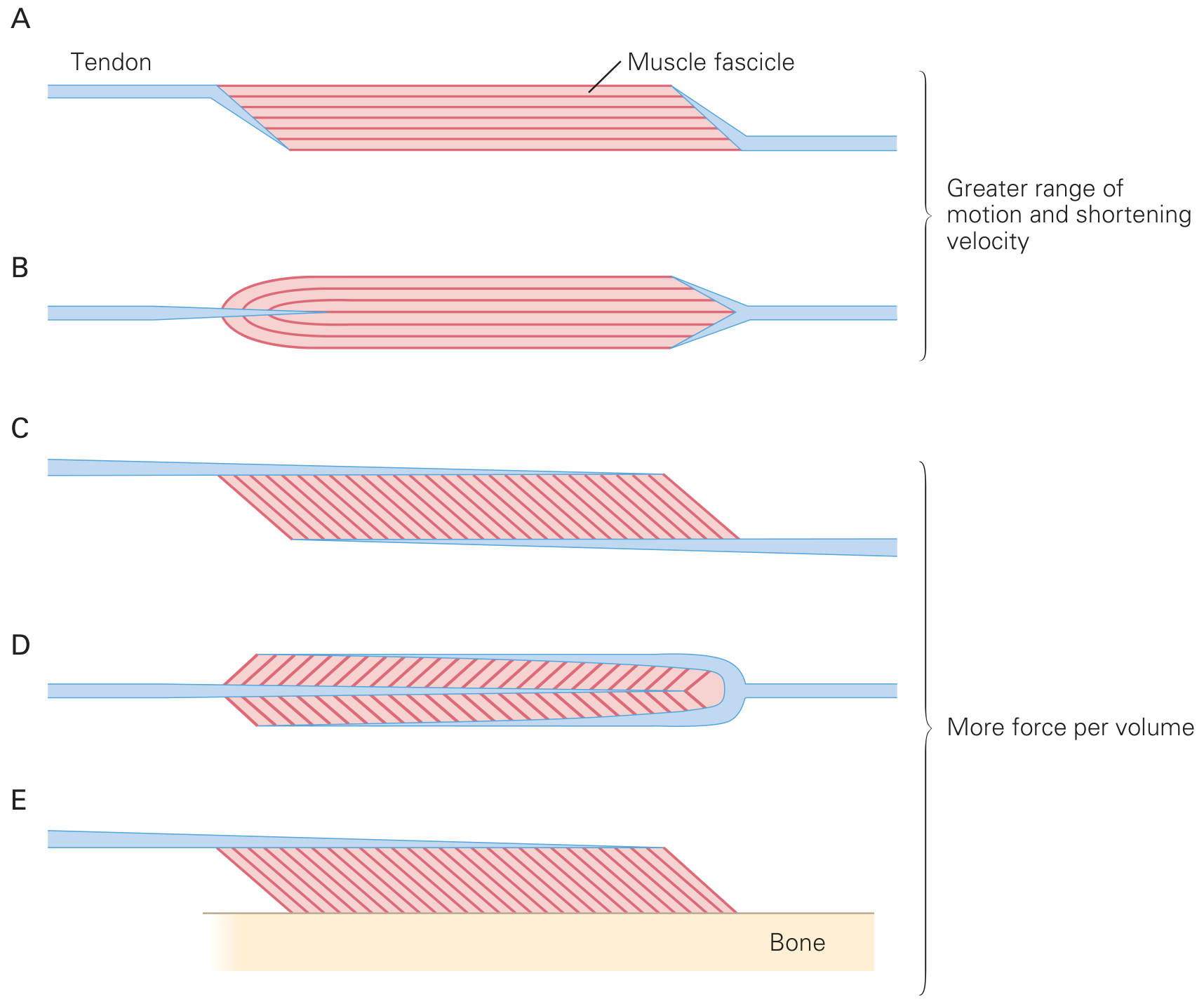
1. Parallel (Non-Pennate) Muscles
Fascicles run parallel to the longitudinal axis (line of pull) of the muscle. These muscles have longer fibers relative to muscle length, giving them a greater range of motion and higher shortening velocity. Force capacity is limited compared to pennate muscles of similar volume.
a. Strap (Ribbon) Muscles
- Flat, ribbon-like; uniform width throughout.
- Fibers run straight along the entire length.
- Example: Sartorius, sternohyoid.
b. Fusiform (Spindle-Shaped) Muscles
- Thick in the belly, tapering to tendons at each end.
- Fibers converge on a central belly.
- Example: Biceps brachii, brachioradialis.
c. Triangular (Fan-Shaped / Convergent) Muscles
- Broad origin, fibers converge toward a narrow insertion point.
- Example: Pectoralis major, temporalis.
d. Quadrilateral Muscles
- Flat, roughly four-sided shape with parallel fibers.
- Example: Pronator quadratus, thyrohyoid.
e. Cruciate Muscles
- Fibers cross each other at an angle.
- Example: Sternocleidomastoid (crossing pattern).
f. Spiral (Twisted) Muscles
- Fibers follow a twisted course between origin and insertion.
- Example: Latissimus dorsi, teres major.
2. Pennate Muscles
Fascicles are attached obliquely to a central tendon (intramuscular tendon), like the barbs of a feather (penna = feather). The pennation angle ranges from ~0° (nearly parallel, e.g., biceps brachii) to ~30° (e.g., soleus). Because more fibers can pack into the same volume at an angle, pennate muscles have a larger physiological cross-sectional area and generate greater maximal force, but with a shorter range of motion and lower shortening velocity than parallel muscles.
a. Unipennate
- Fascicles arise from one side of the tendon only (angled to the tendon on one side).
- Example: Extensor digitorum longus, flexor pollicis longus, semimembranosus.
b. Bipennate
- Fascicles arise from both sides of a central tendon, like a full feather.
- Example: Rectus femoris, flexor hallucis longus, gastrocnemius.
c. Multipennate
- Multiple tendons within the muscle, with fascicles arranged in multiple pennate groups.
- Example: Deltoid, subscapularis.
d. Circumpennate (Radiate)
- Fascicles surround the tendon radially.
- Example: Tibialis anterior (sometimes classified here).
Summary Table
| Type | Sub-type | Fiber Arrangement | Key Property | Example |
|---|---|---|---|---|
| Parallel | Strap | Straight, uniform | Speed & range | Sartorius |
| Fusiform | Converge to belly | Speed & range | Biceps brachii | |
| Triangular | Fan-shaped | Versatile pull directions | Pectoralis major | |
| Quadrilateral | Flat, four-sided | Even force distribution | Pronator quadratus | |
| Spiral | Twisted | Multi-directional | Latissimus dorsi | |
| Pennate | Unipennate | One side of tendon | Force > range | Ext. digitorum longus |
| Bipennate | Both sides of tendon | Greater force | Rectus femoris | |
| Multipennate | Multiple tendons | Maximum force | Deltoid |
Functional Significance
- Force is proportional to the physiological cross-sectional area (number of sarcomeres in parallel). Pennate muscles pack more fibers in parallel, producing more force.
- Range of motion and shortening velocity depend on fiber length (number of sarcomeres in series). Parallel muscles have longer fibers and therefore more speed and excursion.
- This is why the soleus (highly pennate, ~30°) is built for sustained postural force, while the sartorius (strap, nearly 0°) favors large limb excursions.
Sources: Eric Kandel, Principles of Neural Science, 6th Ed.; Imaging Anatomy: Bones, Joints, Vessels and Nerves, Vol. 3
This is a shared conversation. Sign in to Orris to start your own chat.