
Diabetes Mellitus
Reading File
"Diabetes Mellitus" is a broad topic. Let me clarify what you need before diving in.
Asking for Preferences
What aspect of Diabetes Mellitus are you looking for? · What is your purpose?
Everything
Searching sources...
Finding Sources
Finding Sources
Searching PubMed
"diabetes mellitus"[MeSH Terms] AND "management"[MeSH Terms]
Good - found strong textbook sources. Now reading the core DM chapters in parallel.
Reading File
Reading File
Finding Sources
Finding Sources
Excellent - found Harrison's and Guyton content. Now reading the core chapters in parallel.
Reading File
Reading File
Good content from Guyton. Now I need the Harrison's diabetes chapter directly and the complications/management sections.
Finding Sources
Reading File
Now reading the Harrison's DM chapter and the complications section.
Reading File
Reading File
Excellent - very rich content from Harrison's. Now let me read the key sections on diagnosis criteria, chronic complications, and T2DM pharmacotherapy.
Reading File
Reading File
Excellent data. Now I have everything needed. Let me also read the key T1DM/T2DM pathophysiology and diagnosis sections from Harrison's block 45.
Reading File
Now I have comprehensive content. Let me fetch a key image and also read the T1DM/T2DM core pathophysiology section.
Reading File
Loading Image
I now have comprehensive content from multiple authoritative textbooks. Let me compile the full comprehensive review.# Diabetes Mellitus - Comprehensive Review
Sources: Harrison's Principles of Internal Medicine 22E (2025), Guyton & Hall Textbook of Medical Physiology, Creasy & Resnik's Maternal-Fetal Medicine, Textbook of Family Medicine 9e, Tietz Textbook of Laboratory Medicine 7E, Tintinalli's Emergency Medicine
1. Definition & Overview
Diabetes mellitus (DM) is a group of metabolic diseases characterized by hyperglycemia resulting from defects in insulin secretion, insulin action, or both. Chronic hyperglycemia leads to long-term damage, dysfunction, and failure of multiple organ systems - especially the eyes, kidneys, nerves, heart, and blood vessels.
2. Pancreatic Anatomy & Normal Physiology
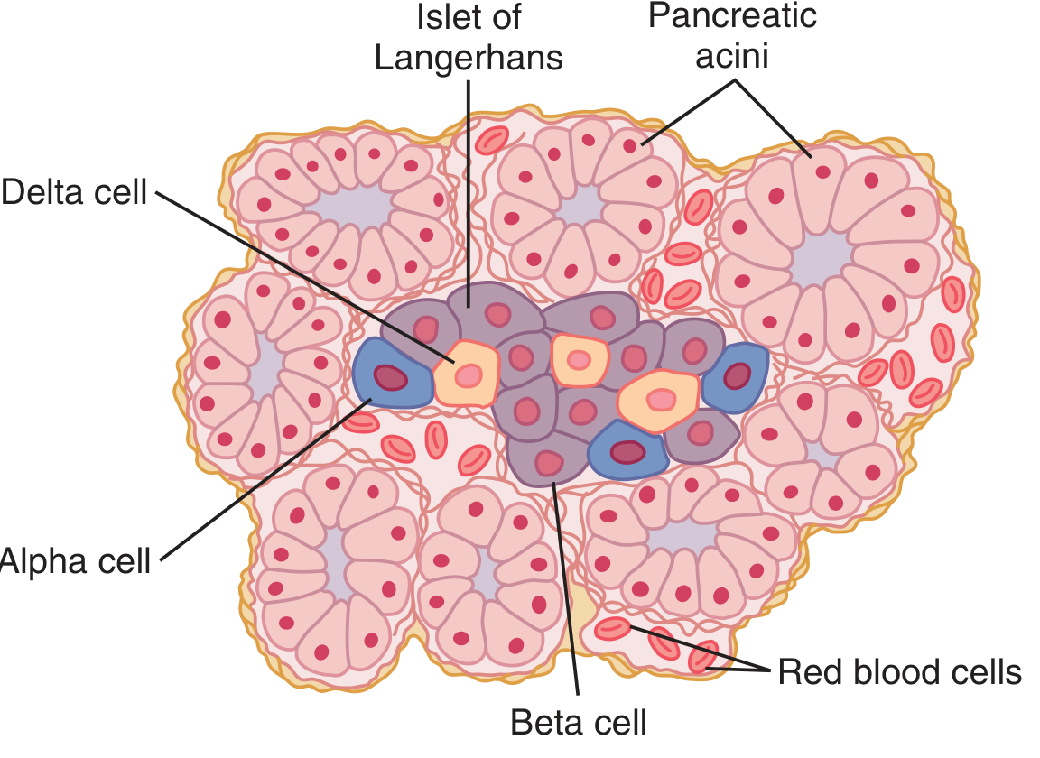
The human pancreas contains 1-2 million islets of Langerhans, each about 0.3 mm in diameter. Cell composition:
| Cell Type | Proportion | Hormone Secreted |
|---|---|---|
| Beta (B) cells | ~60% | Insulin + Amylin (IAPP) |
| Alpha (A) cells | ~25% | Glucagon |
| Delta (D) cells | ~10% | Somatostatin |
| PP cells | Small numbers | Pancreatic polypeptide |
Key regulatory interactions: insulin inhibits glucagon secretion; amylin inhibits insulin secretion; somatostatin inhibits both insulin and glucagon.
Insulin secretion is triggered by energy abundance (especially carbohydrate intake). Its metabolic effects include:
-
Stimulating glycogen synthesis in liver and muscle
-
Inhibiting hepatic gluconeogenesis and glycogenolysis
-
Promoting fatty acid synthesis and fat storage
-
Stimulating protein anabolism
-
Facilitating glucose uptake in peripheral tissues (muscle, fat) via GLUT4 transporters
-
Guyton & Hall Textbook of Medical Physiology, p. 965
3. Classification (ADA)
The ADA classifies DM into four main types:
Type 1 DM (T1DM)
- Due to autoimmune beta-cell destruction leading to absolute insulin deficiency
- Accounts for 5-10% of all DM
- Markers of autoimmune destruction: islet cell autoantibodies, anti-insulin antibodies, anti-GAD65 antibodies, anti-IA2 antibodies
- Genetically susceptible individuals + environmental trigger (enteroviruses implicated)
-
60 genetic loci identified, many implicating the immune system
- No sex difference in incidence
Type 2 DM (T2DM)
- Due to progressive loss of beta-cell insulin secretion on the background of insulin resistance
- Accounts for ~90-95% of all DM
- Strong genetic component; polygenic; influenced by obesity, physical inactivity, age
Gestational DM (GDM)
- Diabetes diagnosed in pregnancy that was not clearly overt diabetes prior to gestation
- Associated with polycystic ovary syndrome, preeclampsia, and pregnancy hypertension
Other Specific Types
-
Monogenic diabetes - MODY (maturity-onset diabetes of the young), neonatal diabetes
-
Exocrine pancreatic disease - cystic fibrosis, pancreatitis, pancreatic cancer
-
Drug/chemical-induced - glucocorticoids, HIV/AIDS treatment, post-organ transplantation
-
Creasy & Resnik's Maternal-Fetal Medicine; Harrison's Principles of Internal Medicine 22E
4. Pathophysiology
Type 1 DM
T1DM is a chronic autoimmune disease in which immune-mediated destruction of (or damage to) beta-cells results in insulin deficiency and hyperglycemia. The process involves T-cell mediated attack on the islets. Without insulin:
- Glycogenolysis and gluconeogenesis are unrestrained → hyperglycemia
- Lipolysis accelerates → free fatty acids → ketone bodies → diabetic ketoacidosis (DKA)
- Protein catabolism increases → wasting
Type 2 DM
Two central defects:
- Insulin resistance - peripheral tissues (muscle, liver, adipose) fail to respond normally to insulin
- Progressive beta-cell dysfunction - beta cells initially hypersecrete insulin to compensate, but eventually fail
Additional contributors include:
- Increased hepatic glucose production
- Impaired incretin effect (reduced GLP-1 response)
- Increased glucagon secretion from alpha cells
- Increased renal glucose reabsorption
- Lipotoxicity and glucotoxicity accelerating beta-cell apoptosis
Insulin Actions on Specific Tissues
Liver:
- Inactivates liver phosphorylase → prevents glycogen breakdown
- Increases glucokinase activity → traps glucose inside hepatocytes
- Activates glycogen synthase → promotes glycogen storage
- Inhibits gluconeogenesis
- Excess glucose converted to fatty acids → VLDL → fat storage
Muscle:
- Increases glucose uptake via GLUT4
- Promotes glycogen synthesis
- Promotes protein synthesis, inhibits protein catabolism
Adipose:
- Promotes fat storage; inhibits hormone-sensitive lipase (lipolysis)
Brain:
-
Little effect on glucose uptake (insulin-independent); however, insulin affects feeding behavior and energy metabolism
-
Guyton & Hall Textbook of Medical Physiology, p. 965-970
5. Diagnosis
Diagnostic Criteria (ADA - Nonpregnant Adults)
| Test | Diabetes | Prediabetes |
|---|---|---|
| Fasting plasma glucose (FPG) | ≥126 mg/dL (7.0 mmol/L) | 100-125 mg/dL (IFG) |
| 2-hr plasma glucose (75g OGTT) | ≥200 mg/dL (11.1 mmol/L) | 140-199 mg/dL (IGT) |
| HbA1c | ≥6.5% (48 mmol/mol) | 5.7-6.4% |
| Random plasma glucose | ≥200 mg/dL + symptoms | - |
- In an asymptomatic patient, two abnormal tests are required to confirm the diagnosis.
- A single test suffices in a symptomatic patient with classic hyperglycemia symptoms.
Key Investigations
- HbA1c - reflects mean glucose over ~3 months; used for both diagnosis and monitoring
- C-peptide - distinguishes residual beta-cell function; low/absent in T1DM
- Autoantibodies (GAD65, IA2, insulin) - confirm autoimmune T1DM
- Fasting lipid profile - dyslipidemia common in T2DM
- Urinary albumin-to-creatinine ratio (UACR) - screening for nephropathy
- eGFR - assess renal function
- Fundoscopy / dilated eye exam - screening for retinopathy
Glycemic Targets
- HbA1c goal: <7% for most patients; individualized based on patient age, comorbidities, hypoglycemia risk
- Fasting glucose: 80-130 mg/dL
- Post-prandial glucose (<2h): <180 mg/dL
6. Management
Goals of Therapy (Harrison's 2025)
- Eliminate symptoms related to hyperglycemia
- Reduce/eliminate long-term microvascular and macrovascular complications
- Allow the patient to achieve as normal a lifestyle as possible
6a. Lifestyle Modifications (All Types)
- Medical nutrition therapy (MNT): reduce refined carbohydrates, increase fiber, limit saturated fats
- Regular aerobic exercise (≥150 min/week of moderate-intensity)
- Weight loss in overweight/obese patients (even 5-10% weight loss significantly improves insulin sensitivity)
- Smoking cessation
- Self-monitoring of blood glucose (SMBG) or continuous glucose monitoring (CGM)
6b. Type 1 DM - Insulin Therapy
Insulin replacement must mimic physiologic insulin secretion: basal + prandial (bolus) coverage.
Insulin Preparations:
| Preparation | Onset | Peak | Duration |
|---|---|---|---|
| Rapid-acting (Aspart, Glulisine, Lispro) | <0.25 h | 0.5-1.5 h | 3-5 h |
| Short-acting (Regular) | 0.5-1.0 h | 2-3 h | 4-8 h |
| Inhaled human insulin | <0.25 h | 1-2 h | 3 h |
| Intermediate-acting (NPH) | 2-4 h | 4-10 h | 10-16 h |
| Long-acting (Glargine, Detemir) | 1-9 h | Flat/minimal | 20-24 h |
| Ultra-long-acting (Degludec) | 1-9 h | Flat | >42 h |
Delivery Systems:
- Multiple daily injections (MDI): most common
- Continuous subcutaneous insulin infusion (CSII/insulin pump)
- Sensor-augmented pump: integrates CGM; suspends infusion when glucose is low or predicted to drop
- Automated insulin delivery (AID/"closed-loop"): pump + CGM + algorithm adjusts basal rate in real time
6c. Type 2 DM - Pharmacotherapy
Step 1: Metformin (first-line, unless contraindicated)
- Mechanism: decreases hepatic glucose production (inhibits mitochondrial complex I), improves peripheral insulin sensitivity
- Benefits: weight-neutral or modest weight loss, low hypoglycemia risk, low cost, cardiovascular neutrality
- Contraindications: eGFR <30 mL/min (risk of lactic acidosis), iodinated contrast (hold temporarily)
Additional agents (based on comorbidities):
| Drug Class | Examples | Key Benefit | Key Risk |
|---|---|---|---|
| GLP-1 receptor agonists | Semaglutide, Liraglutide, Dulaglutide | CV & renal protection, weight loss | Nausea, pancreatitis risk |
| SGLT-2 inhibitors | Empagliflozin, Dapagliflozin, Canagliflozin | CV & renal protection, weight loss, heart failure benefit | UTI/genital infections, euglycemic DKA |
| DPP-4 inhibitors | Sitagliptin, Saxagliptin | Weight neutral, well tolerated | Moderate HbA1c lowering |
| Sulfonylureas | Glipizide, Glyburide, Glimepiride | Effective, inexpensive | Hypoglycemia, weight gain |
| Thiazolidinediones | Pioglitazone | Durable glucose control, NASH benefit | Weight gain, fluid retention, fracture risk, heart failure |
| Insulin | Various | Most potent glucose lowering | Hypoglycemia, weight gain |
| Alpha-glucosidase inhibitors | Acarbose | Post-prandial glucose | GI side effects |
2024 ACP Guideline update: Newer agents (GLP-1 RAs and SGLT-2i) are recommended for T2DM patients with established CVD or high CVD risk, CKD, or heart failure.
- Harrison's Principles of Internal Medicine 22E, p. 3248
7. Acute Complications
7a. Diabetic Ketoacidosis (DKA)
Primarily in T1DM (rarely T2DM, and as euglycemic DKA with SGLT-2 inhibitors).
Pathophysiology:
Relative/absolute insulin deficiency + counterregulatory hormone excess (glucagon, catecholamines, cortisol, GH):
- Glycogenolysis + gluconeogenesis → hyperglycemia
- Lipolysis → free fatty acids → beta-oxidation → ketone bodies (beta-hydroxybutyrate >> acetoacetate)
- Results in high anion-gap metabolic acidosis
Diagnostic Criteria:
- Serum glucose >250 mg/dL (>13.9 mmol/L)
- Ketonemia/ketonuria
- Metabolic acidosis: serum bicarbonate <15-18 mmol/L, pH 6.8-7.3, elevated anion gap
Clinical Features:
| Symptoms | Signs |
|---|---|
| Nausea/vomiting | Tachycardia |
| Thirst/polyuria | Hypotension |
| Abdominal pain | Kussmaul respirations |
| Weakness | Fruity breath (acetone) |
| Altered mental status | Dehydration |
Management:
- IV fluids (0.9% NaCl initially)
- Insulin infusion (regular insulin IV)
- Potassium replacement (hypokalemia risk as acidosis corrects)
- Bicarbonate only if pH <6.9
- Monitor glucose, electrolytes, anion gap
- Treat precipitating cause (infection is most common)
Note: Despite total-body K+ deficit, serum K+ at presentation may be normal/elevated due to acidosis driving K+ extracellularly. Must replace K+ before or with insulin to prevent life-threatening hypokalemia.
7b. Hyperglycemic Hyperosmolar State (HHS)
- Primarily in T2DM, older adults
- Extreme hyperglycemia (often >600 mg/dL), severe dehydration, hyperosmolality (>320 mOsm/kg)
- No significant ketoacidosis (enough residual insulin to suppress ketogenesis)
- Higher mortality than DKA
- Treatment: aggressive fluid replacement, insulin, electrolyte management
7c. Hypoglycemia
- Most common acute complication in treated diabetes
- Symptoms: sweating, tremor, palpitations (adrenergic); confusion, seizures (neuroglycopenic)
- Severe hypoglycemia threshold: glucose <54 mg/dL with symptoms
- Hypoglycemia unawareness: repeated hypoglycemia blunts counterregulatory epinephrine response; common with long-standing DM and autonomic neuropathy
- Treatment: 15-15 rule (15g fast-acting carbs, recheck in 15 min); glucagon kit for severe/unconscious patients
8. Chronic Complications
Diabetes-related complications affect many organ systems. Microvascular complications generally appear after the second decade of hyperglycemia; macrovascular complications (ASCVD) may precede clinical diagnosis.
8a. Microvascular Complications
Diabetic Retinopathy
- Leading cause of new blindness in adults in the USA
- Stages:
- Non-proliferative diabetic retinopathy (NPDR): microaneurysms, hard exudates, dot/blot hemorrhages, cotton-wool spots
- Proliferative diabetic retinopathy (PDR): neovascularization (new vessel formation) - high risk of vitreous hemorrhage and retinal detachment
- Diabetic macular edema (DME): most common cause of visual loss
- Prevention: tight glycemic control, blood pressure control
- Treatment: laser photocoagulation, anti-VEGF injections (bevacizumab, ranibizumab), vitrectomy
Diabetic Nephropathy
- Leading cause of end-stage renal disease (ESRD)
- Pathological hallmark: Kimmelstiel-Wilson nodules (glomerular nodular sclerosis)
- Stages: microalbuminuria (UACR 30-300 mg/g) → macroalbuminuria → declining GFR → ESRD
- Prevention/treatment: tight glycemic control, ACE inhibitors or ARBs (reduce proteinuria and progression), SGLT-2 inhibitors (have independent renoprotective effect), blood pressure control
- 2024 data: Semaglutide shown to reduce progression of CKD in T2DM patients (FLOW trial, NEJM 2024)
Diabetic Neuropathy
- Present in ~50% of individuals with long-standing T1DM or T2DM
- Risk factors: duration of DM, poor glycemic control, higher BMI, smoking, ASCVD, hypertriglyceridemia, hypertension
Forms:
- Distal symmetric polyneuropathy (DSPN) - most common; numbness, tingling, burning starting in feet, spreading proximally ("stocking-glove" distribution); worse at night; on exam: loss of protective sensation (10-g monofilament), reduced vibration sense, absent ankle reflexes
- Autonomic neuropathy: cardiovascular (resting tachycardia, orthostatic hypotension, QTc prolongation, sudden death risk), GI (gastroparesis, diabetic diarrhea), genitourinary (bladder dysfunction, erectile dysfunction), sudomotor (hyperhidrosis upper extremities, anhidrosis feet)
- Mononeuropathy: sudden-onset, single nerve involvement (CN III palsy with pupil sparing, carpal tunnel, femoral neuropathy)
- Polyradiculopathy: Diabetic amyotrophy - severe pain, weakness in thigh and hip muscles
Screening: annually, starting 5 years post-T1DM diagnosis; at time of T2DM diagnosis.
8b. Macrovascular Complications
- Coronary heart disease (CHD): most common cause of death; DM confers 2-4x increased risk
- Peripheral arterial disease (PAD): claudication, critical limb ischemia; major contributor to lower-extremity amputations
- Cerebrovascular disease: stroke risk 2-4x higher
- Heart failure: DM is an independent risk factor; diabetic cardiomyopathy occurs even without coronary disease
Key fact: Women with DM lose their cardioprotective female sex advantage - their CVD rates equal those of diabetic men, and women with DM have a sixfold greater risk of dying from CVD compared to non-diabetic women.
8c. Other Complications
| System | Complication |
|---|---|
| Gastrointestinal | Gastroparesis (delayed gastric emptying), diabetic diarrhea |
| Genitourinary | Neurogenic bladder, erectile dysfunction, recurrent UTIs |
| Dermatologic | Necrobiosis lipoidica, acanthosis nigricans, diabetic dermopathy, poor wound healing |
| Musculoskeletal | Charcot neuroarthropathy (Charcot joint), frozen shoulder, Dupuytren's contracture |
| Foot | Diabetic foot ulcers, osteomyelitis, gangrene, amputation |
| Infection | Increased susceptibility; malignant otitis externa, rhinocerebral mucormycosis, emphysematous pyelonephritis |
9. Diabetic Foot
- Results from neuropathy + peripheral vascular disease + infection
- Loss of protective sensation (from DSPN) leads to unrecognized trauma and ulceration
- Anhidrosis causes dry, cracked skin - portal of entry for infection
- Annual foot exams essential; patient education on foot care
- Offloading (total contact cast) for plantar ulcers
- Revascularization for ischemic ulcers
10. Monitoring & Targets
| Parameter | Target |
|---|---|
| HbA1c | <7% (individualized) |
| Fasting glucose | 80-130 mg/dL |
| Post-prandial glucose | <180 mg/dL |
| Blood pressure | <130/80 mmHg |
| LDL cholesterol | <70 mg/dL (high-risk) |
| Urine albumin/creatinine | <30 mg/g |
Monitoring tools:
- HbA1c: every 3 months if not at target; every 6 months if stable
- SMBG: finger-stick glucose monitoring
- CGM (Continuous Glucose Monitoring): real-time interstitial glucose; time-in-range (TIR) >70% is the current quality metric target
11. Special Populations
Gestational DM (GDM)
- Screening: all pregnant women at 24-28 weeks with 75g OGTT (or 1-hour 50g screen followed by 3-hour diagnostic test)
- Risks: macrosomia, shoulder dystocia, neonatal hypoglycemia, preeclampsia
- Treatment: diet/exercise first; if glucose targets not met, insulin (first-line pharmacotherapy in pregnancy); metformin and glyburide are alternatives but cross the placenta
Type 1 DM in Pregnancy (Pre-gestational)
- Risk of congenital malformations is 2-3x higher; strongly correlated with first-trimester glycemic control (HbA1c)
- Target HbA1c <6.5% pre-conception; <7% during pregnancy
12. Screening & Prevention
- T2DM screening: all adults ≥35 years; younger if overweight/obese + risk factors (family history, GDM history, hypertension, dyslipidemia, PCOS, high-risk ethnicity)
- Prevention of T2DM in prediabetes:
- Intensive lifestyle intervention (Diabetes Prevention Program): reduces T2DM incidence by ~58%
- Metformin: reduces incidence by ~31% (especially effective in <60 years, BMI ≥35, history of GDM)
- Weight loss is the single most effective intervention
Key Summary Table
| Feature | Type 1 DM | Type 2 DM |
|---|---|---|
| Pathogenesis | Autoimmune beta-cell destruction | Insulin resistance + beta-cell dysfunction |
| Age of onset | Usually <30 years | Usually >40 (but increasingly in youth) |
| Body habitus | Usually lean | Usually overweight/obese |
| Ketosis | Prone | Resistant (usually) |
| Insulin required | Always | Eventually |
| Autoantibodies | Present | Absent |
| C-peptide | Low/absent | Normal or elevated (early) |
| First-line Rx | Insulin | Metformin + lifestyle |
| Acute emergency | DKA | HHS |
References: Harrison's Principles of Internal Medicine 22E (2025), Chapter 416-417 | Guyton & Hall Textbook of Medical Physiology, Chapter 79 | Creasy & Resnik's Maternal-Fetal Medicine, Chapter 59 | Textbook of Family Medicine 9e, Chapter 20 | Tintinalli's Emergency Medicine, Chapter on DKA
This is a shared conversation. Sign in to Orris to start your own chat.