
hernia
I'll look up hernia in the medical library for you. Let me read the skill first.
Searching sources...
Reading File
"Hernia" is a broad topic. Let me search for it across the medical library and pull relevant images in parallel.
Finding Sources
Searching Images
hernia types anatomy diagram inguinal umbilical
Good. I have strong surgical textbook sources. Let me read the main hernia chapters from Bailey & Love (the classic surgical reference) and Maingot's Abdominal Operations, plus specific hernia types from Roberts & Hedges.
Reading File
Reading File
Reading File
Excellent content retrieved. Now let me fetch the key anatomical diagram and read the main hernia chapter from Bailey & Love and the comprehensive GI textbook.
Loading Image
Loading Image
Finding Sources
I now have the key diagrams and good textbook content. Let me read the dedicated inguinal hernia section from Bailey & Love (the most authoritative surgical reference) and Schwartz's Principles of Surgery for management details.
Reading File
Reading File
I now have comprehensive, high-quality content from multiple authoritative surgical textbooks. Let me compile the full answer.
Hernia
A hernia is the protrusion of a viscus (or part of a viscus) through a defect in the wall of its containing cavity, typically covered by a peritoneal sac. The abdominal wall is by far the most common site.
Definition & Basic Anatomy
Every hernia has three components:
- Sac - the peritoneal covering
- Contents - bowel (most common), omentum, bladder, ovary
- Coverings - layers of abdominal wall over the sac
Classification by Type
1. Inguinal Hernia (most common overall)
The most common hernia in both sexes, ~10x more frequent in men than women. There are two fundamentally different subtypes:
| Feature | Indirect (lateral/oblique) | Direct (medial) |
|---|---|---|
| Pathway | Through deep inguinal ring, along canal, out superficial ring | Through posterior wall of canal (Hesselbach's triangle) |
| Relation to inferior epigastric vessels | Lateral | Medial |
| Origin | Usually congenital (patent processus vaginalis) | Acquired (weakness of transversalis fascia) |
| Risk of strangulation | Higher | Lower |
| Common in | Young males, premature infants | Older men |
Anatomy of the inguinal canal: The testis descends through the deep inguinal ring (midway between ASIS and pubic tubercle, 2-3 cm above femoral artery), travels medially, and exits via the superficial inguinal ring (an inverted-V defect in external oblique aponeurosis). The conjoint tendon (fused transversus + internal oblique) forms the posterior wall support. The inferior epigastric vessels lie just medial to the deep ring - this is the landmark separating indirect (lateral) from direct (medial) hernias. - Bailey and Love's Short Practice of Surgery, 28th Edition
Pantaloon hernia: A combination of direct + indirect hernias straddling the inferior epigastric vessels. Difficult to reduce manually; often found at surgical exploration. - Roberts and Hedges' Clinical Procedures in Emergency Medicine
2. Femoral Hernia
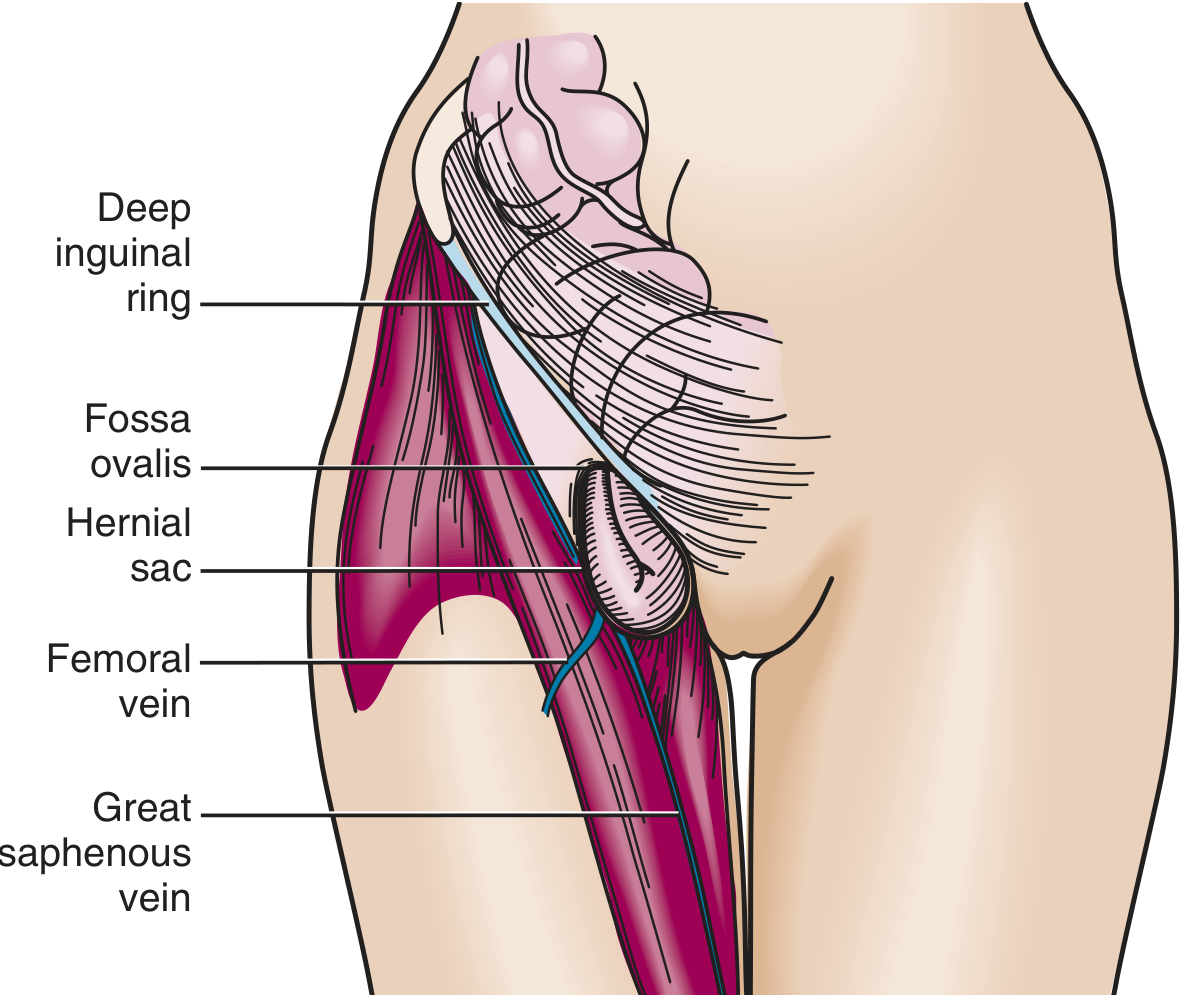
- Protrudes inferior to the inguinal ligament through a defect in transversalis fascia, into the femoral canal (medial to femoral vein, lateral to lacunar ligament)
- Presents as a medial thigh mass below the groin crease
- More common in women
- High incarceration rate (~45%) due to the narrow rigid femoral ring
- Rare in children
- Roberts and Hedges' Clinical Procedures in Emergency Medicine
3. Ventral Hernias (Anterior Abdominal Wall)
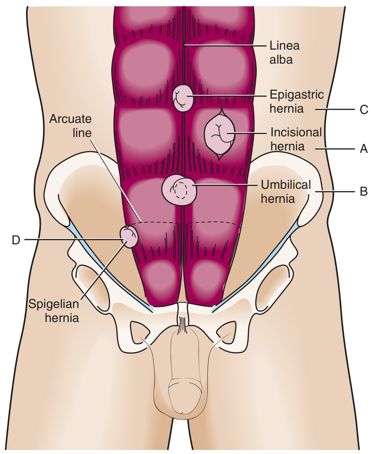
| Type | Location | Key Points |
|---|---|---|
| Incisional (A) | Through prior surgical scar | Occurs in ~1/5 post-surgical patients; recurrence 20-50%; larger defects paradoxically have lower incarceration risk |
| Umbilical (B) | Through fibromuscular ring of umbilicus | Congenital (usually resolves by age 5); acquired in adults with increased intra-abdominal pressure (obesity, ascites, pregnancy); adults have higher incarceration risk |
| Epigastric (C) | Midline through linea alba, above umbilicus | Between xiphoid and umbilicus; found in ~4% of infants |
| Spigelian (D) | Through linea semilunaris, at arcuate line | Lateral to rectus, at/below arcuate line; often interparietal (between muscle layers) so can be missed clinically |
Roberts and Hedges' Clinical Procedures in Emergency Medicine
Complications
| Complication | Description |
|---|---|
| Reducible | Contents can be returned to cavity manually or spontaneously |
| Irreducible (Incarcerated) | Contents cannot be reduced; may obstruct bowel |
| Obstructed | Bowel lumen obstructed without vascular compromise |
| Strangulated | Vascular supply cut off → ischemia → necrosis → surgical emergency |
| Richter's hernia | Only part of the bowel wall caught in the defect; full obstruction may be absent but strangulation risk is high |
Pediatric vs. Adult Hernias
Pediatric (indirect): Caused by failure of closure of the processus vaginalis. All congenital pediatric hernias are indirect by definition. High incarceration risk in infants due to narrow ring. Treatment: herniorrhaphy (closing the patent processus vaginalis). Parent counseling: not caused by child's crying or activity. - Schwartz's Principles of Surgery, 11th Edition
Adult: May be indirect or direct. Requires hernioplasty (reconstruction of the inguinal floor, not just sac ligation).
Management Overview
Conservative: A supportive truss may temporarily manage symptoms in patients unfit for surgery, but is not curative.
Surgical repair options:
- Open (tension-free mesh repair / Lichtenstein): Gold standard for most inguinal hernias. Mesh placed over the posterior inguinal wall.
- Laparoscopic (TEP - totally extraperitoneal / TAPP - transabdominal preperitoneal): Faster recovery, less post-op pain; preferred for bilateral or recurrent hernias.
- Robotic-assisted: Increasingly used, especially for complex repairs.
Emergency surgery is required for strangulation, non-reducible incarceration, or bowel obstruction that cannot be manually reduced. Bowel resection may be needed if necrosis is found.
Key Clinical Pearls
- A cough impulse felt at the superficial inguinal ring is the classic sign of an inguinal hernia.
- Femoral hernias are the most dangerous groin hernia due to their high strangulation rate.
- The Shouldice repair (layered tissue repair without mesh) is the gold standard tissue repair for inguinal hernia.
- In women, always consider an obturator hernia if an elderly woman presents with bowel obstruction and medial thigh pain (Howship-Romberg sign).
- An undescended testis is associated with an ipsilateral patent processus vaginalis and therefore hernia risk. - Bailey and Love's Short Practice of Surgery, 28th Edition
This is a shared conversation. Sign in to Orris to start your own chat.