
ArDs
"acute respiratory distress syndrome" AND management
ARDS acute respiratory distress syndrome chest X-ray bilateral infiltrates diffuse alveolar damage
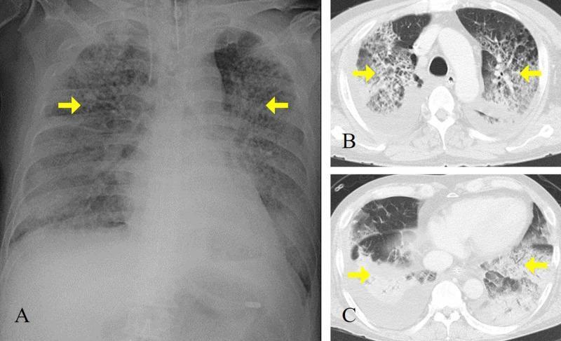
This composite diagnostic image features a chest X-ray (A) and axial CT scans (B, C) of a patient with Acute Respiratory Distress Syndrome (ARDS). (A) An anteroposterior chest radiograph demonstrates diffuse, bilateral alveolar infiltrates and patchy opacification across both lung fields, notably sparing the upper zones, with a normal cardiac silhouette and no signs of cardiomegaly. Yellow arrows highlight dense consolidation in the mid-lung regions. (B, C) Axial CT images at different thoracic levels reveal extensive, bilateral ground-glass opacities and dense consolidation, particularly in the dependent posterior regions of the lower lobes. Associated reticular patterns suggest interstitial involvement. The mediastinal structures and great vessels appear normal, and there is no evidence of significant pleural effusion. The imaging findings are classically consistent with non-cardiogenic pulmonary edema and diffuse alveolar damage typical of severe viral pneumonia (e.g., H1N1 influenza) progressing to ARDS. This material is suitable for training in intensive care medicine and radiology, focusing on the visual differentiation between cardiogenic and non-cardiogenic pulmonary opacities.
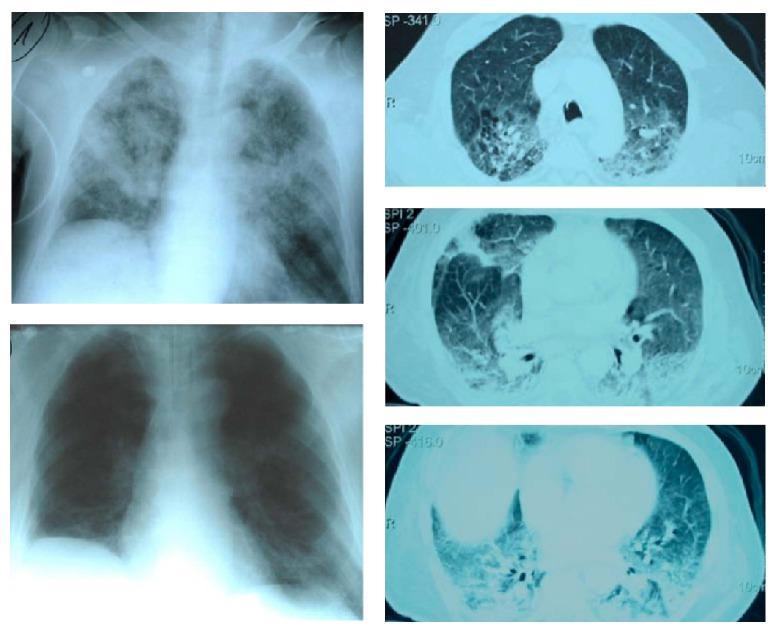
This composite diagnostic image includes chest X-rays and axial CT scans illustrating the progression and resolution of Acute Respiratory Distress Syndrome (ARDS). The top-left anterior-posterior chest X-ray shows diffuse, patchy bilateral alveolar infiltrates and ground-glass opacities in a postoperative patient, characteristic of pulmonary edema. To the right, three axial CT scan slices (cranial to caudal) demonstrate significant bilateral dorsal consolidation and dependent infiltrates with air bronchograms, typical of ARDS morphology. The bottom-left chest X-ray shows the same patient following Continuous Extrathoracic Negative Pressure Ventilation (CENPV), displaying marked regression of the bilateral infiltrates, improved lung aeration, and a reduction in previously noted pleural effusions. The educational focus is on the radiographic monitoring of lung recruitment and the resolution of pulmonary consolidations in critical care. This material is relevant for pulmonary medicine, anesthesia, and intensive care specialties.
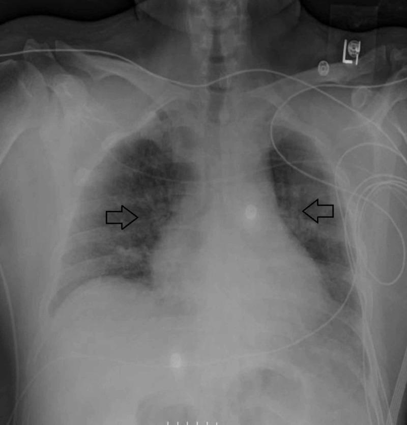
This diagnostic image is an anteroposterior (AP) chest X-ray demonstrating bilateral pulmonary infiltrates in a patient with respiratory failure. The lung fields show diffuse, patchy, and reticular opacities (indicated by horizontal black arrows) consistent with airspace disease and interstitial involvement. The infiltrates appear most prominent in the mid-to-lower lung zones, resulting in increased lung density and haziness that partially obscures normal bronchovascular markings. Several medical devices are visible, including multiple ECG leads and external monitoring tubing overlying the chest, consistent with a patient in an intensive care setting. The mediastinal silhouette and cardiac contours are present, though partially obscured by the adjacent pulmonary opacities. The skeletal structures, including the clavicles and ribs, appear intact. This radiograph serves as a clinical follow-up during intensive management (e.g., ECMO and corticosteroid therapy), illustrating the radiological presentation and interval assessment of diffuse alveolar damage or pulmonary vasculitis manifesting as Acute Respiratory Distress Syndrome (ARDS).
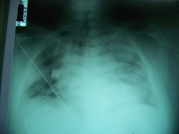
This diagnostic image is an anteroposterior (AP) chest x-ray demonstrating clinical features consistent with Acute Respiratory Distress Syndrome (ARDS). The radiograph reveals diffuse, bilateral pulmonary infiltrates with extensive alveolar opacities and increased density in both lung fields, appearing as a 'white-out' effect that obscures the normal bronchovascular markings. The distribution is patchy and widespread, affecting both central and peripheral regions, indicative of non-cardiogenic pulmonary edema or severe inflammatory consolidation. A prominent medical device is visible: a catheter or pulmonary artery line enters from the left upper thoracic region, traversing diagonally across the right lung field, featuring a distinct coiled (pigtail) component. The image illustrates the radiographic progression of respiratory failure in the context of critical care management, such as that seen in severe pneumonia or systemic inflammatory responses like the Jarisch-Herxheimer reaction.
Acute Respiratory Distress Syndrome (ARDS)
Definition (Berlin Criteria, 2012)
| Criterion | Detail |
|---|---|
| Onset | Acute, within 1 week of a clinical insult or new/worsening respiratory symptoms |
| Chest radiograph | Bilateral opacities not fully explained by effusions, lobar collapse, or nodules |
| Origin of edema | Not primarily explained by cardiac failure/fluid overload (echocardiography may be needed) |
| Oxygenation | PaO2/FiO2 ≤300 mmHg with PEEP ≥5 cmH2O |
- Mild: PaO2/FiO2 200-300 mmHg
- Moderate: PaO2/FiO2 100-200 mmHg
- Severe: PaO2/FiO2 <100 mmHg
Epidemiology
- Annual incidence up to 60 cases per 100,000 population (pre-COVID data)
- Accounts for ~10% of all ICU admissions
- Mortality: ~40% overall; higher in severe ARDS
Etiology
| Direct Lung Injury | Indirect (Extrapulmonary) Injury |
|---|---|
| Pneumonia | Sepsis |
| Aspiration of gastric contents | Severe trauma / multiple bone fractures |
| Pulmonary contusion | Flail chest |
| Near-drowning | Head trauma |
| Toxic inhalation | Burns |
| Multiple transfusions | |
| Drug overdose | |
| Pancreatitis | |
| Post-cardiopulmonary bypass |
80% of cases are due to pneumonia, sepsis (40-60%), aspiration, trauma, or multiple transfusions. Risk is amplified in alcohol abuse, older age, and APACHE II score ≥16 in trauma patients.
Pathophysiology: Three Phases
1. Exudative Phase (Days 1-7)
- Diffuse alveolar damage (DAD): neutrophil influx, release of proteases, reactive oxygen species, and pro-inflammatory cytokines (IL-1, IL-8, TNF-alpha)
- Disruption of alveolar-capillary barrier → protein-rich edema floods alveoli
- Hyaline membrane formation (fibrin + cell debris)
- Surfactant dysfunction → widespread atelectasis
- Result: severe V/Q mismatch and intrapulmonary shunting → refractory hypoxemia
2. Proliferative Phase (Days 7-21)
- Type II pneumocyte proliferation to repair alveolar epithelium
- Organization of intra-alveolar exudate
- Most patients begin recovering, but some progress
3. Fibrotic Phase (>21 days)
- Collagen deposition and fibrosis in some patients
- Reduced lung compliance, increased dead space, pulmonary hypertension
- Associated with prolonged ICU stay and poor prognosis
Clinical Features
- Rapid-onset severe dyspnea and hypoxemia (often within hours of insult)
- Diffuse bilateral crackles on auscultation
- Refractory hypoxemia (does not improve significantly with supplemental O2 alone)
- Reduced lung compliance (stiff lungs)
- Bilateral infiltrates on CXR / ground-glass opacities on CT
Management
Stepwise interventions by severity:
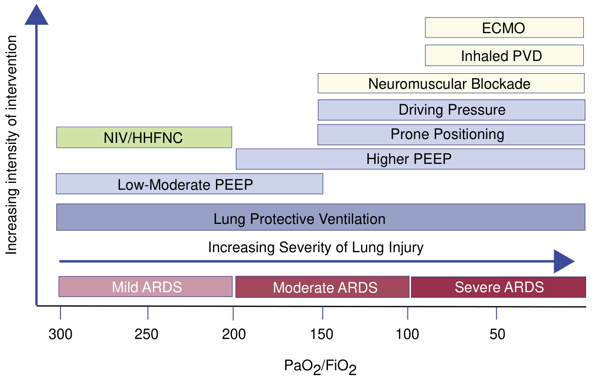
1. Lung-Protective Ventilation (ALL severity)
- Tidal volume (Vt): 6 mL/kg predicted body weight (ARDSnet trial)
- Plateau pressure <30 cmH2O
- Driving pressure <15 cmH2O (Vt / respiratory system compliance)
- Rationale: prevents ventilator-induced lung injury (VILI) from volutrauma and barotrauma
2. PEEP Optimization
- Low-moderate PEEP in mild ARDS
- Higher PEEP (>12-16 cmH2O) in moderate-severe ARDS to recruit collapsed alveoli
3. Conservative Fluid Strategy
- Avoid fluid overload; maintain negative to euvolemic fluid balance after initial resuscitation
- Reduces duration of mechanical ventilation
4. Prone Positioning (Moderate-Severe: PaO2/FiO2 <150)
- Prone ≥16 hours/day improves oxygenation and reduces mortality in severe ARDS
- The landmark PROSEVA trial (Guerin 2013) showed 28-day mortality reduction from 32.8% to 16% in severe ARDS
5. Neuromuscular Blockade (Moderate-Severe)
- 48-hour cisatracurium infusion may improve oxygenation and reduce barotrauma in moderate-severe ARDS
- Note: the 2019 PETAL Network RCT did not confirm mortality benefit vs. light sedation alone
6. Corticosteroids
- A 2026 systematic review/meta-analysis (Soumare et al., Ann Intern Med 2026, PMID 41325621) found systemic corticosteroids may reduce mortality in ARDS and pneumonia, though infectious risk must be considered
- Methylprednisolone is used in some protocols, particularly in fibroproliferative phase
7. Inhaled Pulmonary Vasodilators (Severe)
- Inhaled nitric oxide (iNO) or prostacyclins (aerosolized epoprostenol) improve oxygenation transiently
- No proven mortality benefit; used as rescue or bridge to ECMO
8. ECMO (Severe, refractory)
- Venovenous ECMO (VV-ECMO) for PaO2/FiO2 <80 despite maximal ventilator support
- Last-resort rescue therapy; referral to ECMO centers recommended
Mild ARDS
- NIV (non-invasive ventilation) or high-flow nasal cannula (HFNC/HHFNC) may be trialed
- A 2025 meta-analysis (Alsabri et al., Eur J Pediatr, PMID 40467999) supported early HFNC in respiratory distress
Differential Diagnosis
- Cardiogenic pulmonary edema (elevated PCWP >18 mmHg; echo shows LV dysfunction)
- Acute interstitial pneumonia (AIP/Hamman-Rich syndrome)
- Diffuse alveolar hemorrhage (DAH)
- Cryptogenic organizing pneumonia (COP)
Prognosis and Outcomes
- Mortality ~40% overall; higher with sepsis as precipitant
- Survivors often have long-term sequelae: muscle weakness, cognitive impairment, PTSD, reduced exercise tolerance
- Most lung function recovers to near-normal within 12 months in survivors, but fibrotic cases have persistent restrictive defect
- Harrison's Principles of Internal Medicine 22E (2025), Chapter 312
- Current Surgical Therapy 14e, ARDS Chapter
- Fishman's Pulmonary Diseases and Disorders
- Recent evidence: Soumare et al., Ann Intern Med 2026 (corticosteroids, PMID 41325621)