
Oral pathology
Reading File
The query "Oral pathology" is broad and has many valid directions. Let me clarify before diving in.
Asking for Preferences
What aspect of oral pathology are you interested in?
Asking for Preferences
Which specific lesion or condition?
Odontogenic cysts - great topic. Let me pull comprehensive information from the medical library and recent literature simultaneously.
Finding Sources
Searching PubMed
"odontogenic cysts"[MeSH Terms] AND classification
Good - let me now pull the comprehensive content from the main sections on odontogenic cysts, and also search for the classic types (dentigerous, radicular, OKC).
Reading File
Finding Sources
Excellent - Robbins has a dedicated ODONTOGENIC CYSTS AND TUMORS section. Let me pull that and the Scott-Brown's comprehensive content simultaneously.
Reading File
Reading File
I have rich content. Let me now pull the full Robbins OKC/dentigerous details and the Scott-Brown's dentigerous/radicular/OKC sections, and also fetch the key diagnostic image.
Reading File
Reading File
Loading Image
I now have very comprehensive content. Let me pull a bit more on the OKC and radicular cyst sections from Scott-Brown's before writing the full answer.
Reading File
I now have comprehensive content to write a detailed, well-structured answer. Let me compile it all.
Odontogenic Cysts - Comprehensive Overview
Classification
Odontogenic cysts arise from remnants of the tooth-forming apparatus. They are classified into two broad categories based on pathogenesis:
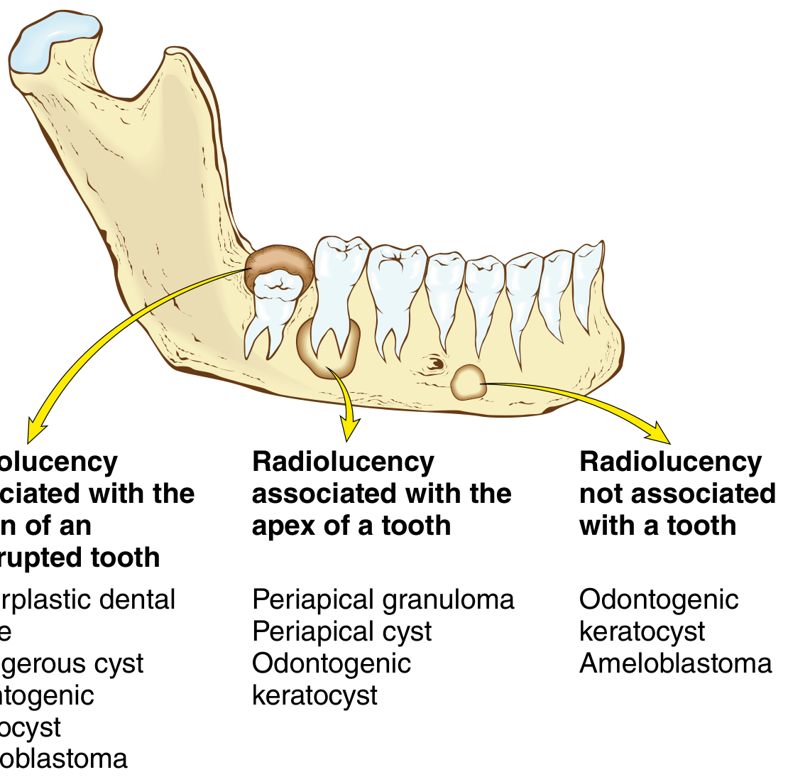
WHO Classification (Table 16.3 - Robbins; Table 25.3 - Scott-Brown's)
| Category | Cyst Type | Subtype |
|---|---|---|
| Inflammatory | Radicular (periapical/dental) cyst | Apical, lateral, residual |
| Inflammatory | Paradental (inflammatory collateral) cyst | - |
| Developmental | Dentigerous (follicular) cyst | Eruption cyst |
| Developmental | Odontogenic keratocyst (OKC) | - |
| Developmental | Gingival cyst of adults | - |
| Developmental | Lateral periodontal cyst | Botryoid variant |
| Developmental | Glandular odontogenic (sialo-odontogenic) cyst | - |
| Developmental | Calcifying odontogenic cyst (Gorlin cyst) | - |
| Developmental | Orthokeratinized odontogenic cyst | - |
Epithelial Origins
Three distinct epithelial remnants persist after odontogenesis and give rise to specific cysts:
- Rests of Malassez - from fragmentation of Hertwig's root sheath at root completion → Radicular cysts
- Reduced enamel epithelium - fusion of inner/outer enamel epithelia covering the unerupted crown → Dentigerous, eruption, and paradental cysts
- Rests (glands) of Serres - remnants of the dental lamina → Lateral periodontal, gingival, and keratocystic odontogenic tumour (OKC)
(Scott-Brown's Otorhinolaryngology, p. 443-444)
Individual Cysts in Detail
1. Radicular Cyst (Periapical / Dental Cyst)
- Most common jaw cyst overall
- Pathogenesis: Pulpitis (usually from caries or trauma) → pulp necrosis → periapical inflammation → stimulation of Rests of Malassez → epithelial proliferation → cyst formation
- Location: Root apex of a non-vital tooth (or lateral root canal in lateral variant; residual cyst remains after tooth extraction)
- Radiology: Unilocular radiolucency at tooth apex, with a radiopaque rim
- Treatment: Root canal treatment (enucleation if residual cyst remains)
- Note: The term "periapical granuloma" is still used despite the fact that the lesion does not show true granulomatous inflammation - it is actually epithelialized in true radicular cysts (Robbins, p. 687)
2. Dentigerous (Follicular) Cyst
- Second most common jaw cyst (accounts for ~15-18% of jaw cysts)
- Pathogenesis: Expansion of the dental follicle around the crown of an unerupted tooth; attached at the cemento-enamel junction (CEJ)
- Commonly associated teeth (in decreasing frequency): mandibular third molars ("wisdom teeth"), maxillary canines, mandibular second premolars, maxillary third molars
- Peak age: 2nd-3rd decades
- Clinical features: Often asymptomatic, found incidentally on X-ray for a missing tooth; may cause swelling, tooth displacement, secondary infection. Can reach up to 5 cm diameter.
- Radiology: Unilocular radiolucency attached to/surrounding the CEJ of an unerupted tooth
- Histology: Thin non-keratinized squamous or cuboidal/columnar lining; focal keratinization and mucous/ciliated cells possible via metaplasia
- Complications: Root resorption of adjacent teeth; transformation to ameloblastoma is a recognized (though rare) complication - ameloblastomas can form in the cyst wall
- Differential diagnosis for larger lesions: OKC, unicystic ameloblastoma
- Eruption cyst variant: Superficially located dentigerous cyst; presents as a bluish gum swelling over an erupting tooth, especially in children; cyclosporin therapy is a predisposing factor
(Scott-Brown's, pp. 444-445; Robbins, p. 686)
3. Odontogenic Keratocyst (OKC) / Keratocystic Odontogenic Tumour (KCOT)
- Derived from: Rests of Serres (dental lamina remnants)
- Important: Previously reclassified by the WHO (2005) as "Keratocystic Odontogenic Tumour (KCOT)" due to aggressive behavior; the 2017 WHO classification reverted to OKC terminology
- Location: Most often in the posterior mandible (angle/ramus region); can occur in a dentigerous relationship or independent of any tooth
- Clinical behavior: Locally aggressive; recurrence rates for inadequately removed lesions can be as high as 60%. About 80% of lesions are solitary.
- Gorlin syndrome (Nevoid Basal Cell Carcinoma Syndrome): Multiple OKCs = strong indicator; caused by germline loss-of-function mutations in PTCH (Patched tumor suppressor gene). Patients with multiple cysts must be evaluated for this syndrome.
- EGFR: Recent studies show EGFR signaling plays a role in OKC behavior and growth (PMID 37314601)
- Radiology: Unilocular or multilocular radiolucency; can mimic dentigerous cyst when associated with a tooth root; may expand along the medullary cavity with little cortical expansion (characteristic)
- Histology: Thin, uniform parakeratinized stratified squamous epithelium (typically 6-8 cell layers thick); flat epithelial-connective tissue interface; prominent palisaded basal cells with hyperchromatic nuclei ("tombstone" appearance); corrugated parakeratotic surface
- Treatment: Excision; curettage and partial excision result in high recurrence. Some advocate marsupialization followed by enucleation. Peripheral ostectomy and Carnoy's solution used adjunctively.
(Robbins, p. 686-688; Scott-Brown's, p. 445; K.J. Lee's Essential Otolaryngology)
4. Lateral Periodontal Cyst
- Derived from Rests of Serres
- Located lateral to the root of a vital tooth (distinguishes it from lateral radicular cyst of non-vital tooth)
- Botryoid variant: Multilocular / polycystic form; higher recurrence rate
5. Glandular Odontogenic Cyst (Sialo-Odontogenic Cyst)
- Rare but locally aggressive developmental cyst
- Contains gland-like structures (mucous cells, ciliated cells) within its lining - hence "glandular"
- Higher recurrence rate; must be distinguished from central mucoepidermoid carcinoma
6. Calcifying Odontogenic Cyst (Gorlin Cyst / CCOT)
- Contains "ghost cells" - keratinized cells lacking nuclei
- May contain calcifications
- Some cases progress to "dentinogenic ghost cell tumor" (solid variant)
- Recent reviews support reclassification into cystic vs. solid forms (PMID 39217025)
7. Orthokeratinized Odontogenic Cyst
- Distinct from OKC; lining shows orthokeratin (not parakeratin)
- Less aggressive than OKC; lower recurrence
- Not associated with Gorlin syndrome
- Important to distinguish histologically from OKC due to different prognosis
Radiology Summary - Differential Approach
| Radiolucency Pattern | Consider |
|---|---|
| Associated with crown of unerupted tooth | Dentigerous cyst, OKC, hyperplastic dental follicle, unicystic ameloblastoma |
| Associated with apex of a (non-vital) tooth | Radicular/periapical cyst, periapical granuloma, OKC |
| Not associated with a tooth | OKC, ameloblastoma, lateral periodontal cyst |
(Robbins, Fig. 16.8)
Key Distinguishing Features at a Glance
| Feature | Radicular | Dentigerous | OKC |
|---|---|---|---|
| Origin | Rests of Malassez | Reduced enamel epithelium | Rests of Serres |
| Category | Inflammatory | Developmental | Developmental |
| Tooth vitality | Non-vital | Unerupted tooth | Any (or independent) |
| Location | Apex | CEJ of crown | Posterior mandible |
| Recurrence | Low | Low | High (up to 60%) |
| Histology | Non-keratinized lining | Thin cuboidal/squamous | Parakeratinized, uniform, palisaded basal layer |
| Gorlin syndrome | No | No | Yes (multiple OKCs) |
Recent Evidence
- A 2024 systematic review and meta-analysis (PMID 38530559) evaluated machine learning accuracy in diagnosing odontogenic cysts and tumors - showing promising diagnostic performance for AI-assisted radiographic classification.
- A 2023 review (PMID 37314601) highlights the significance of EGFR investigation in OKC, with EGFR overexpression contributing to the aggressive behavior and recurrence potential of OKC.
- A 2025 review (PMID 39217025) proposes a novel outlook on classification, diagnosis, and management of calcifying odontogenic cysts.
Sources: Robbins, Cotran & Kumar Pathologic Basis of Disease (pp. 686-688) | Scott-Brown's Otorhinolaryngology Head & Neck Surgery (pp. 443-450) | K.J. Lee's Essential Otolaryngology | Cummings Otolaryngology Head & Neck Surgery
This is a shared conversation. Sign in to Orris to start your own chat.