
Help me write a assignment of Physiology of menstrual cycle in subject of Gynecology and obstetrics
Reading File
Now I'll search the medical library for comprehensive content on the menstrual cycle physiology.
Finding Sources
Finding Sources
Excellent! Found the primary source in Berek & Novak's Gynecology. Now let me read the content comprehensively from multiple sources in parallel.
Reading File
Reading File
Reading File
Excellent content retrieved. Let me now read more detailed content from Berek & Novak's Gynecology and fetch the hormone cycle diagram image.
Reading File
Loading Image
Reading File
I now have very rich content. Let me fetch the comprehensive menstrual cycle diagram from Berek & Novak's and read more detailed sections on follicular development and endometrial changes.
Loading Image
Reading File
I now have comprehensive content from authoritative textbooks. Let me compile the full assignment.
Physiology of the Menstrual Cycle
Subject: Gynecology and Obstetrics
Introduction
The menstrual cycle is a complex, highly orchestrated series of physiological events that prepares the female reproductive system for pregnancy each month. It requires precise coordination between the central nervous system (CNS), the hypothalamus, the pituitary gland, the ovaries, and the uterine endometrium. The average cycle lasts 28 days (normal range: 21-35 days), with 2-6 days of menstrual flow and an average blood loss of 20-60 mL.
Understanding menstrual cycle physiology is foundational to clinical gynecology - disorders of this cycle represent one of the most frequent reasons women seek medical care.
"The reproductive process in women is a complex and highly evolved interaction of many components. The carefully orchestrated series of events that contributes to a normal ovulatory menstrual cycle requires precise timing and regulation of hormonal input from the central nervous system, pituitary gland, and ovary."
- Berek & Novak's Gynecology
Anatomical Structures Involved
The menstrual cycle is governed by a functional axis:
Hypothalamus → Anterior Pituitary → Ovary → Uterus (Endometrium)
1. Hypothalamus
The hypothalamus is a small neural structure at the base of the brain above the optic chiasm, below the third ventricle. It is the master regulator of the reproductive axis. It produces Gonadotropin-Releasing Hormone (GnRH) in a pulsatile fashion - the pulse frequency determines the relative secretion of LH versus FSH from the pituitary.
2. Anterior Pituitary
Under GnRH stimulation, the anterior pituitary secretes:
- Follicle-Stimulating Hormone (FSH) - acts on granulosa cells of ovarian follicles
- Luteinizing Hormone (LH) - triggers ovulation and corpus luteum formation
3. Ovaries
Respond to FSH and LH to produce follicular growth, ovulation, and corpus luteum formation. They secrete:
- Estrogen (Estradiol, E2) - mainly in the follicular phase
- Progesterone (P) - mainly in the luteal phase
- Inhibin A and B - regulate FSH feedback
4. Uterine Endometrium
Acts as a "mirror" of the ovarian cycle, undergoing cyclic proliferative and secretory changes in response to ovarian hormones.
Phases of the Menstrual Cycle
The menstrual cycle is divided into two main phases relative to ovulation:
| Phase | Days (in 28-day cycle) | Dominant hormone |
|---|---|---|
| Follicular (Proliferative) Phase | Days 1-14 | Estrogen |
| Luteal (Secretory) Phase | Days 14-28 | Progesterone |
Ovulation occurs at the midpoint (approximately day 14). The luteal phase has a relatively fixed duration of ~14 days, whereas variations in cycle length are almost entirely due to variations in the follicular phase.
The Endocrine Cycle: Hormonal Variations
The figure below from Berek & Novak's Gynecology shows the cyclic changes in FSH, LH, estradiol (E2), and progesterone (P) throughout the 28-day cycle:
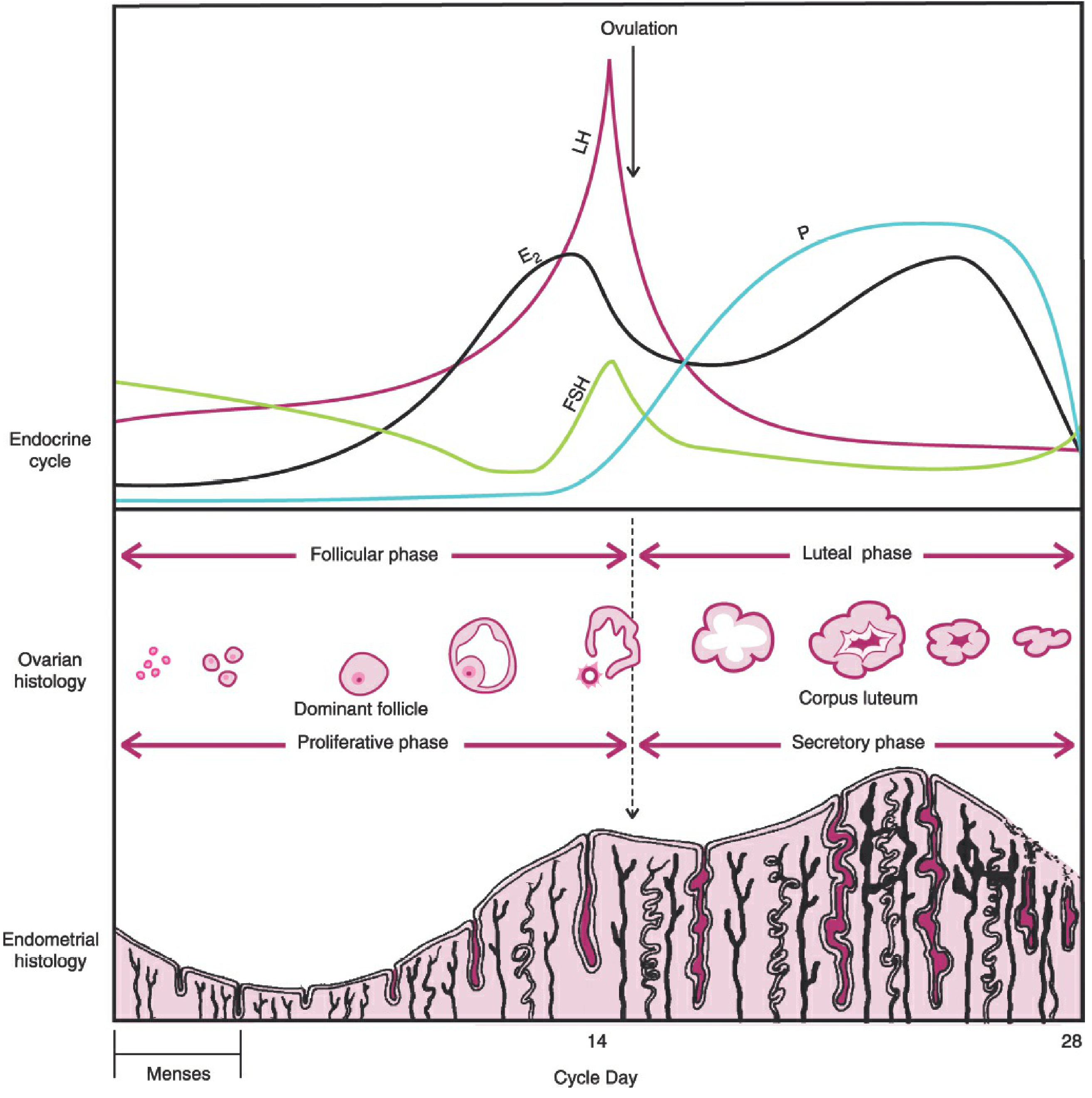
Fig. 7-9 from Berek & Novak's Gynecology: Top panel shows cyclic hormone levels (FSH, LH, E2, Progesterone). Middle panel shows ovarian histology (dominant follicle → ovulation → corpus luteum). Bottom panel shows endometrial histology (proliferative → secretory phases).
Step-by-Step Hormonal Events
-
Early follicular phase (Days 1-5): With demise of the corpus luteum from the previous cycle, estrogen and progesterone fall. This disinhibits the pituitary, and FSH levels begin to rise, recruiting a cohort of ovarian follicles.
-
Mid-follicular phase (Days 6-10): Growing follicles secrete increasing estrogen. Rising estrogen stimulates endometrial proliferation. Rising estrogen and inhibin B provide negative feedback, causing FSH to wane by mid-follicular phase. This ensures only the dominant follicle survives (others undergo atresia).
-
Late follicular phase / Pre-ovulatory (Days 11-13): As the dominant follicle matures, estradiol levels surge dramatically. When plasma estradiol exceeds ~200 pg/mL for a sustained period, the feedback switches from negative to positive - estradiol now stimulates (rather than inhibits) GnRH and LH release. LH receptors appear on granulosa cells under FSH influence.
-
LH Surge and Ovulation (Day 13-14): The positive feedback triggers the LH surge (and a smaller FSH surge). This is the proximate cause of ovulation, which occurs 24-36 hours after the surge begins. The dominant follicle ruptures and releases the mature oocyte.
-
Luteal Phase (Days 14-28): Post-ovulation, the ruptured follicle transforms into the corpus luteum (luteinization), which secretes large amounts of progesterone and some estradiol. Progesterone prepares the endometrium for implantation, increases basal body temperature, and - together with estrogen and inhibin A - suppresses new follicular development through negative feedback.
-
Corpus luteum regression (Days 24-28): If fertilization does not occur, the corpus luteum regresses after ~14 days. Estrogen and progesterone fall sharply, withdrawing support for the endometrium, leading to menstruation and beginning of the next cycle.
Feedback Mechanisms
The diagram below illustrates the switching of feedback control across the three phases:
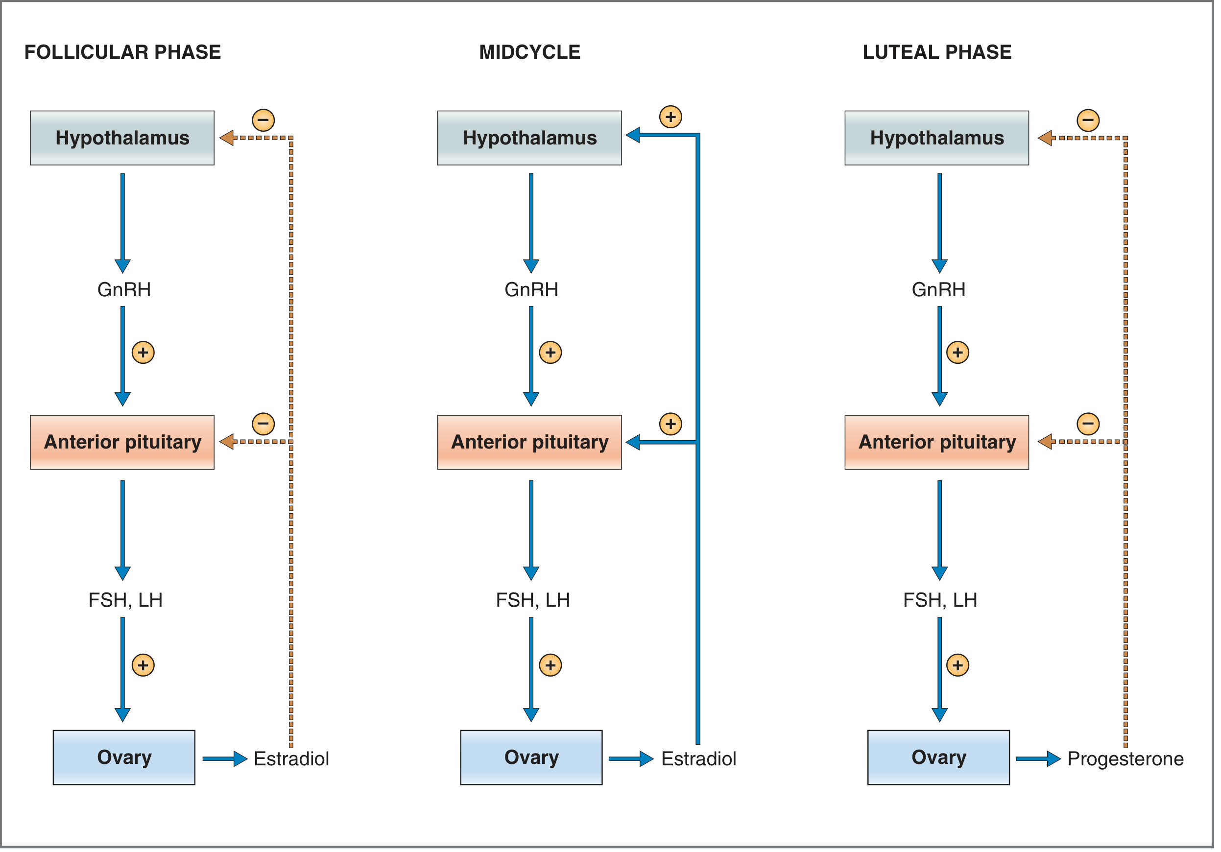
Control of FSH and LH secretion in females. Follicular and luteal phases: negative feedback. Midcycle: positive feedback by estradiol triggers the LH surge. (Costanzo Physiology, 7th Ed.)
| Phase | Feedback Type | Mechanism |
|---|---|---|
| Follicular phase | Negative | Low estradiol suppresses FSH/LH |
| Midcycle | Positive | High estradiol (>200 pg/mL) upregulates GnRH receptors → LH surge |
| Luteal phase | Negative | Progesterone + estrogen + inhibin A suppress gonadotropins |
Ovarian Cycle
1. Follicular Development
The ovary contains a finite pool of primordial follicles established before birth, which declines progressively from ~7 million at 20 weeks gestation to ~300,000 at puberty and ~1,000 at menopause.
Each cycle, FSH recruits a cohort of primary follicles. FSH acts exclusively on granulosa cells (the only ovarian cells bearing FSH receptors), stimulating:
- Granulosa cell proliferation
- Estradiol synthesis
The locally produced estradiol reinforces FSH effects - more cells → more estradiol → more cells. This mutual amplification accelerates growth of one dominant follicle (the Graafian follicle), which produces enough estrogen to suppress FSH and trigger atresia of lesser follicles.
Follicle stages:
- Primordial → Primary → Secondary → Antral (Graafian) follicle
2. Ovulation
The LH surge triggers a cascade:
- Resumption of meiosis I in the oocyte (which had been arrested)
- Proteolytic enzyme release causing follicular wall digestion
- Rupture of the Graafian follicle (~36 hours after LH surge peak)
- Release of the secondary oocyte (arrested at metaphase II) into the peritoneal cavity, where it is captured by the fimbriae of the fallopian tube
3. Meiotic Arrest and Resumption
The primary oocyte is arrested in prophase I of meiosis from fetal life until puberty. After the LH surge, meiosis I resumes and completes, forming the secondary oocyte (with first polar body). The secondary oocyte immediately enters meiosis II but arrests again at metaphase II - meiosis II is only completed upon fertilization by a sperm.
4. Corpus Luteum (Luteal Phase)
After ovulation:
- Granulosa and theca cells of the ruptured follicle undergo luteinization under LH stimulation
- The corpus luteum forms and produces progesterone (predominantly) and estradiol
- Inhibin A is also secreted
- Without hCG from a conceptus, the corpus luteum has a lifespan of ~14 days, then undergoes luteolysis
- If pregnancy occurs, hCG from the trophoblast maintains the corpus luteum until the placenta takes over steroidogenesis (~8-10 weeks)
Cyclic Changes of the Endometrium
The endometrium has two functional zones:
- Decidua functionalis (superficial 2/3): proliferates and is shed monthly - comprises the stratum compactum (superficial) and stratum spongiosum (deeper)
- Decidua basalis (deepest 1/3): does NOT shed; source of endometrial regeneration each cycle
1. Menstrual Phase (Days 1-4)
- Corpus luteum regresses → estrogen and progesterone fall
- Spiral arteries constrict and then dilate → ischemic necrosis of the functionalis
- Endometrial lining is shed along with blood (menses)
- Flow typically lasts 4-5 days
2. Proliferative Phase (Days 5-14)
Corresponds to the follicular phase of the ovarian cycle. Driven by rising estrogen:
- Endometrial thickness increases from 1-2 mm to 8-10 mm
- Glands: initially straight and narrow → become longer and tortuous with pseudostratified epithelium and multiple mitotic figures
- Stroma: dense and compact, sparse vascularity early; increasing stromal edema later
- Spiral arteries elongate
3. Secretory Phase (Days 14-28)
Corresponds to the luteal phase. Driven by progesterone from the corpus luteum:
- Within 48 hours of ovulation: subnuclear vacuoles appear in glandular epithelium (earliest secretory change, best seen around day 16-17)
- Glands become coiled and tortuous, secreting glycogen-rich material
- Stroma: increased edema and vascularity, then undergoes predecidual change (stromal cells enlarge with clear cytoplasm), peak around days 22-24
- Spiral arteries become coiled and prominent ("window of implantation" days 20-24)
- If implantation does not occur → luteal regression → menstruation
Dating the Endometrium (Noyes, Hertig & Rock, 1950)
Histologic dating of the endometrium was described by Noyes, Hertig and Rock in 1950. Key morphologic changes:
| Cycle Day | Morphologic Feature |
|---|---|
| Day 16-17 | Subnuclear vacuoles |
| Day 18-19 | Vacuoles move to supranuclear position |
| Day 20-21 | Secretion into gland lumen |
| Day 22 | Stromal edema |
| Day 23-24 | Spiral artery prominence; predecidual change |
| Day 25-27 | Full predecidual change around arterioles |
| Day 28 | Stromal breakdown; menstruation |
Cervical Changes During the Menstrual Cycle
The cervical mucus changes predictably:
| Phase | Cervical Mucus | Clinical Significance |
|---|---|---|
| Pre-ovulatory (high estrogen) | Thin, watery, "spinnbarkeit" (stretches >6 cm), ferning pattern | Facilitates sperm penetration |
| Post-ovulatory (high progesterone) | Thick, scant, cellular, no ferning | Impedes sperm penetration |
Summary of Menstrual Cycle Regulation
The following table summarizes the key events:
| Event | Day (28-day cycle) | Hormonal Driver |
|---|---|---|
| Menstruation begins | Day 1 | Falling E2 + P |
| FSH rise / follicle recruitment | Days 1-5 | Low E2 disinhibits FSH |
| Dominant follicle selection | Days 5-7 | Rising E2 + inhibin B → falling FSH |
| Endometrial proliferation | Days 5-14 | Rising E2 |
| Estradiol peak (pre-ovulatory) | Day 12-13 | Dominant follicle maturation |
| LH surge | Day 13 | Positive feedback from E2 |
| Ovulation | Day 14 | LH surge (24-36 hrs after onset) |
| Corpus luteum formation | Days 15-20 | LH (luteinization) |
| Progesterone peak | Days 20-22 | Corpus luteum |
| Window of implantation | Days 20-24 | Progesterone/E2 from corpus luteum |
| Corpus luteum regression | Days 24-26 | Luteolysis (if no hCG) |
| E2/P fall → menstruation | Day 28 | Corpus luteum demise |
Two-Cell, Two-Gonadotropin Theory of Estradiol Synthesis
Ovarian estrogen production requires cooperation between theca cells and granulosa cells:
- Theca cells (respond to LH): Convert cholesterol → androgens (androstenedione, testosterone)
- Granulosa cells (respond to FSH): Aromatize androgens to estradiol (via aromatase enzyme)
Neither cell type can produce estrogen alone - hence the "two-cell, two-gonadotropin" model.
Anovulatory Cycles
In anovulatory cycles:
- No dominant follicle matures; no LH surge; no ovulation
- No corpus luteum forms → no progesterone secretion
- Endometrium remains in the proliferative phase (estrogen-stimulated)
- "Breakthrough bleeding" occurs when estrogen levels fall without progesterone support
- Common at menarche, perimenopause, PCOS, and with certain medications
Oral contraceptives exploit this physiology: exogenous estrogen + progestogen suppress GnRH/FSH/LH → prevent follicular development and ovulation.
Clinical Significance
| Condition | Pathophysiology |
|---|---|
| Amenorrhea | Failure at any level of the HPO axis |
| Anovulatory infertility | No LH surge / follicle maturation failure |
| PCOS | Chronic anovulation with excess androgens |
| Luteal phase defect | Inadequate progesterone from corpus luteum |
| Asherman syndrome | Endometrial scarring / loss of basal layer |
| Menorrhagia | Structural or hormonal disruption of cycle |
Conclusion
The menstrual cycle is a masterpiece of biological integration. The hypothalamic-pituitary-ovarian axis, through pulsatile GnRH, governs the sequential secretion of FSH and LH. These gonadotropins drive ovarian folliculogenesis, ovulation, and corpus luteum function. The resulting cyclical changes in estrogen and progesterone coordinate the endometrial proliferative and secretory phases - culminating in either implantation of an embryo or menstruation. Understanding this physiology is the cornerstone of diagnosing and managing nearly all gynecological disorders.
References
- Berek JS, Novak E. Berek & Novak's Gynecology, 16th ed. Chapter 7: Menstrual Cycle Physiology, pp. 264-295.
- Costanzo LS. Physiology, 7th ed. Chapter 10: Reproductive Physiology, pp. 476-481.
- Moore KL, Persaud TVN. The Developing Human: Clinically Oriented Embryology. Chapter 2: Female Reproductive Cycles, pp. 105-106.
- Pawlina W, Ross MH. Histology: A Text and Atlas, 8th ed. Chapter 23: Ovarian and Uterine Cycles.
- Mescher AL. Junqueira's Basic Histology, 17th ed. Chapter on Female Reproductive System: Endometrium and Menstrual Cycle.
- Noyes RW, Hertig AT, Rock J. Dating the endometrial biopsy. Fertil Steril 1950;1:3-25.
Assignment prepared using authoritative medical textbook sources: Berek & Novak's Gynecology, Costanzo Physiology 7th Ed., The Developing Human (Moore & Persaud), Histology: A Text and Atlas (Ross & Pawlina), and Junqueira's Basic Histology.
This is a shared conversation. Sign in to Orris to start your own chat.