
What is karyotyping and what are the steps in it
Karyotyping is a laboratory technique used to visualize and analyze an organism's complete set of chromosomes. A sample of cells is collected, the chromosomes are stained and photographed during cell division (when they are most visible), then arranged in a standardized format for analysis. It is used to detect chromosomal abnormalities like Down syndrome, Turner syndrome, and certain cancers.
Searching sources...
Reading File
Finding Sources
Searching the Web
karyotyping steps procedure chromosomal analysis
Reading File
Finding Sources
Reading File
Reading File
Reading File
Loading Image
Now I have excellent content from multiple authoritative textbooks. Let me compile the full answer.
Karyotyping
What is Karyotyping?
Karyotyping is the process of examining the complete set of chromosomes in a cell - their number, size, shape, and structure. The term comes from the Greek karyon (nucleus). The result is called a karyotype, which is a digital representation of a stained metaphase spread in which chromosomes are arranged in order of decreasing length. Interpreting that image is called the karyotype analysis, while the actual photograph of the stained spread is called a karyogram.
Human somatic cells contain 46 chromosomes - 22 homologous pairs of autosomes plus two sex chromosomes (XX in females, XY in males). Karyotyping is the foundational tool of cytogenetics and can detect chromosomal abnormalities at a resolution of approximately 5-10 megabases (Mb).
- Robbins & Kumar Basic Pathology, p. 123
- Robbins, Cotran & Kumar Pathologic Basis of Disease, p. 161
Steps in Karyotyping
Step 1 - Sample Collection
Cells capable of dividing are collected. Common sources include:
- Peripheral blood (lymphocytes) - most common
- Bone marrow - used in hematological malignancies
- Amniotic fluid - for prenatal diagnosis (via amniocentesis)
- Chorionic villi - prenatal diagnosis via CVS (results faster, 10-14 days for amniotic fluid culture)
- Skin fibroblasts or other tissue biopsies
Step 2 - Cell Culture
The collected cells are cultured in a nutrient medium to stimulate proliferation. Phytohemagglutinin (PHA) is added to peripheral blood cultures to stimulate T-lymphocyte division.
Step 3 - Mitotic Arrest at Metaphase
A mitotic spindle inhibitor, most commonly colchicine (or its derivative colcemid / N-diacetyl-N-methylcolchicine), is added to arrest dividing cells in metaphase - the stage when chromosomes are most condensed and therefore most visible. This produces an accumulation of metaphase cells for analysis.
Step 4 - Hypotonic Treatment
Cells are placed in a hypotonic solution (e.g., 0.075 M KCl). Water enters the cells by osmosis, causing them to swell. This spreads the chromosomes apart within the cell so they do not overlap when the cell bursts.
Step 5 - Fixation
Cells are fixed (usually with Carnoy's fixative - methanol and glacial acetic acid) to preserve chromosome morphology and stop all cellular processes.
Step 6 - Spreading on Slides
The fixed cell suspension is dropped onto glass slides. As the cells burst open on impact, the chromosomes spread out in a single plane - creating a metaphase spread.
Step 7 - Staining and Banding
Several staining methods are used to create distinctive banding patterns on each chromosome:
| Banding Method | Stain Used | Notes |
|---|---|---|
| G banding (Giemsa banding) | Giemsa stain | Most widely used; AT-rich regions stain dark, GC-rich stain light |
| Q banding | Quinacrine (fluorescent) | Fluorescent; useful for Y chromosome |
| R banding | Various | Produces a reverse pattern of G banding |
With standard G banding, 400-800 bands per haploid set can be visualized. If cells are obtained during prophase (more elongated chromosomes), up to 1500 bands per karyotype can be resolved, allowing detection of more subtle abnormalities.
Step 8 - Microscopy and Photography
Metaphase spreads are examined under a microscope and photographed (digitally captured) using a camera attached to the microscope.
Step 9 - Karyogram Construction and Analysis
The chromosome images are cut out (or digitally arranged) and organized into a standardized display - the karyogram. Chromosomes are arranged:
- In homologous pairs
- In order of decreasing length (chromosome 1 is longest; chromosome 21 is actually the shortest, not 22 as was originally thought)
- By arm morphology relative to the centromere:
- Metacentric - roughly equal p and q arms
- Submetacentric - q arm longer than p arm
- Acrocentric - no significant p arm (chromosomes 13, 14, 15, 21, 22 - these encode most rRNAs and are common sites of Robertsonian translocations)
- Sex chromosomes are placed last (XX or XY)
Chromosome Nomenclature
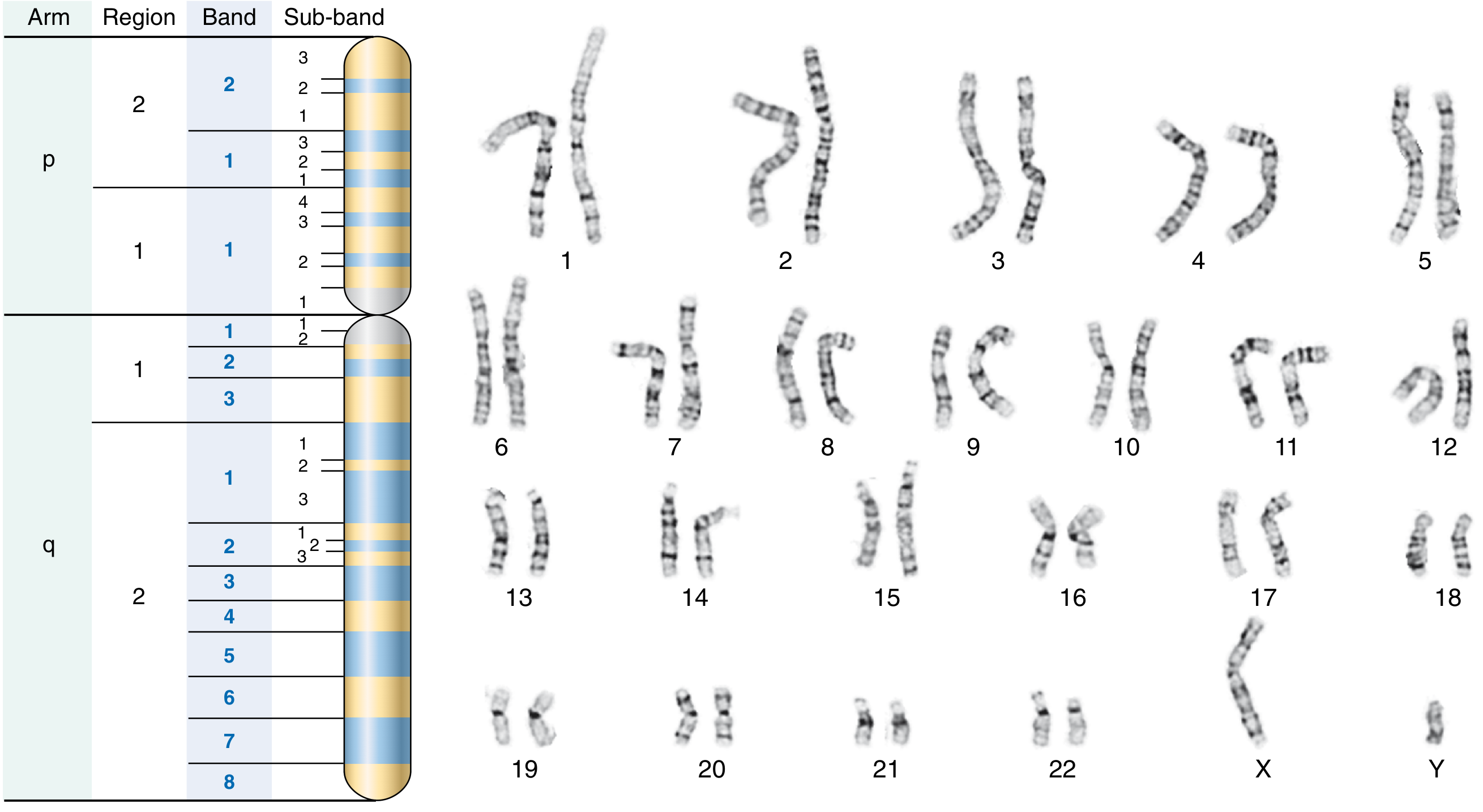
Each chromosome arm is divided into regions, bands, and sub-bands, numbered outward from the centromere:
- p = short arm (from French petit)
- q = long arm
- Notation:
[chromosome][arm][region][band].[subband]
Example: 18q21.3 = chromosome 18, long arm, region 2, band 1, sub-band 3 (where the BCL2 locus resides).
A normal male is written 46,XY; a male with trisomy 21 (Down syndrome) is 47,XY,+21.
Quick Compendium of Clinical Pathology 5th ed., p. 365
G-banded Karyogram (Normal Male, 46,XY)
The image below shows all 22 autosome pairs plus X and Y chromosomes arranged with their characteristic G-banding patterns. On the left is a diagram of chromosome arm/region/band/sub-band nomenclature:
What Karyotyping Can Detect
| Type of Abnormality | Examples |
|---|---|
| Numerical (aneuploidy) | Trisomy 21 (Down), Monosomy X (Turner), XXY (Klinefelter) |
| Structural - deletions | Cri-du-chat (5p-), DiGeorge (22q11) |
| Structural - duplications | Chromosomal segments duplicated |
| Translocations | Philadelphia chromosome t(9;22); Robertsonian translocations |
| Inversions | inv(16) in acute myeloid leukemia |
Limitation: Karyotyping has a resolution limit of ~5-10 Mb and cannot detect smaller variants. For higher resolution (down to 50-200 kb), chromosomal microarray is used. However, karyotyping retains a key advantage: it can detect balanced rearrangements (which microarray cannot, since array only shows the amount of DNA present, not its position).
Robbins & Kumar Basic Pathology; Tietz Textbook of Laboratory Medicine, 7th ed.; Quick Compendium of Clinical Pathology 5th ed.
This is a shared conversation. Sign in to Orris to start your own chat.