
Treatment of ethyl alcohol poisoning
I'll start by reading the medical library skill, then search for relevant content on ethyl alcohol (ethanol) poisoning treatment.
Reading File
Now let me search the library and PubMed in parallel for ethanol poisoning treatment.
Finding Sources
Searching PubMed
"ethanol poisoning" OR "alcohol intoxication" AND treatment management
Finding Sources
Finding Sources
Good - I found relevant sections. Let me read the most relevant ones in parallel.
Reading File
Reading File
Let me read the Lippincott section on acute ethanol intoxication management, and Tintinalli's section on alcohol intoxication.
Reading File
Finding Sources
Excellent - I found two very relevant sections. Let me read them both now.
Reading File
Reading File
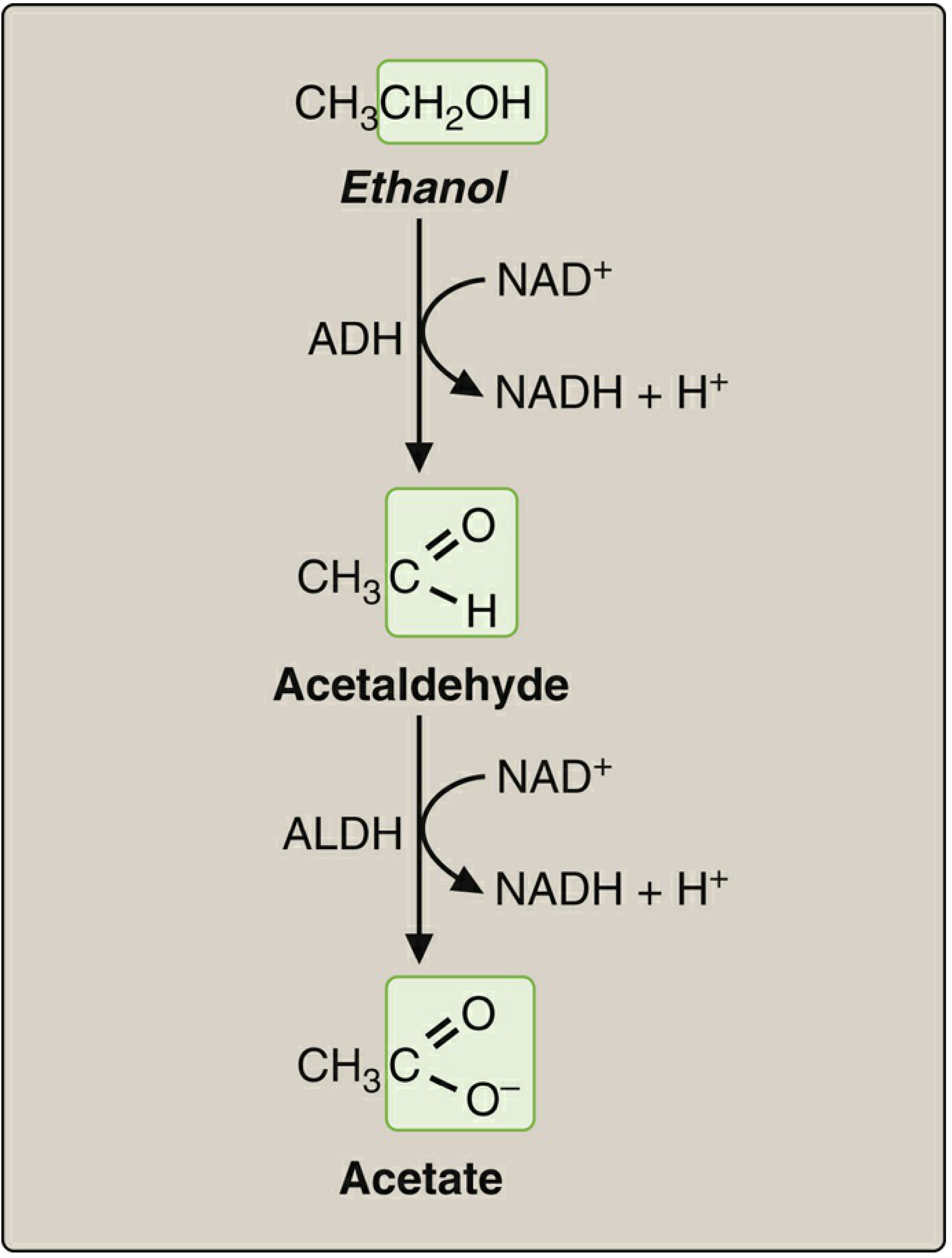
I now have comprehensive information. Let me also fetch the ethanol metabolism pathway diagram for visual clarity.
Loading Image
I now have all the information needed for a comprehensive answer. Here is the full treatment guide for ethyl alcohol (ethanol) poisoning:
Treatment of Ethyl Alcohol (Ethanol) Poisoning
Pathophysiology - Why It Causes Toxicity
Ethanol is metabolized by zero-order kinetics at approximately 15-40 mg/dL/hour - the rate is constant regardless of concentration. It works as a CNS depressant via:
- Enhancement of GABA-A receptor activity (inhibitory)
- Inhibition of NMDA receptor activity (excitatory)
- Resulting in dose-dependent CNS/respiratory depression
Blood Ethanol Concentration (BEC) and Clinical Signs
| BEC (mg/dL) | Clinical Features |
|---|---|
| ~80 | Slurred speech, incoordination, unsteady gait, impaired attention |
| 80-200 | Mood lability, cognitive deficits, aggression, anterograde amnesia ("blackout") |
| >200 | Nystagmus, sedation |
| >300 | Coma, failing vital signs, death |
Note: Tolerant heavy drinkers show markedly less impairment at the same BECs. Co-ingestion of other CNS depressants (benzodiazepines, barbiturates, antihistamines) significantly worsens toxicity at lower BECs.
Treatment of Acute Ethanol Intoxication
Treatment is exclusively supportive - there is no antidote and no safe pharmacological agent that accelerates ethanol metabolism.
1. Airway and Breathing (First Priority)
- Ensure a patent airway; place in lateral decubitus ("recovery position") to prevent aspiration
- If respiratory depression is mild: careful observation is sufficient
- If respiratory depression is severe: endotracheal intubation for airway protection and ventilatory support
- Intubation and mechanical ventilation are unusual for ethanol intoxication alone
2. Gastric Lavage
- Consider if absorption is not yet complete (early presentation) and airway is protected
- Activated charcoal is generally not useful (ethanol is absorbed rapidly and does not bind well)
3. IV Fluids
- IV fluid resuscitation for hypotension and dehydration
- Correct fluid and electrolyte disturbances
4. Glucose
- Check blood glucose; administer dextrose if hypoglycemia is present (ethanol inhibits gluconeogenesis)
5. Thiamine (Vitamin B1) - Mandatory
- Give thiamine before glucose in any patient with altered mental status or suspected chronic alcohol use disorder
- Prevents and treats Wernicke encephalopathy (confusion, ataxia, oculomotor abnormalities)
- Dose for Wernicke: 500 mg IV three times daily for 3-5 days
- All patients with suspected ethanol use disorder should receive thiamine
6. Folate
- Administer folate (ethanol depletes folate stores)
7. Clinical Monitoring
- Serial neurological and vital sign monitoring until clinical sobriety
- Observe for 4-6 hours in the emergency department as ethanol is metabolized
- Do not discharge until the patient has returned to their neurological baseline
8. Hemodialysis (Rarely Required)
- Ethanol is freely miscible with water and can be removed by hemodialysis
- Reserved for extremely high blood alcohol levels or severe cardiovascular compromise
- Note: May precipitate withdrawal in a patient with alcohol use disorder
Differential Diagnosis - Do Not Miss
The following conditions mimic profound ethanol intoxication and must be excluded:
- Hypoglycemia (diabetic coma)
- Other drug intoxication (opioids, benzodiazepines, sedatives)
- Cardiovascular accidents (stroke, TIA)
- Head trauma / skull fracture (especially if the patient fell)
- Diabetic ketoacidosis (can produce similar breath odor to alcohol)
- Metabolic encephalopathy
Blood ethanol levels are necessary to confirm intoxication. Do not rely solely on breath odor - similar odors can occur in DKA or other metabolic acidoses. Serum glucose, metabolic panel, and a low threshold for head CT are appropriate.
Managing Alcohol Withdrawal (Special Consideration)
Patients who are heavy chronic drinkers may transition from intoxication to withdrawal as blood ethanol drops. There are four manifestations:
| Stage | Features |
|---|---|
| Acute uncomplicated withdrawal | Tremulousness, anxiety, nausea, tachycardia, hypertension |
| Alcoholic hallucinosis | Visual/auditory illusions (patient retains reality testing) |
| Withdrawal seizures | May occur even without other withdrawal signs |
| Delirium tremens (DTs) | Dysautonomia, hyperthermia, seizures, agitation, psychosis - life-threatening |
Treatment of withdrawal:
- Benzodiazepines are first-line (symptom-triggered, protocol-based dosing)
- Lorazepam 1-2 mg IV/IM or diazepam 5-10 mg IV, titrated to symptoms
- Phenobarbital is an alternative in refractory cases
- Use symptom-triggered protocols (e.g., CIWA-Ar scale) - fixed-dose schedules are less effective and may cause under- or over-treatment
- Dexmedetomidine is not recommended - does not target GABA/glutamate systems and does not prevent seizures
- High-dose IV thiamine (as above)
Long-Term / Definitive Treatment of Alcohol Use Disorder
After the acute episode, pharmacotherapy to maintain abstinence includes:
| Drug | Mechanism | Notes |
|---|---|---|
| Disulfiram | Inhibits aldehyde dehydrogenase → acetaldehyde accumulation → flushing, nausea, vomiting if alcohol consumed | Acts as aversion therapy |
| Naltrexone | Opioid receptor antagonist | Reduces craving and reward |
| Acamprosate | Modulates glutamate (NMDA) tone | Reduces withdrawal-related dysphoria |
All pharmacotherapy should be combined with psychosocial/behavioral support.
Sources:
- Goodman & Gilman's Pharmacological Basis of Therapeutics, p. 3518-3520
- Washington Manual of Medical Therapeutics, p. 1097
- Lippincott Illustrated Reviews: Pharmacology, pp. 1566-1571
Refer reddy
Reading File
"Refer Reddy" likely means the user wants to see what Jaypee's Textbook of Medical Pharmacology by Remy or more likely K.D. Tripathi or Reddy (a popular Indian pharmacology/medicine author) says about ethanol poisoning. Let me check what's in the library.
Finding Sources
Finding Sources
The library has "The Essentials of Forensic Medicine and Toxicology 36th edition (latest 2026)" - this is the K.S. Narayan Reddy textbook, the most popular forensic medicine and toxicology reference in India! Let me search it for ethyl alcohol poisoning.
Finding Sources
Finding Sources
Finding Sources
Finding Sources
The content is in block6. The ethyl alcohol content starts around line 3911. Let me read the full ethyl alcohol section from Reddy.
Reading File
Reading File
Reading File
Reading File
Reading File
Reading File
I now have all the content from Reddy's textbook on ethyl alcohol poisoning. Let me also fetch the key clinical illustration (Ataxia figure) from the book.
Reading File
I now have the complete content from K.S. Narayan Reddy's textbook. Here is the full, detailed answer:
Ethyl Alcohol Poisoning - As Per Reddy's Forensic Medicine & Toxicology (36th Ed., 2026)
K.S. Narayan Reddy - The Essentials of Forensic Medicine and Toxicology, 36th Edition
Description
Ethyl alcohol is a transparent, volatile, colorless, non-fuming, watery liquid with a characteristic alcoholic odor. It is manufactured by fermentation and distillation of sugar-containing organic grains, jaggery, cane, etc. It is a powerful psychoactive drug.
Forms of Alcohol:
| Form | Description |
|---|---|
| Absolute (anhydrous) alcohol | 99.5% ethyl alcohol |
| Rectified spirit (neutral spirit) | 95% ethyl alcohol |
| Methylated/denatured spirit | Alcohol denatured with methyl alcohol (9.5%) + crude pyridine (0.5 mg%) |
| Standard drink | 10 g of absolute alcohol = 1 unit |
Mechanism of Action
- Higher centers in the cerebrum are depressed first; the primitive hindbrain/respiratory center is inhibited last
- Cerebral forebrain center inhibition causes mood changes like elation and excitation
- Direct toxic effect on cardiac, thyroid, and hepatic cells
- Intracellularly binds with GABA receptor enzyme in brain cells
- Inhibits NMDA (N-methyl-D-aspartate) enzyme - prolonged abuse causes upregulation of this inhibitory response, leading to tolerance and withdrawal signs
Acute Poisoning - Signs and Stages
Signs depend on whether the person is a beginner, occasional, or chronic drinker.
Stage 1 - Excitement / Inebriation / Sobriety
- BAC < 50 mg%
- Mild euphoria, easy socialization, high boasting speech
Stage 2 - Euphoria
- BAC 50-100 mg%
- Decreased attention span and cognitive function
- Loss of inhibitions, talkativeness, sociability
- Nausea, dizziness, palpitations, flushed red face
- Subconjunctival suffusion (red eyes), salivation, dilated pupils
Stage 3 - Excitement
- BAC 100-150 mg%
- Incoordination of muscular movements (ataxia)
- Loss of self-control, slurred speech, delirium
- Sleepiness, disorderly behavior, abusive language
- Alcohol gaze nystagmus
Stage 4 - Coma and Death
- Decreased response to painful stimuli, labored breathing
- Pupillary constriction, brisk reflexes (early)
- Later: loss of consciousness, deep sleep (usually lasts 4-6 hours)
- McEwan's Sign: Painful stimulus (slap on temple) causes pupil to dilate then re-contract
- Very rarely enters deep coma - alcohol alone will not kill unless:
- Asphyxia occurs (aspiration of food bolus - "Café coronary")
- Comorbidities / concomitant diseases
- Exposure to cold while under the influence
BAC decreases by approximately 15 mg%/hour; once BAC falls below 100-150 mg%, the person regains consciousness.
Treatment of Acute Ethyl Alcohol Poisoning (Reddy's)
- Maintain airway - primary priority; prevent aspiration
- Watch renal functions in deeply comatose patients
- Flumazenil may be considered for patients with hepatic encephalopathy
- Vitamin B supplementation - alcoholic addicts have vitamin B (especially thiamine) deficiency
Fatal Dose and Death
- Adults: 5-8 g/kg (6-10 mL/kg) body weight
- Fatal BAC: >500 mg%
- Fatal period: 12-24 hours
- Causes of death:
- Depression of respiratory center (BAC usually >500 mg%)
- Asphyxia - inhalation of food bolus (café coronary)
Autopsy Findings
- Suffused red conjunctiva
- Signs of asphyxia
- Alcoholic fruity odor from body cavities and stomach
- Stomach mucosa congestion
Bedside Tests for Alcohol
| Test | Method | Result |
|---|---|---|
| Ethyl acetate test | 5 mL urine + 1 mL acetic acid + 1 drop H₂SO₄, heat gently for 1 min | Characteristic strong fruity odor of ethyl acetate = positive |
| Woodbury's (Dichromate) test | 10% sodium dichromate + urine | Greenish color change = positive |
| Breath Analyzer (Alcometer/Drunkometer) | Exhaled air passed over conc. H₂SO₄ and dichromate crystals | Greenish color; ratio of 1 mL blood = 2100 mL exhaled air (constant at 34°C) |
Chronic Poisoning / Alcoholism
Types (Jellinek's Classification)
| Type | Description |
|---|---|
| Alpha | Excessive drinking without loss of control or ability to abstain |
| Beta | Excessive drinking without dependence, but with physical complications (cirrhosis, gastritis, pancreatitis) |
| Gamma | Physical dependence, tolerance, inability to control drinking |
| Delta | Inability to abstain, tolerance, withdrawal signs, but quantity can be controlled (wine drinkers) |
| Epsilon | Intermittent / spree drinking |
Systemic Complications of Chronic Alcoholism
| System | Complications |
|---|---|
| GIT | Nausea, vomiting, pancreatitis, gastritis |
| Hepatic | Fatty liver, jaundice, portal hypertension, cirrhosis with ascites |
| CNS | Delirium tremens, Korsakoff's psychosis (amnesia + confabulation - thiamine deficiency), Wernicke's encephalopathy (confusion, ataxia, ophthalmoplegia) |
| CVS | Cardiomyopathy, dysrhythmias, hypertension, Beri-Beri heart, Munich beer heart syndrome (dilated cardiomyopathy) |
| Respiratory | Café coronary (aspiration), alcohol-induced asthma, aspiration pneumonia |
| Endocrine | Males: Gynecomastia, feminization; Females: Amenorrhea, menorrhagia, infertility |
| Nutritional | Anemia, hypoproteinemia, polyneuropathy |
| Psychiatric | Dementia, delusions, hallucinations |
| Fetal | Fetal alcohol syndrome (facial dysmorphism, mental retardation, growth retardation, delayed milestones) |
| Other | Malignancies: colorectal, pancreatic, breast, prostate |
Withdrawal Signs (Timeline)
| Time after cessation | Features |
|---|---|
| 6-8 hours | Common abstinence syndrome: agitation, tremors of digits/limbs, headache, insomnia, nausea |
| 24-36 hours | Alcoholic hallucinations: visual (rarely tactile) |
| 7-48 hours | Rum fits (seizures): tonic-clonic movements with/without loss of consciousness |
| 24-42 hours | Alcoholic ketoacidosis: Associated pancreatitis and gastritis; fat mobilized as energy → ketone bodies |
Medico-Legal Aspects (Drunkenness)
Three grades of medico-legal opinion:
- Signs of consumption - sweating, mild pupillary dilation, conjunctival suffusion, salivation, alcoholic breath odor, increased pulse volume
- Signs of influence - slurred speech, unable to walk straight, cannot touch tip of nose with finger, alcohol gaze nystagmus
- Signs of intoxication - vomiting, constricted pupil, semiconscious/altered sensorium, brisk reflexes or unconscious
Important caveat: These signs may also be seen in epilepsy, viral meningitis, brain tumors, head injury, methyl alcohol poisoning, tranquilizers, sedative-hypnotics, CO poisoning, and other conditions. The examining doctor must rule these out before certifying drunkenness.
Source: The Essentials of Forensic Medicine and Toxicology, 36th Edition (2026) - K.S. Narayan Reddy, pp. 527-529
Treatment of methanol poisoning, refer reddy
Searching Reddy's textbook for methanol poisoning content now.
Finding Sources
The methanol content is around line 4371 in block6. Let me read the full section.
Reading File
Reading File
Reading File
Reading File
I have all the content needed from Reddy. Here is the complete answer:
Methanol (Methyl Alcohol) Poisoning - As Per Reddy's Forensic Medicine & Toxicology (36th Ed., 2026)
K.S. Narayan Reddy - The Essentials of Forensic Medicine and Toxicology, 36th Edition, pp. 530-531
Description / Properties
- Synonyms: Wood alcohol, wood naphtha, pyroxilic acid, Columbian spirit, monohydroxy methane
- Formula: CH₂OH
- Appearance: Colorless, highly polar, flammable liquid with a faintly sweetish odor and bitter taste
Common Uses (Sources of Exposure):
- Carburetor cleaner (20%)
- Antifreeze (10-50%)
- Denatured/methylated spirit (5-10%) - mixed with ethyl alcohol (90-95%)
- Embalming fluid (20%)
- Leather dyes (30%)
- Varnish, shellac, paint remover, windshield cleaner
- Solvent in manufacture of cholesterol, streptomycin, vitamins, hormones
- Nail polish removers, paints (substance abuse among adolescents)
Fatal Dose
- 15-250 mL (average: 70-100 mL)
Mechanism of Action (Metabolism)
- Well absorbed by inhalation, oral, and parenteral routes; peak blood level reached in 30-60 minutes
- Alcohol dehydrogenase (ADH) in liver metabolizes methanol → Formaldehyde → Formic acid
- Formaldehyde and formic acid are highly toxic - they cause metabolic acidosis and neurological symptoms (especially optic nerve and retinal damage)
- Metabolism of methyl alcohol is slow - unmetabolized methanol can be found in blood for up to 1 week
Key point: The latent period before toxicity (18-24 hours) is because metabolites (formic acid) accumulate slowly. This "silent period" is a classic exam point.
Signs and Symptoms of Poisoning
| System | Features |
|---|---|
| General | Vertigo, headache, neck stiffness, vomiting, abdominal pain, general malaise, dizziness, neuritis |
| Ocular (hallmark) | Blurred vision, dimmed vision → Optic atrophy → Blindness (due to retrobulbar degeneration of retina) |
| Metabolic | Metabolic acidosis - usually delayed by 18-24 hours |
| Renal | Renal failure with other organ failure |
| Cardiovascular/CNS | Tachycardia, hypertension, convulsions, coma |
| Residual (if survived) | Putamen nuclear damage → Extrapyramidal movement disorder |
Diagnosis
- High anion gap metabolic acidosis
- Elevated osmolal gap
- Hypophosphatemia
- Elevated CPK and amylase
- Blood methanol level
- Detectable formic acid in blood
- CT/MRI: Evidence of basal ganglia (putamen) degeneration
- Fundoscopy: Retrobulbar edema, hyperemia
- Optical coherence tomography (OCT)
Treatment (Reddy's)
1. Gastric Decontamination
- Stomach wash (gastric lavage) with sodium bicarbonate
2. Antidote - Ethanol (Specific Antidote)
- Ethanol is the specific antidote - both methanol and ethanol compete for the alcohol dehydrogenase (ADH) enzyme, but ethanol has a higher affinity for ADH than methanol
- This competitively blocks the metabolism of methanol, preventing formation of the toxic formaldehyde and formic acid
- Given as whisky, gin, or vodka at 2.5 mg/kg (i.e., approximately 175 mL)
Reddy also provides a dosing table for ethanol therapy (Table 29.4):
| Route | Non-drinker | Chronic drinker |
|---|---|---|
| Intravenous | 0.8-1.3 mL/kg/h | 1.5 mL/kg/h |
| Oral | 0.4-0.7 mL/kg/h | 0.8 mL/kg/h |
3. Hemodialysis
- Most effective treatment to remove methanol and formaldehyde from the body
4. Folinic Acid
- Supplementation is essential in almost all cases
- Dose: 1-2 mg/kg IV
- Folinic acid accelerates the conversion of formate to CO₂ and water, reducing formic acid accumulation
Postmortem / Autopsy Features
- Cyanosis is a prominent feature
- Edema and emphysematous lungs
- Long-standing cases: Basal ganglia degenerative changes (putamen)
- Preservation of viscera for chemical analysis in saturated NaCl solution
Medico-Legal Aspects
- Accidental poisoning is most common - especially via indigenous methylated spirits
- Intentional adulteration of ethyl alcohol with methyl alcohol causes mass deaths - these are called "Liquor Tragedies" (attract significant media and public attention)
- Substance abuse - nail polish removers, paints used as solvents, especially among adolescents
Comparison: Ethanol vs. Fomepizole as Antidote
| Feature | Ethanol | Fomepizole |
|---|---|---|
| Mechanism | Competitive substrate for ADH | Direct ADH inhibitor |
| Status in Reddy's | Specific antidote | "Better alternative" (mentioned for ethylene glycol) |
| Availability | Easily available | More expensive, less available in India |
Note: Fomepizole (4-MP) is mentioned by Reddy as a better alternative in ethylene glycol poisoning and is considered first-line in many modern guidelines, but ethanol remains the classical antidote highlighted in Reddy's text.
Source: The Essentials of Forensic Medicine and Toxicology, 36th Edition (2026) - K.S. Narayan Reddy, Chapter 29 (Alcohols), pp. 530-531
Treatment of carbon monoxide poisoning, refer reddy
Searching Reddy's textbook directly for carbon monoxide poisoning.
Finding Sources
Found it - the dedicated CARBON MONOXIDE section starts at line 4826. Let me read the full section.
Reading File
Reading File
Reading File
Reading File
I have the complete CO poisoning section from Reddy. Here is the full, detailed answer:
Carbon Monoxide (CO) Poisoning - As Per Reddy's Forensic Medicine & Toxicology (36th Ed., 2026)
K.S. Narayan Reddy - The Essentials of Forensic Medicine and Toxicology, 36th Edition, pp. 534-536
Description / Properties
- Synonyms: Carbonic oxide, Carbon oxide, Exhaust gas, Flue gas
- Pure CO is an odorless, colorless, non-irritating gas, lighter than air
- It gives no warning of its presence - this is why it is so dangerous
Important: Domestic gas (LPG - mixture of propane and butane) does not contain CO. Endogenous CO from haem degradation never reaches toxic levels; normal plasma COHb = 1-5%, up to 7-8% in smokers.
Sources
- Incomplete combustion of any fuel - wood, charcoal, gas, kerosene
- Automobile exhaust (most common environmental source)
- Fires (house fires, enclosed spaces)
- Paint remover - especially methylene chloride (metabolized to CO in the body)
- Tobacco smoke - mainstream cigarette smoke contains up to 5% CO by volume
- Shisha/hookah water pipes - CO poisoning reported after prolonged smoking
- Ingestion or inhalation of methylene chloride
Fatal Dose
- COHb level >50-60% is potentially lethal
- A CO concentration of 5,000 ppm in air is lethal after 5 minutes of exposure
Toxicokinetics
- Lungs avidly absorb CO
- CO combines with:
- Hemoglobin: 85% → forms Carboxyhemoglobin (COHb)
- Myoglobin: 15%
- Elimination occurs exclusively through the lungs
Mode of Action (Mechanism)
- CO has an affinity for hemoglobin 230-270 times greater than that of oxygen
- Despite adequate PO₂ in blood, there is reduced arterial oxygen content
- CO causes a leftward shift of the oxyhemoglobin dissociation curve - impairing offloading of oxygen from Hb to tissues
- Net result: decreased oxygen carrying capacity + decreased tissue oxygen delivery
- CO also inactivates mitochondrial cytochrome oxidase - interferes with cellular respiration directly
- Profound hypotension in severe poisoning due to:
- Activation of guanyl cyclase → smooth muscle relaxation
- Displacement of nitric oxide from platelets → vasodilation
Clinical Features
Acute Poisoning
Earliest manifestations are nonspecific and often misdiagnosed (especially in India where awareness is low). Cherry red color of skin/blood is seen in only 2-3% of cases.
Progressing with increasing COHb levels:
| Severity | Features |
|---|---|
| Mild | Headache, nausea, vomiting, dizziness, exertional dyspnea, chest pain, blurred vision, confusion, weakness |
| Moderate | Tachycardia, tachypnea, ataxia, severe headache, syncope, flushing, cyanosis, perspiration, decreased vigilance, diminished manual dexterity, impaired sensorimotor performance, prolonged reaction time, difficulty thinking, tinnitus/roaring in ears, drowsiness, hallucinations, cardiovascular toxicity |
| Severe | Trismus, muscle spasms, convulsions, palpitations, disorientation, ventricular dysrhythmias, hypotension, myocardial damage, skin blisters, pulmonary edema, respiratory failure, involuntary evacuations, coma, collapse, death |
Chronic Poisoning
Headache, dizziness, confusion, intellectual deterioration, weakness, nausea, vomiting, abdominal pain, visual disturbances (homonymous hemianopia, papilledema, scotomata, retinal hemorrhage), paresthesia, hypertension, hyperthermia, cherry red skin, palpitations, aggravation of angina, intermittent claudication, elevated RBC and WBC count, albuminuria, glycosuria.
Differential Diagnosis
Alcoholic intoxication, cardiac arrhythmias, hyperventilation syndrome, cerebrovascular accident, influenza, meningitis, encephalitis, depression, migraine, epilepsy, myocardial infarction, food poisoning, pneumonia.
Diagnosis
1. COHb Level (Co-oximetry)
- Normal: 0-5% (up to 10% in heavy smokers)
- Done spectrophotometrically using a co-oximeter
- Arterial or venous blood (in lithium heparin tube)
2. Pulse Oximetry
- Unreliable in CO poisoning - standard pulse oximetry cannot distinguish COHb from OxyHb (both absorb at similar wavelengths), so SpO₂ will appear falsely normal
3. Arterial Blood Gas (ABG)
- PO₂ is usually normal (CO doesn't reduce dissolved O₂)
- O₂ saturation (calculated) is decreased
- A gap between measured and calculated HbO₂ indicates need for COHb measurement
- Metabolic acidosis is invariably present
4. ECG
- May reveal myocardial damage: ST depression or elevation
5. Chest X-ray
- May show ground-glass appearance, perihilar haze, peribronchial cuffing, intra-alveolar edema (pulmonary edema)
6. Ancillary Investigations
- Elevated serum creatine kinase (CK) and lactate dehydrogenase (LDH)
- Elevated creatinine
- Hypokalemia and hyperglycemia
- Neuropsychometric testing indicated after moderate-to-severe poisoning
Bedside Tests for COHb
| Test | Method | Result |
|---|---|---|
| Dilution test | 1 drop blood + 10-15 mL water; compare with normal | Blood with CO appears pink (normal blood turns red-brown) |
| Ammonium hydroxide test | 0.1 mL blood + 2 mL ammonium hydroxide (0.01 mol/L), vortex 5 sec | Pink tint = COHb present |
| NaOH test | 1 mL blood + 10 mL water + 1 mL 5% NaOH | COHb: straw yellow (<20%) or pink (>20%); Normal HbO: brown |
Treatment
Step 1 - Admission Criteria
Admit all patients with:
- Neurologic signs or symptoms
- Chest pain
- Abnormal ECG
- Metabolic acidosis
- COHb level >20%
Step 2 - Remove from Environment + Oxygen (100%)
- Immediate removal from the contaminated environment
- Administer 100% oxygen through a tight-fitting mask or endotracheal tube
- Continue until COHb falls to 15-20%
- Monitor cardiac and respiratory status continuously
Half-life of COHb: ~5 hours on room air → ~40 minutes on 100% normobaric oxygen → ~20 minutes on 100% oxygen at 2.5 ATA (hyperbaric)
Step 3 - Monitor for Cerebral Edema
- Serial neurological examinations
- CAT scans
- Fundoscopic examination
Step 4 - Correct Metabolic Acidosis
- If metabolic acidosis persists despite correction of hypoxia and adequate fluid resuscitation
- Give IV sodium bicarbonate
Step 5 - Control Convulsions
- IV diazepam and phenytoin in the usual manner
Step 6 - Correct Hypotension
- Adequate fluid resuscitation with crystalloids
Step 7 - Antidote: Hyperbaric Oxygen (HBO) (Specific Antidote)
Several authorities consider HBO to be antidotal in CO poisoning.
| Protocol | Details |
|---|---|
| Initial | 100% oxygen at 3 ATA for 30 minutes |
| Maintenance | 2 ATA for 60 minutes OR until COHb <10% |
| Mechanism | Inhaling O₂ at pressure >1 atmosphere absolute (ATA) |
| Half-life reduction | Normobaric 100% O₂: 40 min → HBO at 2.5 ATA: 20 min |
Autopsy Features
- Cherry red (pink) color of skin - especially in areas of postmortem lividity; in dark-skinned individuals, look at inner lips, nail beds, tongue, palms, soles
- Cutaneous bullae (skin blisters) - sometimes seen at calves, buttocks, wrists, knees
- Cherry pink blood and tissues throughout
- Diluted blood in water held against light appears pink/red (vs. brown in normal blood)
- Pulmonary edema
- White matter of brain is firmer than usual; brain retains shape better after removal
- In delayed deaths: Necrosis and cavitation of basal ganglia - especially globus pallidus and putamen (classic finding)
- Petechiae and ring-shaped hemorrhages in white matter
- Focal myocardial necrosis
- Blood collection: preferably from a peripheral vein; if unavailable, heart blood, body cavities, or bone marrow can be used (preserve with sodium fluoride)
Medico-Legal Importance
- Next to CO₂, CO is the most abundant atmospheric pollutant - progressively increasing
- Accidental - most common; especially in ill-ventilated areas with incomplete combustion
- Suicidal - historically via coal gas ovens in UK; now less common since LPG replaced coal gas (LPG has little/no CO)
- Homicidal - by confining victim in a closed space with a CO source
- Tobacco smoke is an important source of CO contamination
Source: The Essentials of Forensic Medicine and Toxicology, 36th Edition (2026) - K.S. Narayan Reddy, Chapter on Asphyxiant Poisons / Carbon Monoxide, pp. 534-536
This is a shared conversation. Sign in to Orris to start your own chat.