
Ct shoulder indications pathologies etc everything
CT shoulder indications pathologies glenohumeral instability bony Bankart
CT scan shoulder bony Bankart lesion glenoid bone loss
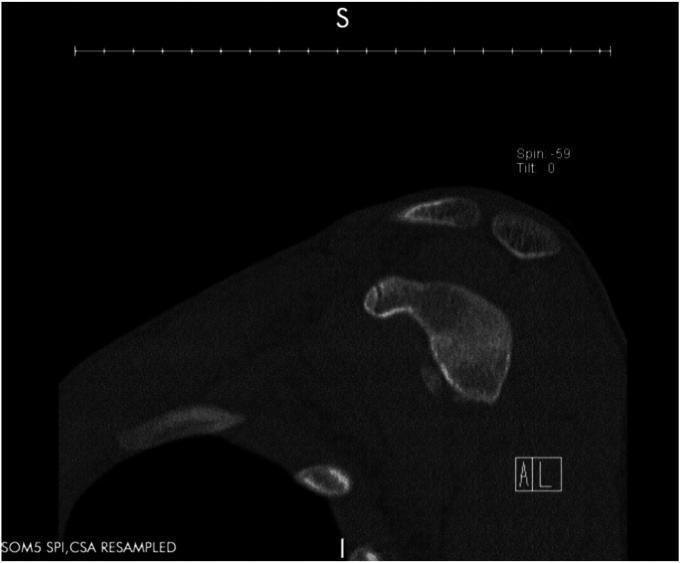
This diagnostic image is a sagittal Computed Tomography (CT) scan of the shoulder region, specifically demonstrating the glenohumeral anatomy. The image focuses on the glenoid fossa of the scapula, showing bone density and morphology. A significant structural abnormality is visible along the anterior-inferior aspect of the glenoid rim. There is a clear discontinuity and irregularity in the cortical bone contour, consistent with a bony Bankart lesion. This finding indicates an avulsion fracture of the anteroinferior glenoid margin, typically resulting from an anterior shoulder dislocation. The acromion and coracoid processes are visible superiorly, providing anatomical orientation. This imaging is clinically significant for evaluating glenoid bone loss and stability in patients with recurrent shoulder instability. The neutral gray tones represent varying tissue densities, with the bright white areas indicating the cortical and trabecular bone of the scapular spine and glenoid.
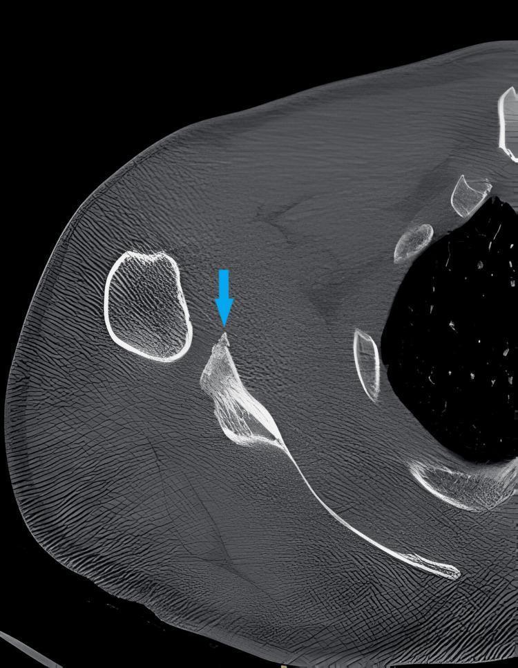
This diagnostic image is an axial Computed Tomography (CT) scan of a right shoulder, focused on the glenohumeral joint and scapula. The scan demonstrates a bony Bankart lesion, which is indicated by a blue arrow. Visually, the lesion manifests as a small, irregular avulsion fracture at the anterior-inferior aspect of the glenoid rim. The detached bony fragment is clearly visible adjacent to the primary margin of the glenoid fossa. This finding is a hallmark of anterior shoulder instability, often occurring during an anterior dislocation where the humeral head impacts and shears off a portion of the glenoid. In this axial view, the relationship between the fractured anterior rim and the main body of the scapula is evident, facilitating the assessment of glenoid bone loss—a critical factor in determining the appropriate surgical intervention, such as a Latarjet procedure or labral repair. The surrounding trabecular bone of the scapula appears intact, and the visualized soft tissue densities provide anatomical context for the shoulder's musculoskeletal architecture.
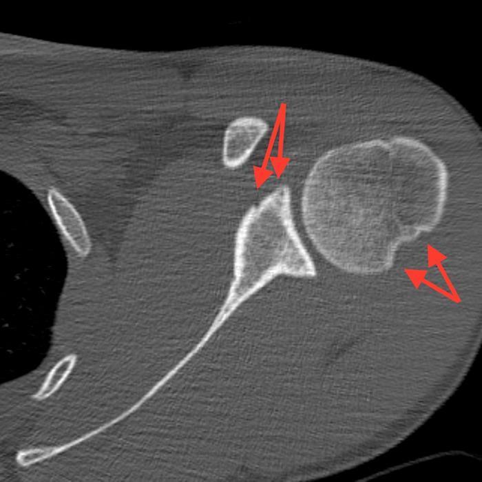
This diagnostic image is an axial cross-section from a non-contrast computerized tomography (CT) scan of the left shoulder. It demonstrates the bony anatomy of the glenohumeral joint, showing findings characteristic of chronic anterior shoulder instability. Two specific pathologies are highlighted with red arrows: 1. A Bankart lesion (bony type) appearing as mild compression or blunting of the anterior glenoid rim, extending approximately 1.1 cm. 2. A Hill-Sachs lesion, seen as a posterolateral compression fracture or 'dent' in the humeral head, measuring roughly 1.4 cm x 0.3 cm. The CT scan effectively illustrates these reciprocal injuries typically caused by recurrent anterior dislocations, where the humeral head impacts against the anterior glenoid. This content is suitable for orthopedic and radiology education, focusing on diagnostic markers of shoulder instability and surgical planning considerations such as the degree of bone loss.
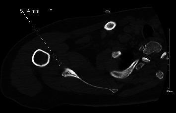
This diagnostic image is an axial Computed Tomography (CT) scan of the right shoulder in a postreduction state. The imaging demonstrates the transverse anatomy of the glenohumeral joint, including the humeral head and the glenoid process of the scapula. A key pathological finding is the presence of multiple small, discrete bony fragments measuring approximately 5.14 mm (annotated with a measurement line) located at the anterior-inferior aspect of the glenoid rim. These findings are diagnostic of a bony Bankart lesion, which represents an avulsion fracture of the glenoid rim typically occurring during an anterior shoulder dislocation. The humeral head is currently reduced within the glenoid fossa, though the presence of these fragments indicates significant joint instability and bone loss. This image is used in orthopedics and radiology to evaluate glenoid integrity and the necessity for surgical stabilization, such as a Latarjet procedure, in the context of recurrent shoulder instability.
Hill-Sachs lesion CT axial humeral head
**Imaging Modality:** Computed Tomography (CT)
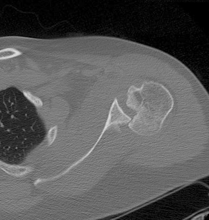
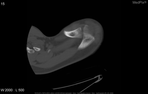
This diagnostic image is an axial Computed Tomography (CT) scan of the right shoulder, illustrating a posterior locked fracture-dislocation. The key visible pathology is a significant impaction fracture on the anterior-medial aspect of the humeral head, clinically identified as a reverse Hill-Sachs lesion (or McLaughlin lesion). The humeral head is displaced posteriorly relative to the glenoid fossa, resulting in an articular incongruity where the humeral head is 'locked' behind the posterior glenoid rim. The CT scan demonstrates the disruption of the normal rounded contour of the humeral head, showing an approximately 35-40% articular surface defect. The scapular spine and the glenoid fossa are visible, with the latter appearing relatively intact. This finding is a classic orthopedic manifestation of a posterior shoulder dislocation, often occurring as a complication of high-energy trauma or seizures. The image serves as an educational tool for identifying humeral head defects and assessing glenohumeral alignment in orthopedic radiology.
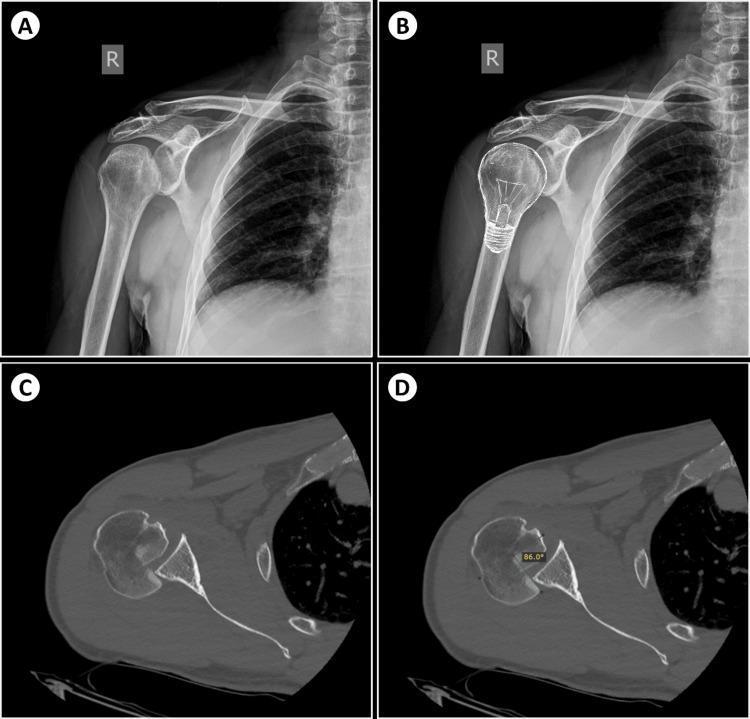
This composite diagnostic image illustrates the radiologic signs of a posterior shoulder dislocation and an associated reverse Hill-Sachs lesion in the right shoulder. Panels A and B present an anteroposterior (AP) X-ray. Panel A shows the 'light-bulb sign,' where internal rotation of the humerus causes the humeral head to appear symmetric and rounded, resembling a light bulb. Panel B includes a graphic overlay of a light bulb to highlight this configuration. Panels C and D provide axial CT scan slices of the same joint. Panel C confirms the posterior displacement of the humeral head relative to the glenoid fossa and reveals a synchronous reverse Hill-Sachs lesion, characterized by an impaction fracture on the anteromedial aspect of the humeral head. Panel D demonstrates a quantitative assessment of the defect using the Cicak method, showing a large impaction fracture involving approximately 50% of the articular surface with an angular measurement of 86.0 degrees. This visual set is an educational resource for identifying subtle radiographic indicators of posterior shoulder instability and associated bony defects.
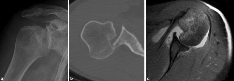
This composite diagnostic image illustrates the radiological findings of an acute locked posterior shoulder dislocation and its subsequent reduction across three modalities. (a) An anteroposterior shoulder X-ray demonstrates an abnormal glenohumeral relationship, often characterized by the 'light bulb sign' due to internal rotation of the humeral head. (b) An axial CT scan reveals a clear posterior dislocation where the humeral head is displaced behind the glenoid fossa. A prominent impaction fracture on the anterior aspect of the humeral head, known as a 'reverse Hill-Sachs defect' or 'McLaughlin lesion,' is visible. (c) An axial T2-weighted MRI scan shows the joint following reduction. The humeral head is realigned with the glenoid, but diagnostic markers remain, including the persistent humeral head impaction and a posterior Bankart lesion (avulsion of the posterior labrum). These images provide educational value in identifying the bony and soft tissue sequelae of posterior glenohumeral instability, categorized under the ABC classification for shoulder injuries.
CT of the Shoulder: Indications, Technique, and Pathologies
Why CT is used (role relative to MRI/US)
- It is the gold standard for detecting and quantifying bone loss (glenoid and humeral head) in instability, with quantitative accuracy for glenoid bone loss superior to plain radiographs and MRI.
- It is preferred when MRI is contraindicated (pacemakers, certain implants, severe claustrophobia, metal hardware causing artifact) - CT arthrography can substitute for MR arthrography to assess the labrum, capsule, and rotator cuff in these patients - Campbell's Operative Orthopaedics, and Rheumatology (Firestein & Kelley).
- It gives superior cortical/trabecular bone detail for fracture characterization, comminution, and surgical planning (3D reconstructions), which plain films and MRI cannot match.
Main indications
-
Glenohumeral instability work-up
- Quantifying glenoid bone loss (e.g., "inverted pear" glenoid, bony Bankart fragment size) - critical for deciding between arthroscopic Bankart repair vs. bone-block procedures (Latarjet).
- Measuring Hill-Sachs lesion depth/size and determining if it is "engaging."
- Assessing glenoid version and morphology.
-
Fracture characterization and surgical planning
- Complex scapular and glenoid fractures (superior, anterior rim, inferior, transverse patterns) - CT (with 3D reconstruction) is considered essential because plain radiographs alone are inadequate to define fracture anatomy - Rockwood and Green's Fractures in Adults, 10th ed.
- Proximal humerus fractures with articular extension, head-split fractures, and assessment of humeral head vascularity/displacement.
- Complex clavicle fracture variants and associated injuries.
- Locked fracture-dislocations (e.g., posterior dislocation with reverse Hill-Sachs/McLaughlin lesion).
-
CT arthrography (contrast injected into the joint)
- Used when MRI is contraindicated, to evaluate labral tears, rotator cuff integrity, capsular volume, and postoperative shoulders (e.g., after instability or cuff repair) - Goldman-Cecil Medicine; Rheumatology, 2-Volume Set.
-
Rotator cuff evaluation (secondary role)
- CT is not first-line for soft tissue (MRI/US are more sensitive for partial-thickness tears), but it is used to grade fatty infiltration/muscle atrophy of the cuff musculature (Goutallier classification, originally described on CT and modified by Fuchs et al.) - Campbell's Operative Orthopaedics.
- Helpful preoperatively/postoperatively to assess structural integrity after cuff repair when MRI is not feasible.
-
Glenohumeral arthritis / arthropathy
- Assessing glenoid bone stock, wear pattern (concentric vs. posterior erosion), and version before shoulder arthroplasty planning.
- When MRI is contraindicated, CT arthrography is a reasonable next-best option for evaluating cartilage and soft tissue in arthropathy - Campbell's Operative Orthopaedics.
-
Postoperative assessment
- Hardware position, fusion/nonunion after fracture fixation, and cuff repair integrity via CT arthrography.
Key pathologies visualized on CT
| Pathology | CT Findings |
|---|---|
| Bony Bankart lesion | Avulsion fracture fragment of the anteroinferior glenoid rim, from anterior dislocation; CT reliably quantifies fragment size and resultant glenoid bone loss (surgically relevant threshold often cited around >20-25% loss). |
| Hill-Sachs lesion | Posterolateral compression/impaction defect of the humeral head from impact against the glenoid rim during anterior dislocation; CT assesses depth/size and "engaging" risk. |
| Reverse Hill-Sachs (McLaughlin) lesion | Anteromedial humeral head impaction fracture seen with posterior dislocation, often with the "light bulb sign" on radiographs and locked posterior dislocation on CT. |
| Glenoid fractures | Classified by location - superior, anterior rim, inferior, transverse/through the body - best delineated with 3D CT reconstruction. |
| Complex scapular fractures | Body, neck, glenoid, and combined acromion/glenoid fracture patterns; CT clarifies fracture lines poorly seen on plain film. |
| Proximal humerus fractures | Comminution, articular surface involvement, head-splitting patterns, displacement of tuberosities. |
| HAGL/BHAGL lesions | Humeral avulsion of the glenohumeral ligament; the "bony" variant (BHAGL) with an avulsed bone fragment can be identified on CT, though soft-tissue component is better seen on MRI. |
| Rotator cuff fatty atrophy | Goutallier grading of fatty infiltration in cuff muscle bellies (supraspinatus, infraspinatus, subscapularis) on axial CT - prognostic for repair outcomes. |
| Glenohumeral arthritis | Joint space narrowing, osteophytes, glenoid erosion pattern, humeral head flattening - relevant to arthroplasty planning. |
| Calcific tendinopathy, os acromiale, AC joint pathology | Bony contour and calcific deposits well shown on CT bone windows. |
Practical summary of when to order CT vs. MRI
- Bone-dominant question (fracture pattern, glenoid/humeral bone loss in instability, surgical planning, arthroplasty templating) -> CT (± 3D reconstruction).
- Soft-tissue-dominant question (labral tear, cuff tear characterization, capsule/ligament injury) -> MRI (or MR arthrography) is first-line; CT arthrography is the substitute when MRI is contraindicated.
- Combined bone + soft tissue need with MRI contraindication -> CT arthrography.
Can you give ct findings of the above pathologies you mentioned
CT findings glenoid bone loss pico method percentage measurement en face view
CT Findings of Shoulder Pathologies (Detailed)
1. Bony Bankart Lesion
- Discontinuity/irregularity of the anteroinferior glenoid cortex with a displaced or non-displaced bone fragment
- Fragment can range from a small chip to a large "inverted pear" glenoid deformity
- Best assessed on axial slices and 3D surface-rendered en face reformats with the humeral head digitally subtracted
- Quantification (surgically important - determines Bankart repair vs. Latarjet):
- Glenoid width method: compare missing anterior width (A) to the diameter of the best-fit inferior circle of the glenoid (B); bone loss = A/B x 100%
- Pico method: CT-based area/surface calculation (mm² or % surface loss) using en face 3D reformats - considered accurate in most CT studies, though a 2023 study flagged it as having a high false-negative rate for grafting decisions
- Clinical thresholds: >20-25% glenoid width loss = bone reconstruction procedure (Latarjet/bone graft) indicated; 10-20% = borderline, individualized; <40% of patients have some bone loss at first dislocation, rising to ~85% with recurrent dislocations
2. Hill-Sachs Lesion
- Wedge-shaped/concave cortical depression or "dent" at the posterosuperolateral humeral head, from impaction against the anterior glenoid during dislocation
- Measured for size, depth, and position on axial CT
- Engaging vs. non-engaging ("on-track" vs "off-track") - glenoid track concept:
- Glenoid track (GT) width is calculated from glenoid measurements
- Hill-Sachs interval (HSI) = width of the Hill-Sachs defect + bone bridge width between rotator cuff footprint and lateral margin of the lesion
- If HSI > GT -> lesion is "off-track"/engaging -> higher risk of recurrent instability and arthroscopic repair failure
- If HSI < GT -> "on-track"/non-engaging -> lower risk
- Example from literature: Hill-Sachs width to footprint of 23.1 mm vs glenoid track of 20.1 mm = off-track, high engaging risk
3. Reverse Hill-Sachs (McLaughlin) Lesion
- Impaction fracture on the anteromedial humeral head from posterior dislocation
- Associated with posterior dislocation: humeral head displaced posteriorly, "locked" behind the posterior glenoid rim
- Defect size quantified as % of articular surface involved (reported ranges of 35-50% in locked dislocations) and by angular measurement methods (e.g., Cicak method)
- Companion radiographic clue: "light bulb sign" on AP view (symmetric, rounded humeral head from fixed internal rotation) - confirmed and better characterized on axial CT
4. Glenoid Fractures
- Ideberg-Goss classification (originally radiograph-based, refined with CT):
- Type I: anterior/posterior rim avulsion fracture
- Type II: transverse/oblique fracture through the glenoid fossa exiting inferiorly
- Type III: fracture exiting superiorly, often through the scapular notch/coracoid base
- Type IV: fracture extending through the glenoid into the scapular body/medial border
- Type V: combination of types with additional fracture lines
- CT findings: fracture line direction and comminution, articular step-off/gap, associated scapular body extension, involvement of coracoid/acromion
- 3D CT is considered necessary because plain radiographs underestimate fracture complexity and displacement
5. Complex Scapular Fractures
- Fracture lines through body, neck, glenoid, and/or acromion, frequently comminuted
- 3D CT reconstructions used to define fracture separation (e.g., total glenoid fracture separated at the surgical neck)
- Assessment of glenopolar angle, medialization/displacement, and associated clavicle or acromioclavicular injury
6. Proximal Humerus Fractures
- Defines articular surface involvement, head-splitting patterns, comminution, and displacement of greater/lesser tuberosities not well seen on plain film
- Assesses humeral head vascularity risk indirectly via displacement pattern and metaphyseal head extension (medial hinge integrity)
- Used to refine Neer-type classification when radiographs are equivocal
7. HAGL / Bony HAGL (BHAGL)
- Bony variant: small avulsed bone fragment from the anatomic neck of the humerus at the inferior glenohumeral ligament attachment
- Cortical irregularity along the humeral neck; the soft-tissue ligament avulsion itself is not visualized on CT (requires MRI/MR arthrography) - CT mainly detects the associated bony fragment
8. Rotator Cuff Fatty Infiltration (Goutallier Classification - originally CT-based)
- Grade 0: Normal muscle, no fatty streaks
- Grade 1: Some fatty streaks within muscle
- Grade 2: Fatty infiltration present but less than muscle (fat < muscle)
- Grade 3: Fat and muscle in equal amounts
- Grade 4: More fat than muscle
- Higher grades (3-4) correlate with poor repair outcomes and are often a relative contraindication to attempted repair; classification later adapted for MRI (Fuchs modification) but originally described on CT densitometry
9. Glenohumeral Arthritis / Arthropathy
- Joint space narrowing, subchondral sclerosis, osteophyte formation
- Glenoid wear pattern and version - critical for arthroplasty planning: concentric wear vs. posterior/biconcave erosion (Walch classification uses CT axial images at the glenoid center)
- Humeral head flattening or subluxation relative to glenoid
- Loose bodies, subchondral cysts
10. Calcific Tendinopathy / Os Acromiale / AC Joint Pathology
- Calcific tendinopathy: discrete hyperdense calcific deposits within the cuff tendon substance (usually supraspinatus), well seen on bone windows
- Os acromiale: unfused ossification center at the acromion tip, seen as a lucent line with smooth cortical margins (distinguishes from fracture)
- AC joint: osteophytes, joint space widening/narrowing, distal clavicle osteolysis
- Glenoid bone loss >20-25% -> bone grafting procedure (Latarjet) generally indicated
- Glenoid bone loss 10-20% -> borderline/individualized decision
- Hill-Sachs "off-track" (HSI > glenoid track) -> engaging, higher failure risk with soft-tissue-only repair
- Goutallier grade 3-4 fatty infiltration -> poor prognosis for cuff repair