
All about EXTRA OCULAR Muscle
extraocular muscles anatomy diagram orbit
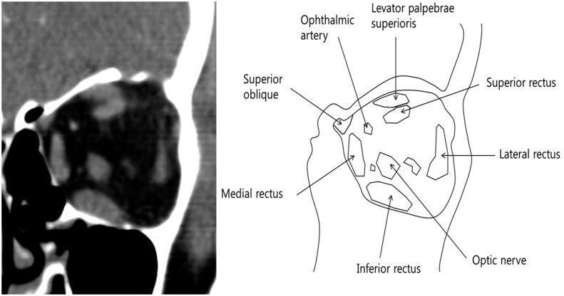
This composite educational graphic features a contrast-enhanced computerized tomography (CT) scan and a corresponding anatomical schematic of the left orbit in the coronal plane. The CT image displays the soft tissue window, highlighting the retrobulbar space and extraocular muscles (EOM) within the orbital fat. The accompanying diagram provides labeled identification of the orbital contents, including the superior rectus, inferior rectus, medial rectus, and lateral rectus muscles. Additionally, the schematic delineates the superior oblique, levator palpebrae superioris, ophthalmic artery, and the central optic nerve. The visual focus is on the cross-sectional morphology of the EOMs, particularly demonstrating a larger cross-sectional area of the inferior rectus compared to the superior rectus. This comparison is relevant for assessing Thyroid Eye Disease (TED) or other orbital pathologies involving muscle hypertrophy. The material is designed for intermediate to advanced medical education, specifically within ophthalmology and radiology, to teach orbital anatomy and the diagnostic interpretation of CT scans for muscle-sparing or involvement patterns.
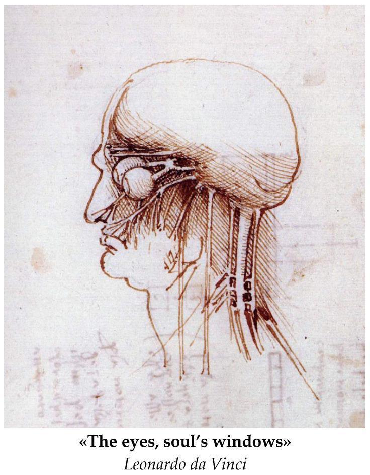
Anatomical Diagram: A historical medical illustration by Leonardo da Vinci, depicting a right lateral profile of the human head and neck. The drawing serves as an early study of gross anatomy and neuroanatomy. It features a detailed representation of the orbit, showing the eyeball as a spherical structure with attached extraocular muscles or neural pathways extending posteriorly toward the brain. The cranium is outlined, with fine hatching and cross-hatching used to provide topographical depth to the skull and facial features. The nasal cavity and oral region are visible in profile. Extending inferiorly from the base of the skull through the neck are several vertical, parallel structures representing major vessels (carotid arteries/jugular veins) and possibly nerves or muscular bundles. The illustration highlights the early Renaissance focus on understanding the relationship between surface anatomy and underlying physiological structures, particularly the visual system's connection to the central nervous system.
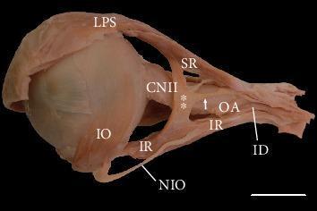
This clinical photograph displays a lateral view of a dissected human left orbit, demonstrating a rare anatomical variation of the extraocular muscles. The primary finding is a variant muscular slip (marked with double asterisks) forming a bridge between the superior rectus (SR) and the inferior rectus (IR) muscles. This bridge is located lateral to the optic nerve (CNII). Additionally, a specific tendinous band (marked with a white arrow) is visible arising from this muscular bridge and attaching posteriorly to the common tendinous ring (Annulus of Zinn). Key neurovascular and muscular structures identified include the levator palpebrae superioris (LPS), the inferior oblique (IO), the inferior branch of the oculomotor nerve (ID), and the nerve to the inferior oblique (NIO). Notably, the NIO is shown piercing the fibers of the inferior rectus. The lateral rectus muscle has been removed to expose these deep orbital structures. This specimen illustrates potential sources of diagnostic confusion in orbital imaging and relevant anatomy for ophthalmologic surgery and strabismus management.
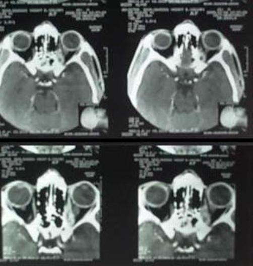
This diagnostic image consists of axial slices from a computerized tomography (CT) scan of the orbits. The imaging demonstrates the anatomy of the ocular globes, extraocular muscles, and posterior orbital structures. In the left orbit, there is significant thickening of the medial rectus and superior rectus muscles. Within these thickened muscles, multiple small, well-defined, non-enhancing hypodense lesions are visible, particularly in the posterior aspect. One prominent lesion within the superior rectus muscle displays a central hyperdense dot, characteristic of a scolex, which is highly suggestive of orbital cysticercosis. The pathology is seen to approximate the optic canal, potentially causing compression or thickening of the optic nerve. This case illustrates a classic radiological presentation of parasitic infection localized to the extraocular muscles, resulting in inflammatory myositis and associated clinical symptoms such as ptosis and restrictive ocular motility. The scan is a key educational tool for distinguishing orbital inflammatory conditions and infectious myositis in ophthalmology and radiology.
Extraocular Muscles - Complete Overview
Anatomical Overview
- Extrinsic (extraocular) muscles - move the eyeball or raise the eyelid
- Intrinsic muscles - inside the eyeball (ciliary, sphincter pupillae, dilator pupillae)
The 7 Extrinsic Muscles - Origin, Insertion, Innervation, Action
| Muscle | Origin | Insertion | Nerve | Action |
|---|---|---|---|---|
| Levator palpebrae superioris | Lesser wing of sphenoid, anterior to optic canal | Anterior surface of superior tarsal plate; few fibers to skin + conjunctival fornix | CN III (superior branch) | Elevates upper eyelid |
| Superior rectus | Superior part of common tendinous ring (Annulus of Zinn) | Anterior half of eyeball, superiorly | CN III (superior branch) | Elevation, adduction, intorsion |
| Inferior rectus | Inferior part of Annulus of Zinn | Anterior half of eyeball, inferiorly | CN III (inferior branch) | Depression, adduction, extorsion |
| Medial rectus | Medial part of Annulus of Zinn | Anterior half of eyeball, medially | CN III (inferior branch) | Adduction |
| Lateral rectus | Lateral part of Annulus of Zinn (bridges superior orbital fissure) | Anterior half of eyeball, laterally | CN VI (Abducens) | Abduction |
| Superior oblique | Body of sphenoid, superomedial to optic canal | Outer posterior quadrant, superior surface | CN IV (Trochlear) | Depression, abduction, intorsion |
| Inferior oblique | Medial floor of orbit, posterior to orbital rim (maxilla, lateral to nasolacrimal groove) | Outer posterior quadrant, inferior surface | CN III (inferior branch) | Elevation, abduction, extorsion |
Memory mnemonic for innervation: LR₆SO₄ - Lateral Rectus = CN VI, Superior Oblique = CN IV, all others = CN III
The Common Tendinous Ring (Annulus of Zinn)
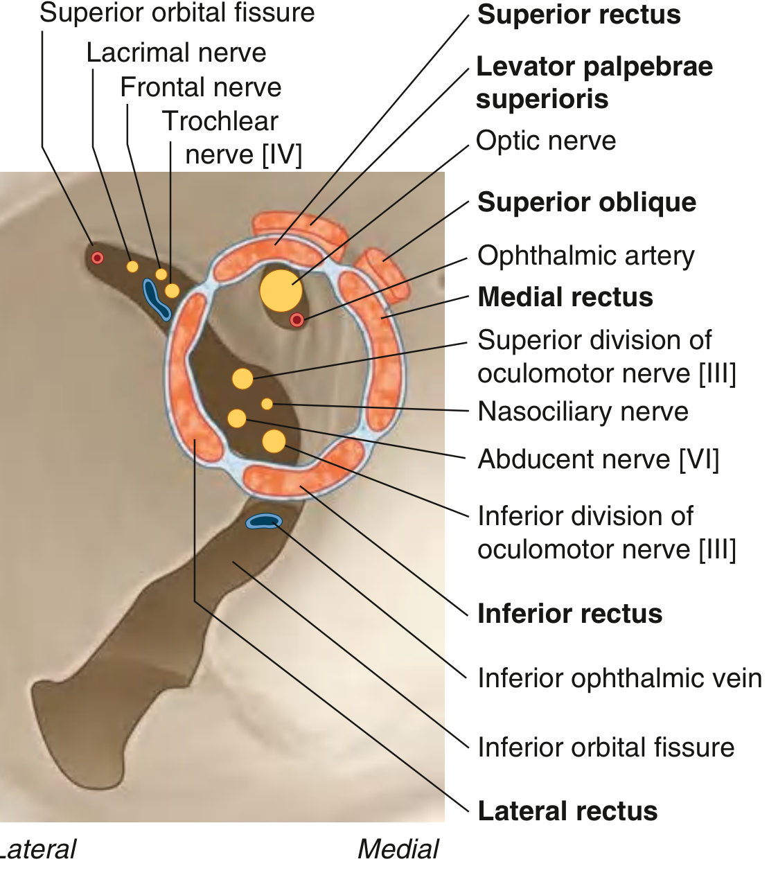
Agonist-Antagonist Pairs
- Medial rectus ↔ Lateral rectus (horizontal rotation)
- Superior rectus ↔ Inferior rectus (primarily vertical + torsional)
- Superior oblique ↔ Inferior oblique (primarily vertical + torsional)
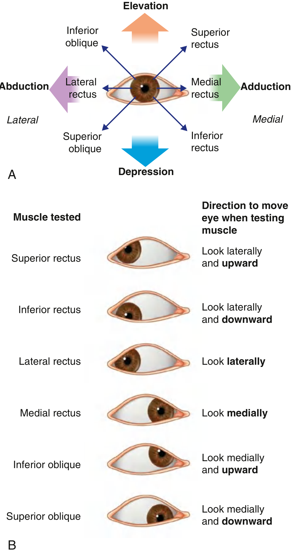
Actions of Individual Muscles
Key points about the obliques:
- The oblique muscles insert posterior to the equator of the globe - this is why their actions are counterintuitive (superior oblique depresses, inferior oblique elevates)
- The rectus muscles insert anterior to the equator
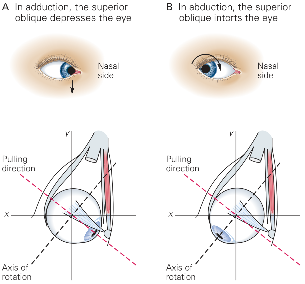
- The superior oblique tendon passes through the trochlea (a fibrocartilaginous pulley on the superomedial orbital rim), making the trochlea its functional origin
- The superior oblique's action depends on eye position:
- In adduction → primarily depresses the eye
- In abduction → primarily intorts the eye
Movements of the Eyeball
| Movement | Description |
|---|---|
| Elevation | Pupil moves superiorly |
| Depression | Pupil moves inferiorly |
| Abduction | Pupil moves laterally |
| Adduction | Pupil moves medially |
| Intorsion (internal rotation) | Top of pupil rotates toward the nose |
| Extorsion (external rotation) | Top of pupil rotates toward the temple |
Conjugate Eye Movements - Yoke Muscles
| Right eye muscle | Left eye muscle | Gaze direction |
|---|---|---|
| Right lateral rectus | Left medial rectus | Gaze right |
| Right medial rectus | Left lateral rectus | Gaze left |
| Right superior rectus | Left inferior oblique | Right-upgaze |
| Right inferior rectus | Left superior oblique | Right-downgaze |
| Right superior oblique | Left inferior rectus | Left-downgaze |
| Right inferior oblique | Left superior rectus | Left-upgaze |
Clinical Testing of Individual Muscles
| Muscle | Clinical test position |
|---|---|
| Superior rectus | Abduct eye, then look up |
| Inferior rectus | Abduct eye, then look down |
| Superior oblique | Adduct eye, then look down |
| Inferior oblique | Adduct eye, then look up |
| Lateral rectus | Look laterally |
| Medial rectus | Look medially |
Cranial Nerve Palsies - Clinical Correlations
CN III (Oculomotor) Palsy
- Supplies SR, IR, MR, IO, levator palpebrae, and pupillary constrictor
- Complete CN III palsy → "down and out" eye + complete ptosis + fixed dilated pupil
- Pupil-sparing CN III palsy is typically microvascular (diabetes, hypertension) - the pupillomotor fibers run on the outside of the nerve and are spared with ischemic injury
- Pupil-involving CN III palsy = posterior communicating artery aneurysm until proven otherwise (surgical emergency)
- Aberrant regeneration follows trauma/compression - misdirected fibers cause lid elevation on downgaze (pseudo-Graefe sign) or adduction
CN IV (Trochlear) Palsy
- Innervates the superior oblique only
- Only cranial nerve that decussates before leaving the brainstem (exits dorsally from the midbrain)
- Palsy causes hypertropia + excyclotorsion - patients complain of vertical diplopia worse on reading/descending stairs
- Classic feature: head tilt test (Parks-Bielschowsky) - tilting the head toward the affected side worsens vertical diplopia; tilting away alleviates it
- Most common cause of isolated vertical diplopia; commonly idiopathic microvascular or post-head trauma (free edge of tentorium impinges on nerve)
CN VI (Abducens) Palsy
- Innervates the lateral rectus only
- Palsy → esotropia + horizontal diplopia, worse on ipsilateral gaze
- Abducens nucleus lesion (vs fascicle): nuclear lesion affects interneurons projecting via MLF to contralateral medial rectus → complete ipsilateral horizontal gaze palsy (not just one eye)
- Foville syndrome (dorsal pons): lateral gaze palsy + ipsilateral facial palsy + contralateral hemiparesis
- Millard-Gubler syndrome (ventral pons): lateral rectus weakness alone + facial palsy (fascicle involved, not nucleus)
- The long subarachnoid course of CN VI makes it susceptible to raised ICP (false localizing sign - bilateral abducens palsy)
- Gradenigo's syndrome: mastoiditis → petrous apex involvement → abducens palsy + ipsilateral facial pain + deafness
Special Features
Levator Palpebrae Superioris
- Most superior muscle in the orbit
- Contains smooth muscle fibers (Superior Tarsal Muscle / Müller's muscle) on its inferior surface
- These smooth fibers are innervated by postganglionic sympathetic fibers from the superior cervical ganglion
- CN III loss → complete ptosis; sympathetic loss (Horner's syndrome) → partial ptosis
Fascial Sheaths and Check Ligaments
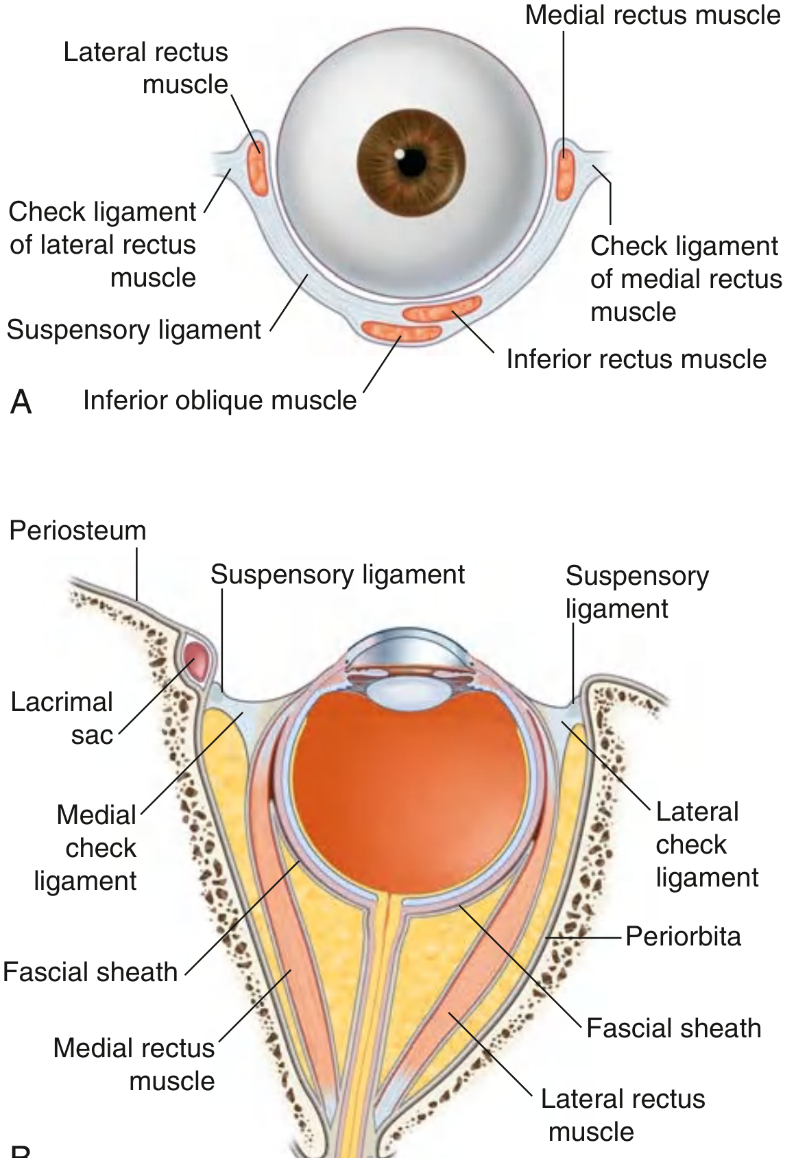
- Each EOM is enclosed in a fascial sheath that fuses with the fascial sheath of the globe (Tenon's capsule)
- Where these sheaths thicken, they form check ligaments that limit overaction of each muscle
- The medial and lateral check ligaments are especially strong; together with inferior ligamentous extensions they form the suspensory ligament of Lockwood which supports the globe
Soft-Tissue Pulleys
- Modern understanding: each EOM has a dual insertion - part of the muscle inserts on a soft-tissue pulley in the orbit, through which the rest of the muscle passes to reach the globe
- These pulleys change the muscle's pulling direction as the eye moves, stabilizing ocular rotations
Blood Supply
Summary Table
| Nerve | Muscles supplied | Palsy causes |
|---|---|---|
| CN III (Oculomotor) | SR, IR, MR, IO, Levator palpebrae, ciliary & sphincter pupillae | Down-and-out eye, ptosis, mydriasis; aneurysm (pupil-involving), microvascular ischemia (pupil-sparing) |
| CN IV (Trochlear) | Superior oblique | Vertical diplopia, worse reading; head tilt; trauma, microvascular |
| CN VI (Abducens) | Lateral rectus | Esotropia, horizontal diplopia; raised ICP, pontine lesions, Gradenigo's |