
analyse_media
Reading File
Finding Sources
Finding Sources
Reading File
Reading File
Loading Image
Reading File
Here is a thorough clinical analysis of the wound shown in your video.
Clinical Analysis of the Wound
What the Video Shows
The video captures a close-up of a chronic skin ulcer on the lower extremity, featuring:
- A deep, circular/oval-shaped open wound
- Bright red, beefy, moist granulation tissue filling the wound bed
- Raised, thickened, pale/white wound margins (suggestive of epibole - rolled edges that can stall healing)
- A faint hyperpigmented ring in the surrounding skin (prior inflammation or resolved larger lesion)
- Dry, slightly flaky surrounding skin with sparse hair - consistent with a lower leg/ankle location
Differential Diagnosis
Based on the appearance, the most likely causes are (in order of probability on the lower leg):
| Type | Distinguishing Features |
|---|---|
| Venous stasis ulcer (most common, 45-60% of leg ulcers) | Medial lower leg/above medial malleolus; shallow; fibrinous yellow base; background of stasis dermatitis and hyperpigmentation |
| Diabetic ulcer | Often on the foot/plantar surface; deep; neuropathy history; small vessel disease |
| Arterial/ischemic ulcer | Painful; punched-out edges; necrotic base; pale/necrotic rather than red granulation; absent pulses |
| Pressure sore | Over bony prominences (sacrum, heel); in bedridden patients |
The bright red granulation tissue in this wound is a positive finding - it indicates active healing with adequate blood supply. However, the raised, rolled (epibolic) wound edges are a concern because they prevent epithelial migration across the wound bed, potentially stalling closure.
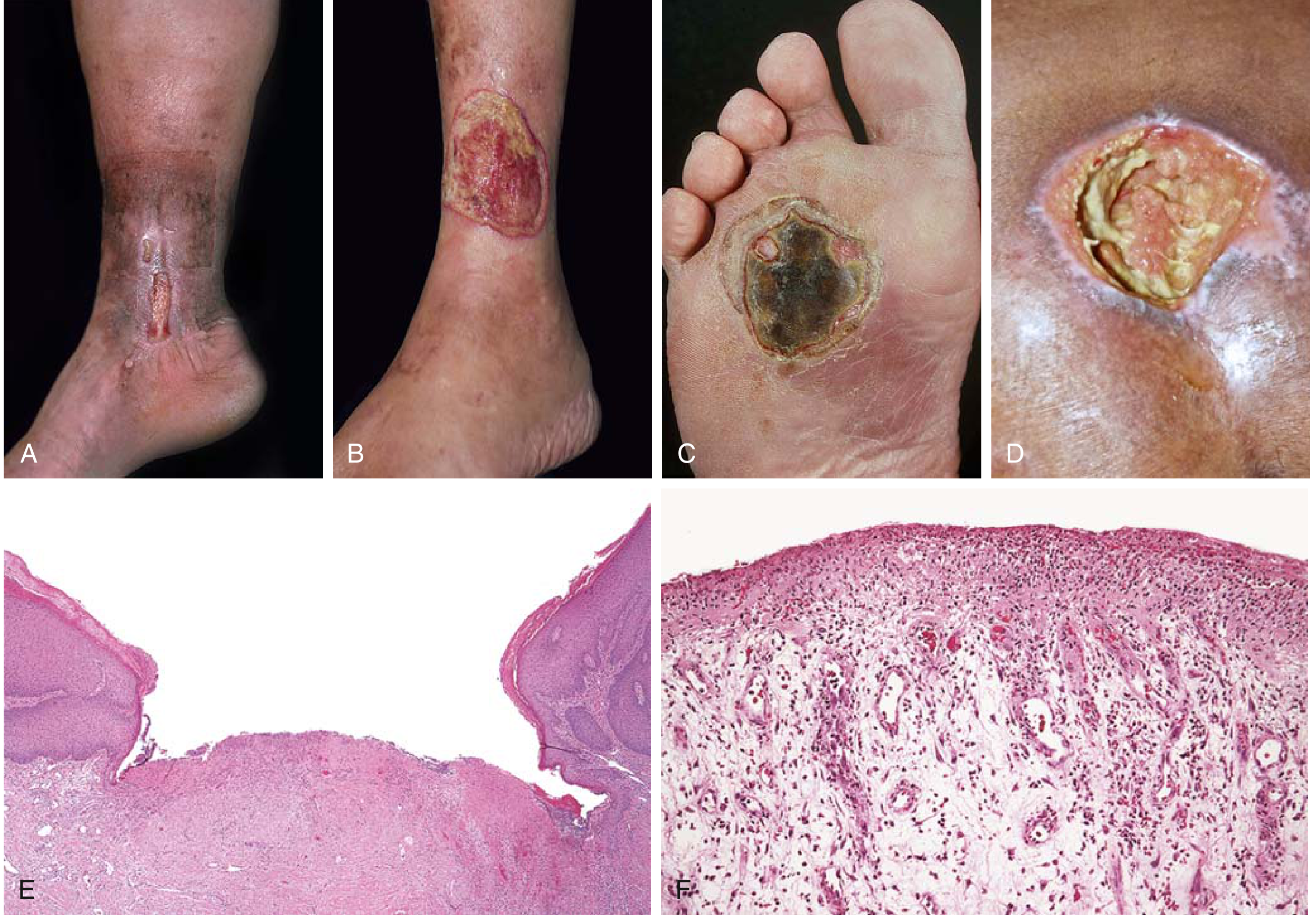
According to Robbins & Kumar Basic Pathology: Venous leg ulcers develop most often in elderly people as a result of chronic venous hypertension (from varicose veins or congestive heart failure), resulting in poor oxygen delivery. Diabetic ulcers of the lower extremities are characterized by epidermal ulceration and extensive granulation tissue in the underlying dermis (Fig. 2.25, p. 73).
Here are reference images comparing the four main types of chronic lower extremity ulcers:
Wound Healing Status
The wound appears to be healing by secondary intention (open wound filling from the base up), currently in the proliferative phase characterized by granulation tissue formation. Key observations:
- Positive sign: Bright red/beefy granulation = good vascularity and active tissue repair
- Concern: Rolled/thickened wound edges (epibole) = epithelial cells cannot migrate across; this is a known cause of stalled chronic wounds
- Surrounding skin changes: Dryness, flaking, and hyperpigmentation suggest chronic venous/lymphatic insufficiency or repeated inflammation
Recommended Clinical Workup
If evaluating this patient, standard next steps would include:
- Ankle-Brachial Index (ABI) - to rule out arterial disease before applying compression
- History: Diabetes, varicose veins, prior DVT, heart failure, hypertension
- Venous duplex ultrasound if venous insufficiency suspected
- Wound biopsy if the lesion is in an atypical location, does not respond to therapy, or malignancy is suspected
General Management Principles
Per Andrews' Diseases of the Skin (p. 981):
- Venous ulcers: Leg compression (40 mmHg graduated compression stockings or multilayer bandaging) is the cornerstone of treatment; elevation; moisture-retentive dressings
- Debridement of epibolic/thickened wound edges to stimulate re-epithelialization
- Moist wound environment - once the wound is clean and granulating, maintain moisture without disturbing healing tissue
- Treat underlying cause (venous insufficiency, glycemic control in diabetes, etc.)
- Topical agents such as silver sulfadiazine, cadexomer iodine, or growth factor preparations may be used for non-healing wounds
Important disclaimer: This is a visual/educational analysis only. A definitive diagnosis requires a full clinical history, physical examination, vascular studies, and potentially wound biopsy. Please consult a wound care specialist, vascular surgeon, or dermatologist for proper evaluation and management.
This is a shared conversation. Sign in to Orris to start your own chat.