
Explain large intestine with specimen for presentation
Reading File
Now let me search the medical library for large intestine anatomy, histology, and specimen content in parallel.
Finding Sources
Finding Sources
Excellent results. Now let me read the most comprehensive sources in parallel - Gray's Anatomy for the gross anatomy/specimen content, Junqueira's for histology, and Sabiston for surgical anatomy.
Reading File
Reading File
Reading File
Good material gathered. Now let me read more histology details and fetch the key images for the presentation.
Reading File
Reading File
Loading Image
Loading Image
Excellent! I have great anatomy and histology images. Now let me fetch the colon mucosa histology image and read more on blood supply and functions.
Loading Image
Reading File
Finding Sources
I now have all the key content needed. Let me read the blood supply and functions content from Yamada's and Gray's to complete the picture.
Reading File
Reading File
I now have comprehensive content. Let me compile the full presentation-ready explanation.
The Large Intestine - Complete Presentation
Overview
The large intestine extends from the distal end of the ileum to the anus, approximately 1.5 m in length in adults with a diameter of 6-7 cm (wider than the small intestine). Its primary roles are water/electrolyte absorption and converting undigested material into feces.
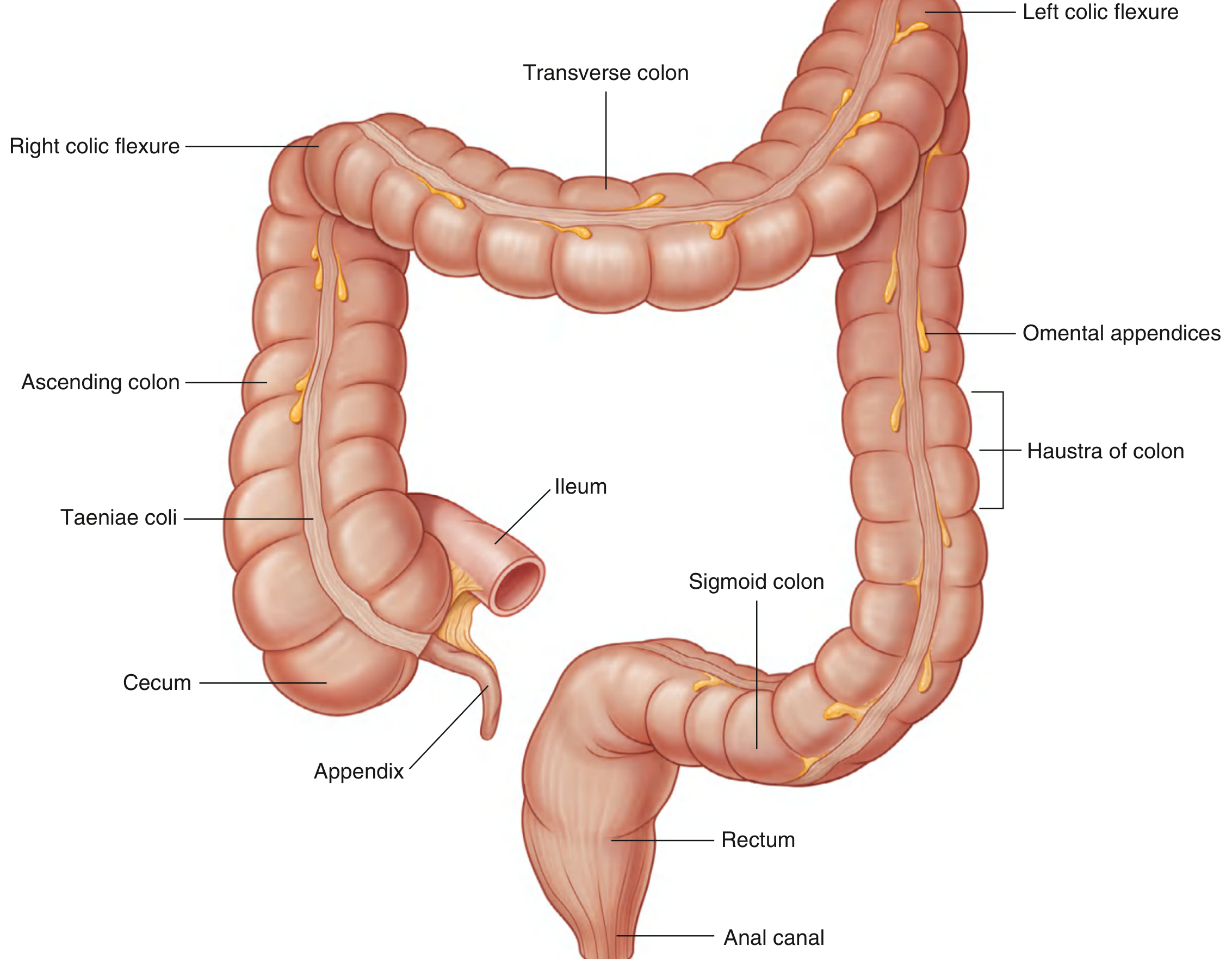
Parts / Segments
The large intestine is divided into:
| Segment | Position | Peritoneal Relation |
|---|---|---|
| Cecum | Right iliac fossa | Intraperitoneal |
| Ascending colon | Right flank → right hypochondrium | Retroperitoneal |
| Right colic (hepatic) flexure | Inferior to right lobe of liver | - |
| Transverse colon | Right hypochondrium → left hypochondrium | Intraperitoneal |
| Left colic (splenic) flexure | Inferior to spleen | - |
| Descending colon | Left flank | Retroperitoneal |
| Sigmoid colon | Pelvis | Intraperitoneal |
| Rectum | Posterior pelvic wall | - |
| Anal canal | Perineum | - |
Characteristic External Features (Specimen Identification)
These four features distinguish the large intestine from the small intestine on a specimen and are your key identification points:
1. Taeniae Coli
Three flattened bands of longitudinal smooth muscle running along the colon. The outer longitudinal muscle layer is NOT circumferential - it condenses into these three distinct ribbon-like bands. They lead directly to the base of the appendix, useful as a landmark. They are absent in the rectum, where they spread out to form a complete longitudinal layer.
2. Haustra of the Colon (Sacculations)
Outpocketings or sacculations in the colon wall produced by the contraction of taeniae coli. They give the colon its characteristic "puckered" appearance, visible both on specimen and on barium enema X-ray. Haustra are absent in the rectum and sigmoid colon.
3. Omental Appendices (Appendices Epiploicae)
Small, peritoneal-covered accumulations of fat attached along the colon. These pendulous adipose protuberances hang off the serosal surface. They are a hallmark of the colon on specimen examination.
4. Large Internal Diameter
Clearly wider lumen than the small intestine (6-7 cm vs. ~2.5 cm), helping immediate identification.
Specimen tip: When shown an intestinal specimen, look for haustra + taeniae coli + omental appendices = large intestine. If these are absent and villi are present = small intestine.
Cecum and Appendix
The cecum is the saccular beginning (avg. diameter 7.5 cm, length 10 cm), located in the right iliac fossa. It is completely covered by peritoneum (intraperitoneal). The ileocecal valve connects the ileum to the cecum and prevents backflow.
The appendix is a narrow blind-ended tube attached to the posteromedial wall of the cecum where the three taeniae coli converge. Its position varies:
- Retrocecal - most common (~65%)
- Pelvic/descending
- Subcecal
- Pre-ileal / post-ileal
McBurney's point - surface projection of the appendix base: junction of lateral and middle 1/3 of a line from the anterior superior iliac spine to the umbilicus. Pain here = appendicitis.
Blood Supply
Arterial Supply
-
Superior mesenteric artery (SMA) - supplies right side of colon (to proximal 2/3 of transverse colon):
- Ileocolic artery → cecum, appendix, ascending colon
- Right colic artery → ascending colon
- Middle colic artery → transverse colon
-
Inferior mesenteric artery (IMA) - supplies left side (distal transverse to rectum):
- Left colic artery → descending colon
- Sigmoid arteries → sigmoid colon
- Superior rectal artery → rectum
Marginal artery of Drummond - runs along the mesenteric border of the entire colon, forming an important anastomotic arcade connecting SMA and IMA territories.
Griffiths' critical point - the watershed/vulnerable zone at the splenic flexure between SMA and IMA territories; most susceptible to ischemia.
Histology (Microscopic Specimen)

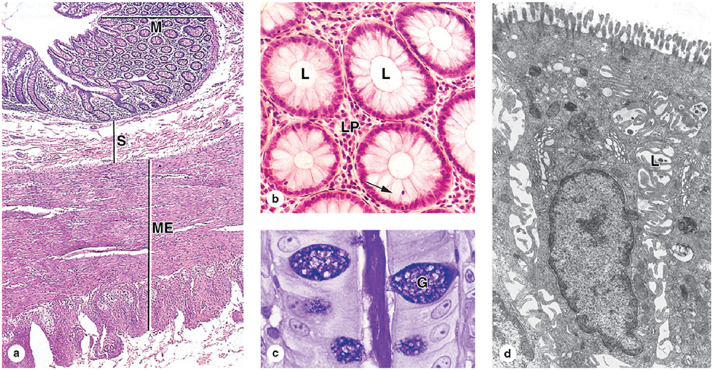
Wall Layers (from lumen outward):
1. Mucosa
- Lining: simple columnar epithelium with abundant goblet cells (increase in number from cecum to rectum - provide lubrication)
- No villi (unlike small intestine) - a key histological point
- No major folds except in rectum
- Intestinal glands (crypts of Lieberkühn) run the full depth of the mucosa - lined by absorptive colonocytes and goblet cells
- Colonocytes have short, irregular microvilli and dilated intercellular spaces - indicating active fluid absorption
- Stem cells located in the bottom 1/3 of each crypt
- Lamina propria is rich in MALT (mucosa-associated lymphoid tissue) due to the large bacterial load of the colon
2. Submucosa
- Dense irregular connective tissue, well-vascularized
- Meissner's nerve plexus present
3. Muscularis Externa
- Inner circular layer (complete)
- Outer longitudinal layer = condensed into three taeniae coli (not a complete layer)
- Auerbach's (myenteric) nerve plexus between the two layers
- Local contractions of taeniae coli produce haustra
4. Serosa
- Peritoneum covering intraperitoneal segments
- Characterized by omental appendices (adipose pendulous masses)
- Retroperitoneal segments have adventitia instead
Rectum and Anal Canal
The rectum is 10-15 cm long; it lacks haustra, and the taeniae coli merge to form a complete longitudinal muscle coat. It stores feces prior to defecation.
The anal canal (3-4 cm long) has:
- Anal columns of Morgagni - longitudinal folds of mucosa
- Anal sinuses - between columns
- At the pectinate (dentate) line: mucosa transitions from simple columnar (rectum) to stratified squamous epithelium
- Internal anal sphincter - involuntary, smooth muscle (thickened circular layer)
- External anal sphincter - voluntary, striated muscle
- Rich venous plexus in submucosa (when dilated = hemorrhoids)
Functions Summary
| Function | Mechanism |
|---|---|
| Water absorption | Colonocytes absorb Na⁺ (active), water follows osmotically; ~1.5 L/day absorbed |
| Electrolyte absorption | Na⁺, Cl⁻ absorption; K⁺, HCO₃⁻ secretion |
| Fecal formation | Compaction of indigestible material |
| Mucus secretion | Goblet cells lubricate passage |
| Microbial fermentation | Gut microbiome ferments undigested carbohydrates → short-chain fatty acids |
| Defecation reflex | Rectal distension triggers coordinated sphincter relaxation |
Clinical Correlations (for Specimen/Presentation Context)
Colorectal Cancer - Adenocarcinoma arising from benign adenomatous polyps; most common in rectum, sigmoid, and descending colon. Low-fiber diet prolongs mucosal contact with fecal carcinogens. Screened by colonoscopy and fecal occult blood testing.
Appendicitis - Obstruction of appendix lumen by fecalith or lymphoid hyperplasia → bacterial overgrowth → wall necrosis → perforation. Classic: periumbilical pain migrating to McBurney's point + fever + nausea.
Diverticulosis - Herniations of mucosa/submucosa through muscular wall, especially in sigmoid colon. Associated with low-fiber diet and increased intraluminal pressure.
Ischemic Colitis - Occurs at watershed zones (splenic flexure - Griffiths' point); reduced perfusion through SMA/IMA.
Hemorrhoids - Dilated rectal venous plexus sinuses; internal (above pectinate line, painless) vs. external (below pectinate line, painful).
Sources
- Gray's Anatomy for Students (pp. 379-382)
- Junqueira's Basic Histology - Text and Atlas, 17e (pp. 793-797)
- Histology: A Text and Atlas with Correlated Cell and Molecular Biology
- Yamada's Textbook of Gastroenterology, 7th ed. (pp. 184-185)
- Sabiston Textbook of Surgery
For 1st year
Asking for Preferences
Which 1st year course is this presentation for? · What format do you need?
Mbbs/bams
Asking for Preferences
What format do you need?
Perfect. For MBBS/BAMS 1st year Anatomy, the focus is on gross anatomy (dissection hall level), histology, relations, blood supply, nerve supply, and applied anatomy - all exam-oriented. Let me pull the most detailed content from the textbooks.
Reading File
Finding Sources
Reading File
Reading File
Finding Sources
Reading File
Reading File
Loading Image
Reading File
Loading Image
Loading Image
I now have an actual specimen photograph showing TC (teniae coli), HC (haustra coli), and OA (omental appendices) - perfect for a 1st year student. Let me read the nerve supply and get the histology microslide photo too.
Reading File
Loading Image
Loading Image
Here are your complete 1st Year MBBS/BAMS Anatomy Study Notes on the Large Intestine - structured exactly as it appears in university exams and dissection guides.
LARGE INTESTINE
1st Year MBBS/BAMS - Anatomy Notes
1. INTRODUCTION
- The large intestine extends from the ileocecal junction to the anus
- Length: approximately 1.5 m (about 5 feet)
- Diameter: 6-7 cm (wider than small intestine)
- Main functions: absorption of water and electrolytes, formation and storage of feces
2. PARTS OF LARGE INTESTINE
| # | Part | Location |
|---|---|---|
| 1 | Cecum | Right iliac fossa |
| 2 | Appendix | Attached to posteromedial cecum |
| 3 | Ascending colon | Right flank (retroperitoneal) |
| 4 | Right colic (hepatic) flexure | Below right lobe of liver |
| 5 | Transverse colon | Crosses abdomen (intraperitoneal) |
| 6 | Left colic (splenic) flexure | Below spleen |
| 7 | Descending colon | Left flank (retroperitoneal) |
| 8 | Sigmoid colon | Pelvis (intraperitoneal) |
| 9 | Rectum | Posterior pelvic wall |
| 10 | Anal canal | Perineum |
3. DISTINGUISHING FEATURES (SPECIMEN EXAM FAVOURITE)
These 4 features distinguish the large intestine from the small intestine on a cadaveric specimen.
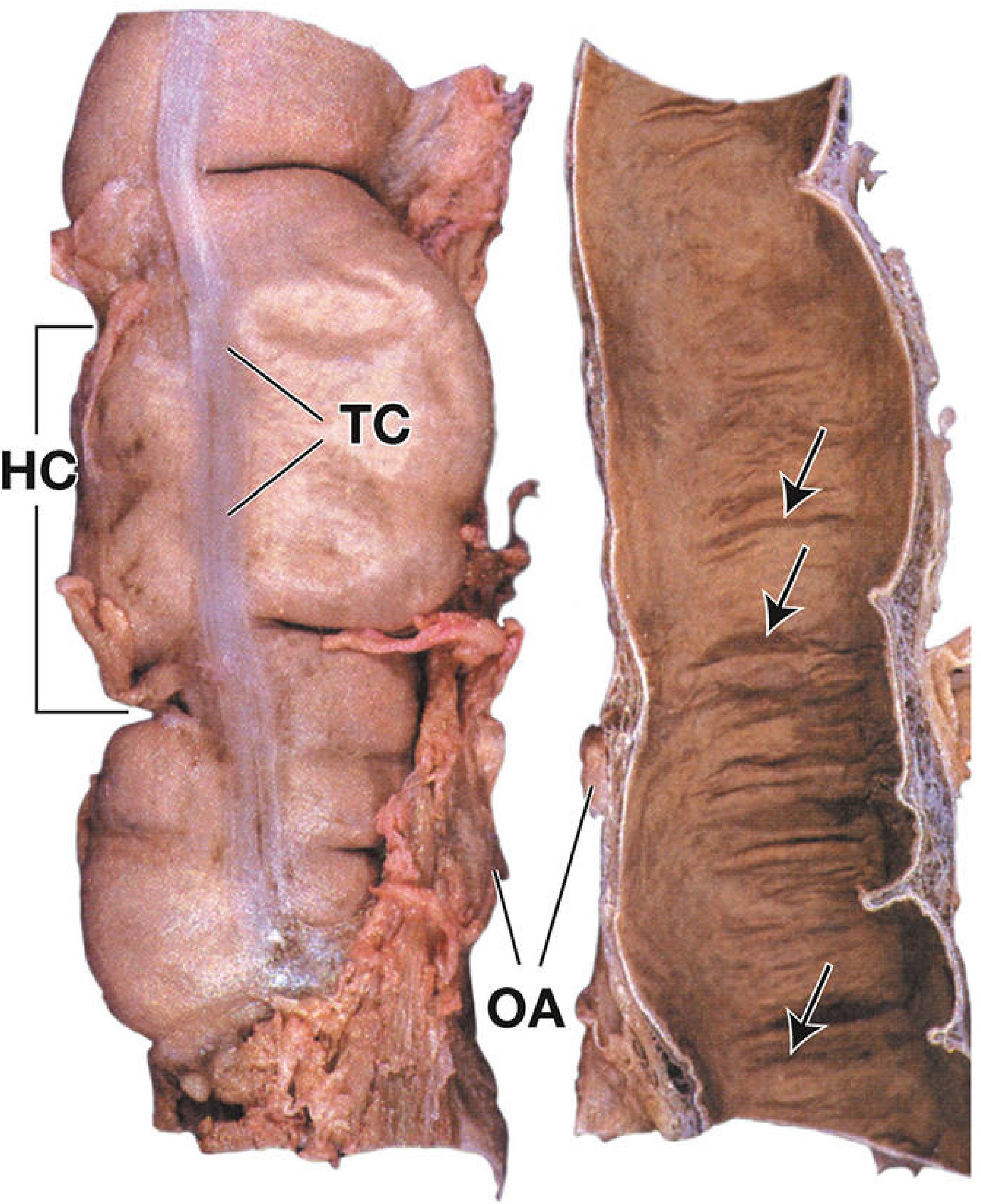
3.1 Taeniae Coli (Teniae Coli)
- Three flat ribbon-like bands of longitudinal muscle
- Run along the length of the cecum and colon
- Absent in the rectum, anal canal, and appendix (where longitudinal muscle forms a complete coat)
- All three taeniae converge at the base of the appendix - a surgical landmark
3.2 Haustra Coli (Sacculations)
- Bulging outpocketings between the taeniae
- Produced by contraction of the taeniae coli
- Give the colon its characteristic puckered appearance on gross specimen and barium enema X-ray
- Absent in the rectum and sigmoid colon
3.3 Omental Appendices (Appendices Epiploicae)
- Small, fatty, peritoneal-covered projections attached along the colon
- Contain fat; visible on serosal surface
3.4 Large Lumen
- Wider than the small intestine (no villi, no circular folds / plicae circulares)
ACTUAL SPECIMEN PHOTOGRAPH:
4. CECUM AND APPENDIX
Cecum
- Saccular beginning of large intestine
- Location: right iliac fossa
- Size: ~7.5 cm diameter, ~10 cm length
- Intraperitoneal (covered by peritoneum on all sides, but no mesentery)
- The ileocecal valve (at the ileocecal junction) prevents backflow of feces into the ileum
Appendix (Vermiform Appendix)
- Narrow, blind-ended tube attached to the posteromedial wall of the cecum
- Where all 3 taeniae coli converge - useful to locate it surgically
- Contains lymphoid tissue in its walls (part of MALT)
- McBurney's Point = base of appendix on the surface = junction of lateral and middle 1/3 of a line from the anterior superior iliac spine (ASIS) to the umbilicus
Positions of appendix (in order of frequency):
- Retrocecal - most common (~65%)
- Pelvic/Descending - 2nd most common
- Subcecal
- Pre-ileal
- Post-ileal
5. COLON - PERITONEAL RELATIONS
| Segment | Peritoneal Status | Mesentery |
|---|---|---|
| Ascending colon | Retroperitoneal (secondarily) | None |
| Transverse colon | Intraperitoneal | Transverse mesocolon |
| Descending colon | Retroperitoneal (secondarily) | None |
| Sigmoid colon | Intraperitoneal | Sigmoid mesocolon |
Key points:
- Right colic (hepatic) flexure - just inferior to the right lobe of liver
- Left colic (splenic) flexure - just inferior to the spleen; higher and more posterior than right; attached to the diaphragm by the phrenicocolic ligament
- Paracolic gutters - depressions lateral to ascending and descending colon; allow fluid spread within the peritoneal cavity
6. BLOOD SUPPLY
6.1 Arterial Supply
| Segment | Artery | From |
|---|---|---|
| Cecum + Appendix | Anterior/Posterior cecal artery, Appendicular artery | Ileocolic artery (SMA) |
| Ascending colon | Ileocolic + Right colic artery | Superior Mesenteric Artery (SMA) |
| Transverse colon | Middle colic artery | SMA |
| Descending colon | Left colic artery | Inferior Mesenteric Artery (IMA) |
| Sigmoid colon | Sigmoid arteries | IMA |
| Rectum | Superior rectal (IMA) + Middle rectal (Internal iliac) + Inferior rectal (Internal pudendal) | Multiple |
Important concepts:
- Marginal artery of Drummond - anastomotic arcade running along the mesenteric border of the whole colon, connecting SMA and IMA territories
- Griffiths' critical point - watershed zone at the splenic flexure (between SMA and IMA); most vulnerable to ischemia
6.2 Venous Drainage
- Mirrors the arterial supply
- Right colon → Superior Mesenteric Vein → Portal vein
- Left colon/rectum → Inferior Mesenteric Vein → Portal vein
- Lower rectum/anal canal → Internal iliac vein → IVC (portosystemic anastomosis)
7. LYMPHATIC DRAINAGE
- Lymphatics follow the arteries
- Right colon → Superior mesenteric nodes
- Left colon → Inferior mesenteric nodes
- Rectum → Internal iliac nodes
8. NERVE SUPPLY
| Segment | Sympathetic | Parasympathetic |
|---|---|---|
| Cecum to splenic flexure (midgut) | Greater/Lesser splanchnic → Superior mesenteric plexus | Vagus nerve (CN X) |
| Splenic flexure to rectum (hindgut) | Lumbar splanchnic → Inferior mesenteric plexus | Pelvic splanchnic nerves (S2, S3, S4) |
| Anal canal (below pectinate line) | Pudendal nerve (somatic) | - |
- Sympathetic: inhibits peristalsis, causes vasoconstriction
- Parasympathetic: stimulates peristalsis and glandular secretion
- The pectinate/dentate line is the key nerve supply boundary in the anal canal
9. RECTUM AND ANAL CANAL
Rectum
- Begins at the rectosigmoid junction (~vertebra S3)
- Length: ~12-15 cm
- No haustra, no taeniae (longitudinal muscle forms complete coat)
- No mesentery (retroperitoneal)
- Contains transverse rectal folds (Houston's valves) - support the fecal column
Anal Canal
- Length: 3-4 cm
- Pectinate (Dentate) Line - crucial landmark dividing upper and lower anal canal:
| Feature | Above Pectinate Line | Below Pectinate Line |
|---|---|---|
| Embryological origin | Endoderm (hindgut) | Ectoderm (proctodeum) |
| Epithelium | Simple columnar | Stratified squamous |
| Arterial supply | Superior rectal artery | Inferior rectal artery |
| Venous drainage | Portal system (internal hemorrhoids) | Systemic (external hemorrhoids) |
| Lymph drainage | Internal iliac nodes | Superficial inguinal nodes |
| Nerve supply | Autonomic (visceral pain - poorly localized) | Somatic - Pudendal nerve (sharp/precise pain) |
| Sensation | Poorly felt | Exquisitely painful |
Sphincters of anal canal:
- Internal anal sphincter - involuntary, smooth muscle (thickened circular muscle)
- External anal sphincter - voluntary, striated muscle; supplied by pudendal nerve (S2, S3, S4)
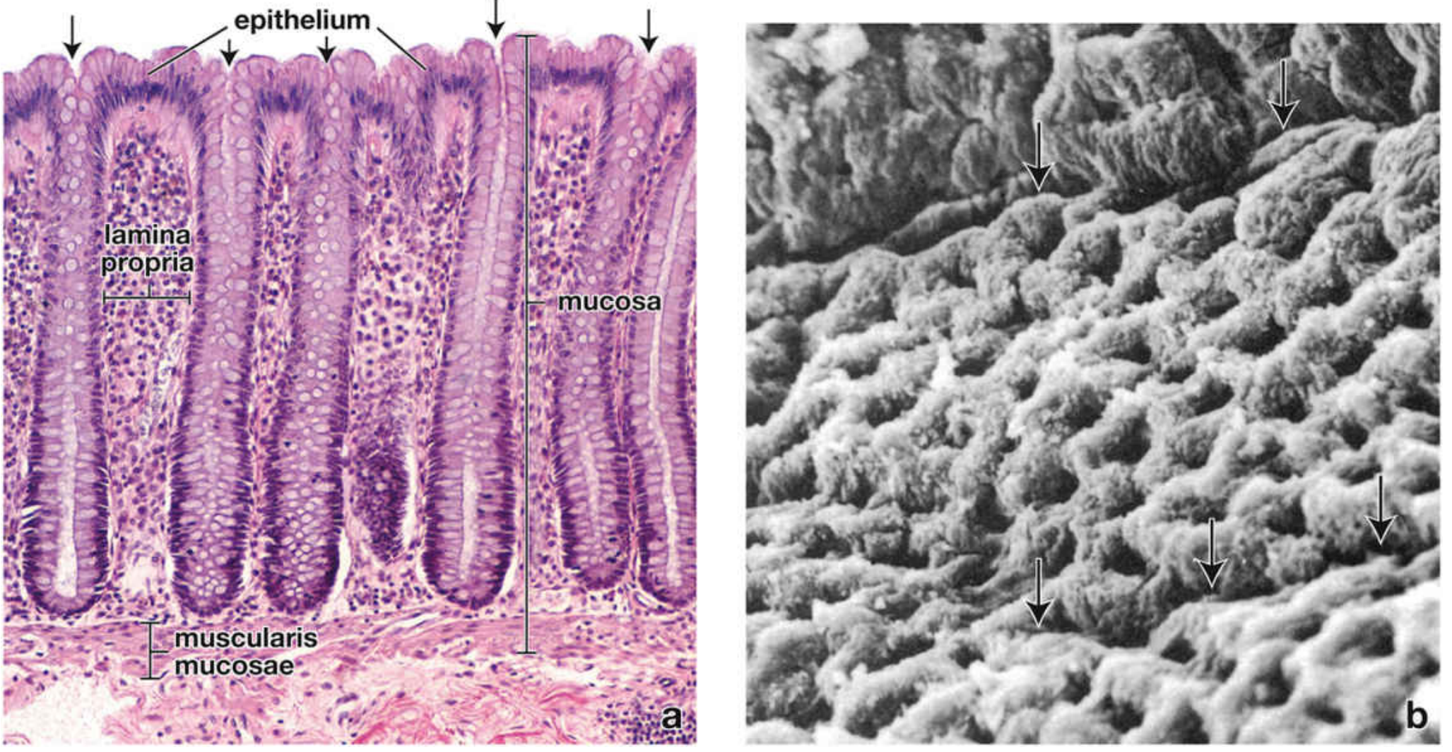
10. HISTOLOGY (MICROSCOPIC ANATOMY)
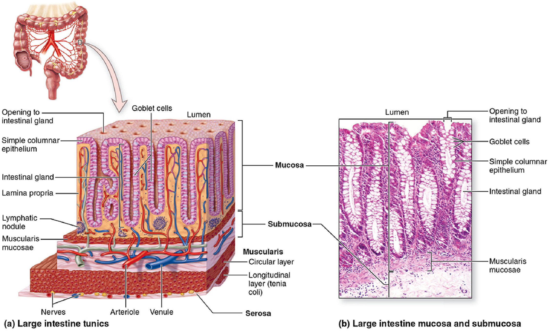
Layers of Large Intestine Wall (Lumen → Outside):
Layer 1 - Mucosa:
- Epithelium: Simple columnar with numerous goblet cells (increase from cecum to rectum)
- NO villi (unlike small intestine) - key exam point
- NO plicae circulares (no circular folds)
- Contains Crypts of Lieberkühn (straight, unbranched tubular glands running through full thickness of mucosa)
- Colonocytes (absorptive cells): short irregular microvilli, dilated intercellular spaces - active water absorption
- Lamina propria: rich in MALT (mucosa-associated lymphoid tissue) due to large intestine's heavy bacterial load
- Muscularis mucosae: thin smooth muscle layer
Layer 2 - Submucosa:
- Dense irregular connective tissue
- Well-vascularized
- Contains Meissner's plexus (submucosal nerve plexus)
Layer 3 - Muscularis Externa:
- Inner circular layer (complete)
- Outer longitudinal layer - condensed into 3 taeniae coli (NOT a complete layer - key feature)
- Auerbach's plexus (myenteric nerve plexus) between the two layers
Layer 4 - Serosa/Adventitia:
- Intraperitoneal portions: serosa with omental appendices (fat-filled protrusions)
- Retroperitoneal portions: adventitia (no peritoneal covering)
11. FUNCTIONS SUMMARY
| Function | Detail |
|---|---|
| Water absorption | ~1.5 L/day absorbed; mainly in right colon |
| Electrolyte balance | Na⁺ absorbed, K⁺ secreted |
| Feces formation | Compaction of residue |
| Mucus secretion | Goblet cells lubricate fecal passage |
| Microbial fermentation | Gut bacteria produce short-chain fatty acids, vitamin K, vitamin B12 |
| Defecation | Rectal distension → internal sphincter relaxation → voluntary external sphincter control |
12. APPLIED ANATOMY (Exam Favourite Questions)
A. Appendicitis
- Obstruction of appendix lumen (by fecalith or lymphoid hyperplasia) → bacterial proliferation → wall necrosis → possible perforation
- Pain: starts periumbilical (visceral, colicky) → migrates to McBurney's point after 6-10 hours (somatic, constant)
- Retrocecal position: can mimic hip pathology (psoas irritation)
- Treatment: Appendectomy
B. Colorectal Cancer
- Adenocarcinoma from adenomatous polyps in mucosal epithelium
- Most common in rectum, sigmoid, descending colon
- Risk factors: low-fiber diet, prolonged fecal contact with mucosa
- Screening: colonoscopy, fecal occult blood test
C. Hemorrhoids
- Dilatation of rectal venous plexus in anal submucosa
- Internal hemorrhoids: above pectinate line → painless (visceral innervation), bleed bright red
- External hemorrhoids: below pectinate line → painful (somatic innervation), thrombose
- Causes: low-fiber diet, constipation, straining, prolonged sitting
D. Hirschsprung's Disease (Congenital Megacolon)
- Absence of Auerbach's plexus (ganglion cells) in a segment of colon/rectum
- Affected segment fails to relax → functional obstruction → proximal colon dilates
- Presents in neonates as failure to pass meconium
E. Diverticulosis
- Outpouchings of mucosa/submucosa through weak points in the muscular wall
- Most common in sigmoid colon
- Caused by increased intraluminal pressure from low-fiber diet
F. Ischemic Colitis
- Affects watershed zones particularly the splenic flexure (Griffiths' point) and sigmoid colon
- Occurs when SMA/IMA flow is compromised
QUICK REVISION POINTS FOR VIVA
- Length of large intestine = 1.5 m
- Three features visible on specimen = Taeniae coli, Haustra coli, Omental appendices
- Appendix position - most common = Retrocecal
- McBurney's point = Lateral 1/3 junction on ASIS-umbilicus line
- Watershed zone of colon = Splenic flexure (Griffiths' critical point)
- Marginal artery = Marginal artery of Drummond
- Parasympathetic to right colon = Vagus nerve; to left colon = Pelvic splanchnic nerves (S2, S3, S4)
- Large intestine histology vs small intestine: No villi, No plicae circulares, has Crypts of Lieberkühn, many goblet cells
- Pectinate line importance = embryological, vascular, neural, lymphatic boundary
- Internal sphincter = involuntary smooth muscle; External sphincter = voluntary striated muscle (pudendal nerve)
Sources: Gray's Anatomy for Students | Junqueira's Basic Histology 17e | Histology: A Text and Atlas (Pawlina) | Yamada's Textbook of Gastroenterology | Sabiston Textbook of Surgery
This is a shared conversation. Sign in to Orris to start your own chat.