
Centrosome and centriole
centrosome centriole structure diagram
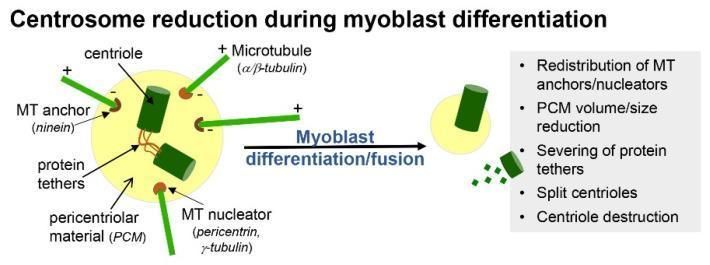
This medical illustration depicts the process of centrosome reduction during human myoblast differentiation and fusion. The diagram is divided into two states: pre-differentiation and post-differentiation. On the left, a mature centrosome in a myoblast is shown. It consists of a pair of cylindrical centrioles connected by protein tethers and embedded within a large, yellow sphere of pericentriolar material (PCM). Microtubules (α/β-tubulin) are shown radiating outward with defined polarity (+/-), anchored by ninein and nucleated by complexes containing pericentrin and γ-tubulin within the PCM. A central arrow labeled 'Myoblast differentiation/fusion' leads to the right side, which illustrates the structural disassembly of the centrosome. Key changes noted include: a significant reduction in PCM volume and size, the redistribution of microtubule (MT) anchors and nucleators to other cellular compartments, and the severing of protein tethers. This results in split centrioles and the eventual destruction of the centriole barrels, facilitating a transition to non-centrosomal microtubule organization in mature myotubes.
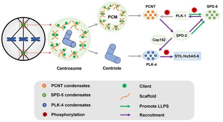
A pathophysiology diagram illustrating the role of liquid-liquid phase separation (LLPS) in centrosome biogenesis and maturation. The illustration depicts a centrosome within a mitotic cell, transitioning into its primary components: a pair of perpendicular centrioles and the surrounding pericentriolar material (PCM). The PCM is shown as a biomolecular condensate composed of 'scaffold' and 'client' proteins. A molecular pathway detailed on the right maps the interactions between key proteins. It demonstrates that the kinase PLK-1 phosphorylates PCNT and SPD-5 (facilitating LLPS), while SPD-2 acts as a central scaffold that recruits PLK-1, Cep152, and the STIL/HsSAS-6 complex. Additionally, SPD-2 is shown to promote the LLPS of SPD-5. Below this, a separate pathway shows PLK-4 condensates recruiting the STIL/HsSAS-6 effector to promote centriole replication. This diagram highlights how the formation of membrane-less organelles through protein phosphorylation and recruitment drives the organization of the centrosome during the cell cycle.
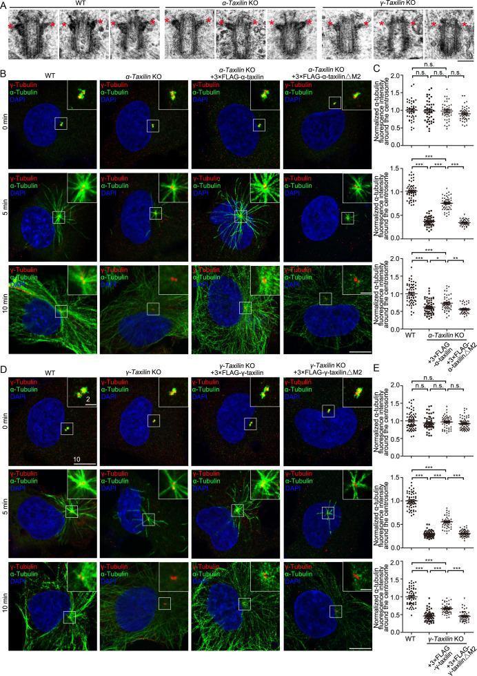
This composite educational image illustrates the role of ̑-taxilin and ̳-taxilin in centrosome structure and microtubule organization in RPE-1 cells. Section A displays high-magnification Transmission Electron Microscope (TEM) images of the mother centriole. Wild-type (WT) cells show prominent subdistal appendages (SDAs, marked with red stars), which appear significantly reduced in size and complexity in ̑-taxilin and ̳-taxilin knockout (KO) models. Sections B and D present time-course confocal microscopy (0, 5, and 10 minutes) of microtubule reformation post-cold depolymerization. Immunostaining identifies ̑-tubulin (green), ̳-tubulin (red), and DNA (blue). The images demonstrate that both KO phenotypes significantly impair microtubule aster formation compared to WT. Rescue experiments show that full-length taxilin proteins restore microtubule nucleation, while M2 deletion mutants fail to do so. Accompanying dot plots (C and E) provide quantitative statistical analysis of ̑-tubulin fluorescence intensity, confirming that taxilin depletion compromises the microtubule-organizing center (MTOC) function at the centrosome.
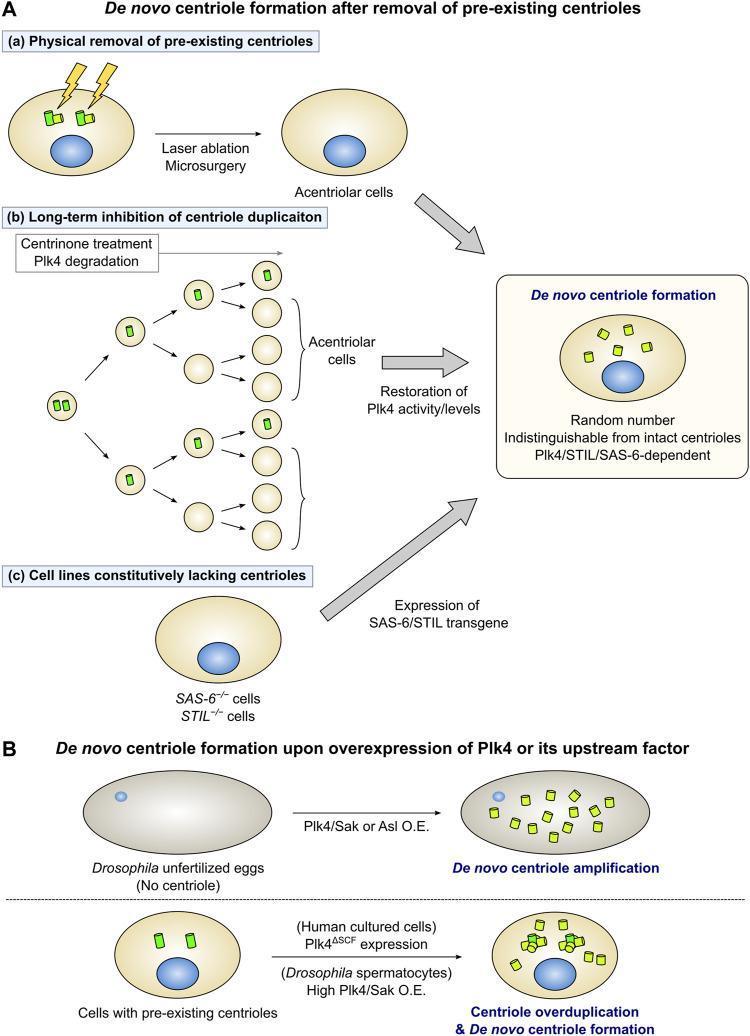
This medical scientific diagram illustrates the mechanisms of de novo centriole formation and centriole overduplication in cellular biology. Section A details pathways leading to de novo formation after centriole removal: (a) physical removal via laser ablation or microsurgery resulting in acentriolar cells, (b) biochemical depletion using centrinone (Plk4 inhibitor) or Plk4 degradation during cell cycling, and (c) genetic induction via SAS-6 or STIL transgene expression in knockout cell lines. All pathways converge on the formation of a random number of new centrioles, a process dependent on Plk4, STIL, and SAS-6 proteins. Section B compares centriole biogenesis under overexpression conditions. In naturally acentriolar Drosophila unfertilized eggs, overexpression of Plk4/Sak or Asl triggers de novo centriole amplification. In contrast, in cells with pre-existing centrioles (e.g., human cultured cells or Drosophila spermatocytes), overexpression of Plk4 mutants (Plk4ΔSCF) or high Plk4/Sak levels leads to both canonical centriole overduplication and independent de novo centriole formation. The diagram highlights key signaling pathway components essential for centrosome biogenesis and cellular inheritance.
Centrosome and Centriole
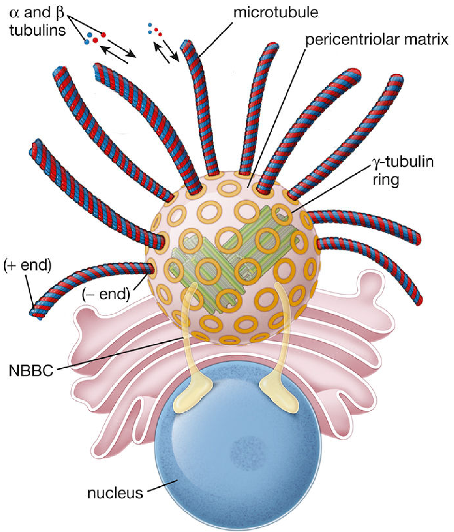
The Centrosome
- A pair of centrioles (oriented at right angles to each other)
- Pericentriolar material (PCM) - an amorphous, electron-dense "cloud" of more than 200 proteins surrounding the centrioles
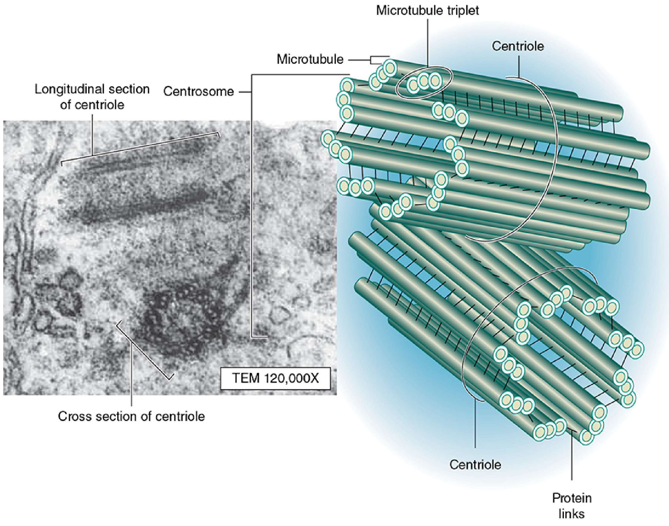
The Centriole - Structure
| Feature | Details |
|---|---|
| Dimensions | ~0.2 μm diameter, 0.3-0.5 μm length |
| Wall | 9 triplets of microtubules (9 × 3 arrangement) |
| Triplet orientation | Parallel to the long axis, running in slightly twisted bundles |
| Microtubule composition | A-tubule (complete ring, 13 protofilaments) + B and C tubules (C-shaped, share walls with neighbors) |
| C-tubule | Shortest of the three |
| Lumen - distal | Contains centrin (20-kDa Ca²⁺-binding protein) |
| Lumen - proximal | Lined by γ-tubulin (template for triplet arrangement) |
| Other proteins | δ-, ε-, ζ-, η-tubulin; pericentrin; protein p150 |
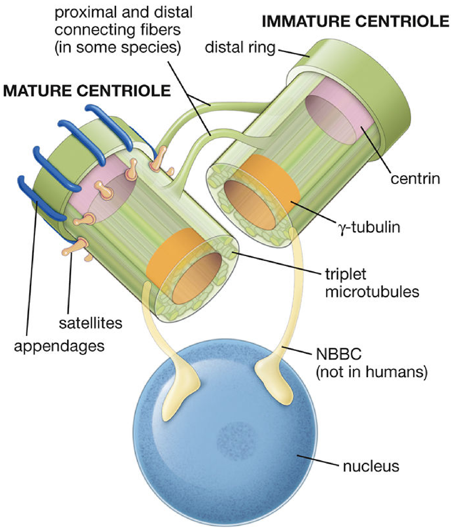
- Mother centriole (mature): bears distal appendages (also called subdistal appendages) and satellites
- Daughter centriole (immature): lacks these appendages
Functions of the Centrosome/Centriole
1. Microtubule Organization (Interphase)
2. Mitotic Spindle Formation
- Astral microtubules radiate from each centriole in a star-like pattern - they establish the axis of the mitotic spindle
- In cells lacking centrioles, mitosis still proceeds but only kinetochore microtubules form - the spindle is anastral and misoriented (anastral bipolar spindle)
3. Basal Body Formation (Ciliogenesis)
- Centriolar pathway (~5%): new centrioles bud from an existing mature centriole
- Acentriolar (de novo) pathway (~95%)**: centrioles form de novo from fibrous granules around structures called deuterosomes
Centrosome Duplication and the Cell Cycle
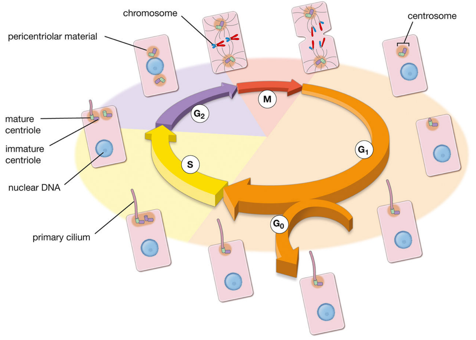
- After mitosis, the cell has one centrosome with one centriole pair
- During S phase (simultaneous with DNA replication): the centrosome duplicates - each old centriole nucleates a new daughter centriole perpendicular to itself
- By G2/early mitosis: two complete centrosomes with two centriole pairs exist
- During mitosis: the centrosomes separate to opposite poles and organize the mitotic spindle
Centrosome vs. Basal Body vs. Centriole - Summary Table
| Structure | Location | Composition | Function |
|---|---|---|---|
| Centrosome | Cytoplasm, near nucleus | 2 centrioles + PCM | MTOC; spindle pole organization |
| Centriole | Within centrosome | 9 triplet microtubules | Structural core of centrosome; templates basal bodies |
| Basal body | Beneath cilia/flagella | 9 triplet microtubules (like centriole, no central pair) | Organizes axoneme of cilia/flagella |
The Ciliary Axoneme vs. the Centriole
Clinical Relevance
- Ciliopathies (e.g., primary ciliary dyskinesia, Bardet-Biedl syndrome, polycystic kidney disease): defects in basal body/centriole function disrupt ciliogenesis. The primary cilium, formed from the mother centriole, serves as a sensory organelle that receives both mechanical and chemical signals.
- Cancer: centrosome amplification (extra centrosomes) causes multipolar mitoses, chromosomal instability, and is found in most human tumors. Driven largely by PLK4 overactivation.
- Microcephaly: mutations in centrosome/MTOC proteins (e.g., ASPM, CDK5RAP2, pericentrin) impair neural progenitor division, leading to primary microcephaly.
- Junqueira's Basic Histology, 17e, pp. 123-125
- Histology: A Text and Atlas (Eroschenko), 8e, pp. 212-217
- Ganong's Review of Medical Physiology, 26e, p. 51
- Guyton and Hall Textbook of Medical Physiology, p. 86
How do centrosomes duplicate during the cell cycle?
Centrosome Duplication During the Cell Cycle
The Key Diagram
Step-by-Step: The Duplication Cycle
1. Post-Mitosis / G1 - One centrosome, asymmetric pair
- One mature (mother) centriole - has satellites and distal appendages, generated at least two cell cycles earlier
- One immature (daughter) centriole - lacks these structures, generated in the previous cycle
2. G1/S Transition - Disengagement and licensing
- The two centrioles in the pair split (disengage) - the orthogonal linkage between parent and daughter is broken
- This disengagement is the critical licensing step - it ensures each centriole can template only one new daughter
3. S Phase - Procentriole formation
- A small mass of fibrillar/granular material appears at the proximal lateral end of each original centriole
- These fibrous granules coalesce into dense spherical structures called deuterosomes
- From each deuterosome, a procentriole (bud) grows outward at a right angle to the parent centriole
- Microtubules begin assembling in the procentriole in a defined sequence:
- First: a ring of 9 single tubules
- Then: doublets (A + B)
- Finally: triplets (A + B + C)
4. S through G2 - Maturation and elongation
- Each parent-daughter pair matures and elongates during S and G2
- The pairs migrate around the nucleus, remaining linked by the pericentriolar material
- By late G2, the cell has two complete centrosomes, each with a mature + immature centriole pair
- Proteins required for mitosis accumulate; the pericentriolar material expands ("centrosome maturation") under the control of PLK1 (polo-like kinase 1) and Aurora A kinase
5. M Phase - Separation and spindle pole formation
- At the onset of mitosis (G2/M), the two centrosomes separate and move to opposite sides of the nucleus
- Each centrosome becomes a spindle pole, nucleating:
- Kinetochore microtubules (attach to chromosomes)
- Astral microtubules (establish spindle axis and orient the cell)
- Interpolar microtubules (maintain spindle structure)
- Following cytokinesis, each daughter cell receives one centrosome and the cycle resets
Summary Table
| Cell Cycle Phase | Centrosome Event |
|---|---|
| Post-M / G1 | One centrosome (mature + immature centriole); primary cilium forms from mother centriole |
| G1/S boundary | Centriole pair disengages (licensing); cyclin E-CDK2 activated; nucleophosmin/B23 phosphorylated |
| S phase | Procentriole buds from proximal end of each parent; 9 singlets → doublets → triplets; simultaneous with DNA replication |
| S - G2 | Procentrioles elongate and mature; two centrosomes now present; PLK1/Aurora A drive centrosome maturation |
| M phase | Centrosomes separate to opposite poles; mitotic spindle forms; each daughter cell inherits one centrosome |
Why Once Per Cycle?
- Histology: A Text and Atlas (Eroschenko), pp. 217-219
- Junqueira's Basic Histology, 17e, pp. 167-168
- Ganong's Review of Medical Physiology, 26e, p. 51The image shows Section 8.5.1 Cell Membrane from an NCERT Biology textbook (Class 11, Chapter 8: Cell - The Unit of Life). Let me give you a thorough explanation of everything on that page.
Cell Membrane - Complete Notes (NCERT Class 11, Section 8.5.1)
1. History - How Was the Structure Discovered?
- Detailed structure could only be studied after the advent of the electron microscope in the 1950s
- Simultaneously, chemical studies on human red blood cells (RBCs/erythrocytes) provided key compositional data
- These two lines of evidence together let scientists deduce the possible structure of the plasma membrane
- The RBC membrane became the model system because RBCs have no nucleus or organelles - the membrane is essentially all you get when you lyse them, making chemical analysis clean
2. What Is the Cell Membrane Made Of?
Approximate composition (Guyton & Hall):
| Component | % by weight |
|---|---|
| Proteins | ~55% (RBC: 52%) |
| Phospholipids | ~25% |
| Cholesterol | ~13% |
| Other lipids | ~4% |
| Carbohydrates | ~3% |
3. The Phospholipid Bilayer
| Part | Chemistry | Behavior in water |
|---|---|---|
| Polar head (phosphate group) | Hydrophilic | Attracted to water ("water-loving") |
| Fatty acid tails (2 hydrocarbon chains) | Hydrophobic | Repelled by water ("water-fearing") |
- The polar heads face outward (toward extracellular water and intracellular water)
- The hydrophobic tails face inward, shielded from water by facing each other
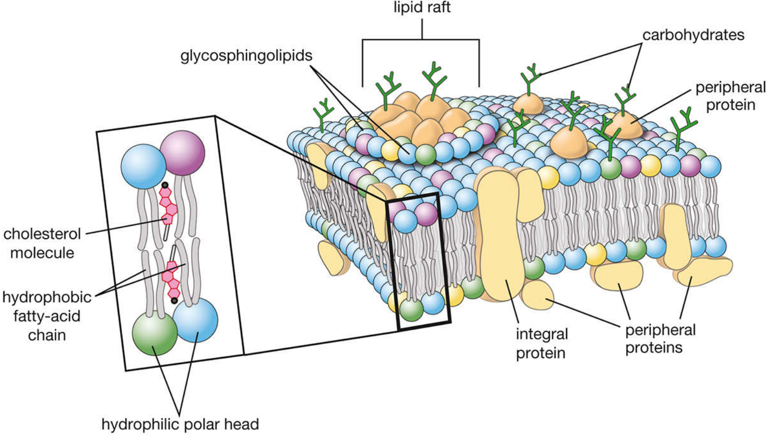
4. Cholesterol
- The membrane also contains cholesterol (in addition to phospholipids)
- Cholesterol molecules slot into the gaps between phospholipids on both sides of the bilayer
- Functions:
- Regulates membrane fluidity - prevents the membrane from becoming too rigid at low temperatures or too fluid at high temperatures
- Controls permeability to water-soluble substances
- Helps maintain membrane integrity
5. Membrane Proteins
A. Peripheral Proteins
- Lie on the surface of the membrane (either extracellular or intracellular face)
- Do not penetrate the lipid bilayer
- Attached via ionic (electrostatic) interactions, mainly with integral proteins
- Can be removed with salt solutions (mild extraction)
- Example functions: cytoskeletal anchoring, signal transduction on the cytoplasmic face
B. Integral Proteins
- Partially or totally buried within the membrane
- Held in place by hydrophobic interactions between their nonpolar amino acid regions and the fatty acid core
- Require detergents to extract (must disrupt lipids)
- Many are transmembrane proteins - they span the entire bilayer (sometimes multiple times = multipass proteins)
- Example functions: ion channels, transporters, receptors, enzymes
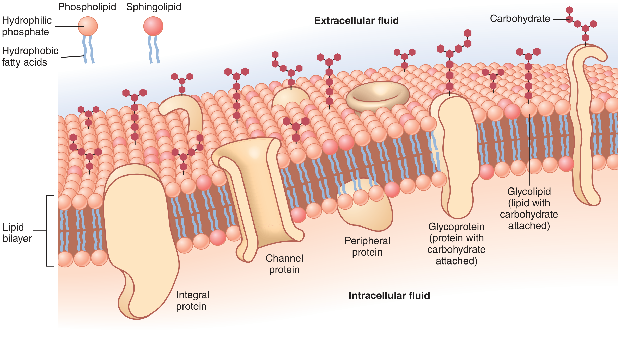
6. Carbohydrates (Sugar Chains - visible in your textbook diagram)
- The diagram at the bottom of your page labels Sugar and shows branching carbohydrate chains projecting from the outer surface
- These carbohydrates attach to:
- Proteins → form glycoproteins
- Lipids → form glycolipids
- Together they form the glycocalyx (cell coat) on the extracellular surface
- Functions: cell recognition, cell adhesion, receptor sites for hormones, immune identity (e.g., ABO blood groups)
7. The Fluid Mosaic Model
| Feature | Meaning |
|---|---|
| Fluid | The phospholipid bilayer behaves as a 2D liquid - lipids and many proteins can move laterally within their leaflet |
| Mosaic | Proteins are scattered throughout the bilayer like tiles in a mosaic - they are not evenly distributed |
8. Why the Bilayer Arrangement Matters (Permeability)
| Substance | Can cross? | Why |
|---|---|---|
| O₂, CO₂, lipid-soluble molecules | Yes, freely | Dissolve in the hydrophobic core |
| Water | Slowly (or via aquaporins) | Small, uncharged, but polar |
| Ions (Na⁺, K⁺, Cl⁻) | No, not directly | Charged - repelled by hydrophobic core |
| Glucose, amino acids | No, not directly | Large polar molecules |
| Integral channel/transport proteins | Yes, via protein channels | Provide a hydrophilic pathway |
Quick Summary for Exams
- Cell membrane = phospholipid bilayer + proteins + carbohydrates + cholesterol
- Phospholipids: polar heads outward, hydrophobic tails inward
- Cholesterol: fills gaps, regulates fluidity
- Peripheral proteins: surface only, removed by salt
- Integral proteins: embedded/spanning, removed by detergent
- Carbohydrates on outer face only → glycocalyx (glycoproteins + glycolipids)
- Overall model = Fluid Mosaic Model (Singer & Nicolson, 1972)
- Histology: A Text and Atlas (Eroschenko), pp. 133-135
- Junqueira's Basic Histology, 17e, pp. 73-75
- Guyton and Hall Textbook of Medical Physiology, pp. 12-14
Are you only for medical student
What I can help with:
- 🔬 Science - biology, chemistry, physics, astronomy (yes, including medical/health topics)
- 📐 Math - from basic arithmetic to calculus, statistics, proofs
- 💻 Technology & Coding - programming, debugging, software concepts
- 📜 History & Geography - events, civilizations, maps, politics
- 📚 Literature & Languages - analysis, grammar, writing, translation
- 💰 Finance & Economics - concepts, taxes, investing basics
- ⚖️ Law & Policy - general concepts and explanations
- 🎓 Exam Prep - any subject, any level (school, college, competitive exams)
- 📄 Documents - creating Word files, PDFs, spreadsheets, presentations
- 🖼️ Images - analyzing photos you upload, generating images, finding diagrams
- 🔍 Research - searching the web, summarizing articles, finding sources
Why medical topics come up a lot:
Ncert ?
How I help with NCERT:
Example: "Explain NCERT Class 11 Biology - Cell Cycle and Cell Division"
Subjects I cover for NCERT:
| Class | Subjects |
|---|---|
| 6-10 | Science, Maths, Social Science, English, Hindi |
| 11-12 | Physics, Chemistry, Biology, Maths, Economics, History, Political Science, Geography, Accountancy |