
Give mbbs notes on preconceptional and antenatal care
Reading File
Reading File
Finding Sources
Finding Sources
Searching PubMed
"Preconception Care"[MeSH Terms] AND "Prenatal Care"[MeSH Terms]
Let me read the Park's sections.
Reading File
Reading File
Reading File
Reading File
Reading File
Reading File
Reading File
Reading File
Reading File
Reading File
Reading File
Finding Sources
Searching the Web
WHO 2016 antenatal care recommendations 8 contacts positive pregnancy experience; India Pradhan Mantri Surakshit Matritva Abhiyan
Reading File
Reading File
Preconceptional and Antenatal Care: MBBS Notes
Part A. Preconceptional care
Definition: Health interventions given to women and couples before conception. They identify and modify biomedical, behavioural and social risks to a future pregnancy. Interventions need to start well before conception to improve neonatal and stillbirth outcomes (Park's Textbook of Preventive and Social Medicine, Pre-conception and antenatal care). Preconception care is an integral part of prenatal care and allows early risk identification and treatment (Textbook of Family Medicine 9e, Preconception Counseling).
Why it matters: Organogenesis is largely complete by 8 to 10 weeks, often before the woman knows she is pregnant. Damage from teratogens, uncontrolled diabetes or folate deficiency has already occurred by the time antenatal care starts.
Components
| Domain | Key points |
|---|---|
| History | Age, obstetric history (abortions, stillbirth, preterm birth, congenital anomaly, PIH), medical and surgical history, drugs, family history, consanguinity |
| Folic acid | 0.4 mg (400 mcg) daily for all women of reproductive age, started before conception. This reduces neural tube defects by about 50%. Use 2 to 4 mg daily for women with a previous NTD child or a family history, starting at least 1 month before conception and continuing through the first 3 months (Textbook of Family Medicine 9e). Some Indian guidelines use 4 to 5 mg for high-risk women. |
| Immunization | Rubella or MMR, varicella and hepatitis B before pregnancy. Live vaccines are contraindicated in pregnancy, so advise avoiding conception for 1 month after a live vaccine. Tetanus (Td) should be up to date. |
| Screening for infections | HIV, syphilis, hepatitis B, STIs and TB |
| Chronic disease optimisation | Diabetes: tight control and a normal HbA1c before conception, to lower the risk of anomalies and miscarriage. Hypertension: switch ACE inhibitors and ARBs. Epilepsy: use the lowest effective dose, preferably monotherapy, and avoid valproate. Thyroid disease, anaemia and cardiac disease also need review. |
| Drug review | Stop or substitute teratogens: warfarin, isotretinoin, ACE inhibitors, statins, methotrexate, valproate, thalidomide |
| Lifestyle | Stop smoking, alcohol and recreational drugs. Keep BMI normal, eat a balanced diet and exercise. Avoid occupational and radiation hazards. |
| Genetic counselling | Maternal age 35 or older, previous affected child, thalassaemia or haemoglobinopathy carrier screening, family history of chromosomal or single-gene disorders, consanguinity |
| Psychosocial | Screen for intimate partner violence and depression, and review whether the pregnancy is planned and wanted |
| Contraception and spacing | Advise an interpregnancy interval of at least 24 months after a live birth to reduce preterm birth and low birth weight |
Counselling for the cardiac patient (Creasy & Resnik's Maternal-Fetal Medicine): Pregnancy may be safe, uncomfortable, high risk or contraindicated. Contraindicated conditions include:
- Severe pulmonary hypertension.
- Dilated cardiomyopathy with EF below 40 to 45%.
- Severe symptomatic valvular stenosis or regurgitation.
- Connective tissue disease with a severely enlarged aorta.
- Certain mechanical valves.
Repaired lesions need 6 to 12 months for recovery and review before pregnancy.
Smoking: Quitting before or in early pregnancy significantly reduces preterm birth, low birth weight and infant mortality. Offer intensive behavioural support rather than minimal advice (Textbook of Family Medicine 9e).
Part B. Antenatal care (ANC)
Definition: Care provided by skilled health professionals to pregnant women to ensure the best health for mother and baby. It has three components: risk identification, prevention and management of pregnancy-related or concurrent disease, and health education and promotion. ANC reduces maternal and perinatal morbidity and mortality, directly through detection and indirectly through identifying high-risk women and referring them (Park's).
Objectives (Park's)
- Promote, protect and maintain maternal health.
- Detect "high-risk" cases and give them special attention.
- Foresee and prevent complications.
- Remove anxiety and dread associated with delivery.
- Reduce maternal and infant mortality and morbidity.
- Teach the mother elements of child care, nutrition, personal hygiene and environmental sanitation.
- Sensitise the mother to family planning, including advice for those seeking MTP.
- Attend to the under-fives accompanying the mother.
Antenatal visits
Ideal schedule: Monthly for the first 7 months, fortnightly in the 8th month, then weekly. This is usually not feasible in India, so the minimum target is 4 visits (Park's):
| Visit | Timing |
|---|---|
| 1st | Within 12 weeks, as soon as pregnancy is suspected, for registration and first check-up |
| 2nd | 14 to 26 weeks |
| 3rd | 28 to 34 weeks (ideally with the PHC medical officer) |
| 4th | 36 weeks to term |
- Registration within 12 weeks is the primary responsibility of the ANM. Village Health Nutrition Days are used for this.
- Name-based tracking of pregnant women is used so that missed visits are followed up.
- WHO 2016 raised the recommendation to a minimum of 8 contacts, because more contacts are linked to fewer stillbirths and perinatal deaths (WHO ANC guideline). Park's still lists 4 visits as the Indian minimum, so write 4 for Indian exams and mention the WHO 8 as an update.
Reasons for early detection of pregnancy (Park's):
- Plan adequate care.
- Record LMP and calculate EDD.
- Assess baseline health (BP, weight, Hb).
- Detect complications early and refer.
- Confirm whether the pregnancy is wanted, and refer for safe abortion if not.
- Build rapport with the woman.
Expected pregnancies in an area: Expected live births = (birth rate × population) / 1000. Add 10% to allow for abortions and stillbirths (Park's).
First visit
1. History (Park's)
- Confirm the pregnancy.
- Previous pregnancy complications.
- Current medical, surgical or obstetric conditions.
- LMP and EDD. Naegele's rule: add 9 months and 7 days to the first day of the LMP.
- Danger symptoms: fever, persistent vomiting, bleeding or discharge, breathlessness, generalised swelling, severe headache with blurred vision, burning micturition, reduced fetal movements.
- Systemic illness: hypertension, diabetes, heart disease, TB, renal disease, epilepsy, asthma, jaundice, malaria, RTI/STI, HIV.
- Family history of hypertension, diabetes, TB, thalassaemia, twins or congenital malformation.
- Drug allergies and habit-forming drugs.
2. General examination: Pallor (every visit, with Hb correlation), pulse, respiratory rate, BP, weight, height, oedema, breasts, and teeth or oral hygiene. Height of 140 cm or less in a primigravida is a risk factor.
3. Abdominal examination
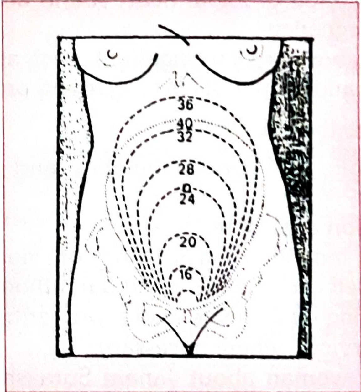
Fundal height at various weeks (Park's, Fig. 1)
- Fundal height: 12 weeks, just palpable above the symphysis. 20 weeks, at the lower border of the umbilicus. 36 weeks, at the xiphisternum. Express gestation in completed weeks.
- Fetal heart sounds: Audible by stethoscope after about 20 weeks (Park's says after the 6th month). Normal rate is 120 to 140 per minute.
- Fetal movements: Felt by the examiner at 18 to 22 weeks.
- Fetal parts: Felt at about 22 weeks. After 28 weeks the head, back and limbs can be distinguished.
- Lie and presentation: Relevant only after 32 weeks.
- Suspect multiple pregnancy if the uterus is larger than the dates.
- Look for an abdominal scar.
4. Gestational age assessment: Early first-trimester ultrasound (CRL) is the gold standard. "Best obstetric estimate" combines LMP and ultrasound (Park's).
5. Investigations (Park's)
- Sub-centre: pregnancy test, Hb, urine albumin and sugar, rapid malaria test.
- PHC/CHC/FRU: blood group and Rh, VDRL/RPR, HIV, blood sugar, HBsAg, malaria test.
- Standard additions in practice: urine culture for asymptomatic bacteriuria, thyroid function, and GTT at 24 to 28 weeks (earlier if high risk). Rubella and TORCH testing is not routine.
- Ultrasound: Dating and viability scan at 6 to 12 weeks. NT scan at 11 to 14 weeks. Anomaly (level II) scan at 18 to 20 weeks. Growth scans if indicated.
Essential components of every visit (Park's): history, weight, BP, respiratory rate, pallor and oedema, abdominal palpation with fetal heart auscultation, and Hb plus urine albumin and sugar.
Interventions
| Intervention | Details |
|---|---|
| Iron and folic acid | Indian programme: 1 tablet daily (100 mg elemental iron + 500 mcg folic acid), started after the first trimester (about 14 to 16 weeks) for at least 180 days. Park's lists folic acid in the first trimester and IFA from 12 weeks. Anaemia is Hb below 11 g/dL, and treatment is higher-dose iron. |
| Tetanus immunisation | TT/Td: 2 doses 4 weeks apart. If vaccinated in a previous pregnancy within 3 years, give a single booster. Td has replaced TT in India. |
| Calcium | 500 mg twice daily from the second trimester (MoHFW guideline, recalled from memory). |
| Deworming | Albendazole 400 mg once in the second trimester (MoHFW guideline). |
| Malaria prophylaxis | In endemic zones, per NVBDCP |
| Other | Referral where needed, home visit, information on JSY and other schemes |
Prenatal advice (Park's)
- Diet: Pregnancy needs about 60,000 kcal extra overall, and lactation about 550 kcal a day. Normal weight gain is 9 to 11 kg. Poor Indian women gain an average of 6.5 kg. Poor maternal iron stores lead to infant anaemia. (Standard figures for Indian women are about +350 kcal and +25 g protein a day in pregnancy.)
- Hygiene, rest and exercise: Bathe daily and wear clean clothes. Sleep 8 hours, plus at least 2 hours of rest after the midday meal. Prevent constipation with leafy vegetables, fruit and fluids, and avoid castor oil purgatives. Light household work is fine, but heavy manual labour late in pregnancy is not.
- Smoking: Babies are about 170 g lighter. Perinatal mortality is 10 to 40% higher. Nicotine causes uterine vasoconstriction and placental insufficiency.
- Alcohol: Increases miscarriage risk and causes fetal alcohol syndrome (IUGR, developmental delay).
- Drugs: Avoid non-essential drugs. Park's examples:
- Thalidomide (limb defects, worst at 4 to 8 weeks).
- LSD (chromosome damage).
- Streptomycin (8th nerve damage).
- Iodides (congenital goitre).
- Corticosteroids (growth impairment).
- Sex hormones (virilism).
- Tetracyclines (bone growth and tooth enamel).
- Pethidine in labour (neonatal respiratory depression).
- Radiation: Avoid X-rays, especially in the first trimester. Intrauterine X-ray exposure is associated with leukaemia and microcephaly.
- Coitus: Restrict in the last trimester.
- Danger signs: Vaginal bleeding, severe headache or blurred vision, convulsions, severe pallor or breathlessness, fever, leaking liquor, reduced fetal movements, swelling of face and hands.
- Birth preparedness: Institutional delivery, transport, blood donor, funds. Counsel on breastfeeding, newborn care and postpartum contraception.
Risk approach: "high-risk" pregnancy (Park's)
The central purpose of ANC is to identify high-risk women early and arrange skilled care for them, while still caring for everyone. The risk factors are:
- Elderly primigravida (30 years or older).
- Short-statured primigravida (140 cm or less).
- Malpresentation (breech, transverse lie).
- Antepartum haemorrhage or threatened abortion.
- Pre-eclampsia or eclampsia.
- Anaemia.
- Twins or hydramnios.
- Previous stillbirth, IUD or manual removal of placenta.
- Previous abortion.
- Elderly grand multipara.
- Prolonged pregnancy (14 days past EDD).
- Previous caesarean or instrumental delivery.
- Medical disease: cardiac, renal, diabetes, TB, liver disease, malaria, epilepsy, asthma, HIV, RTI/STI.
- Treatment for infertility.
- Three or more consecutive spontaneous abortions.
Records and home visits (Park's)
- The Mother and Child Protection Card is given to the woman and carried to every visit and to delivery.
- At least one home visit by the female health worker or public health nurse. Home visiting is the "backbone of MCH services."
Indian programmes linked to ANC
- JSY (2005): Cash incentive for institutional delivery.
- JSSK (2011): Free services and referral transport for mothers and sick infants.
- Pradhan Mantri Surakshit Matritva Abhiyan (PMSMA): Free specialist ANC check-up on the 9th of every month (recalled from memory).
- Name-based tracking of mothers and children.
Quick revision
- Preconception: folic acid 0.4 mg (4 to 5 mg or 2 to 4 mg for high risk), rubella vaccine, optimise chronic disease, stop teratogens, genetic counselling, an interpregnancy gap of 24 months or more.
- Indian minimum is 4 ANC visits (12 weeks, 14 to 26, 28 to 34, 36 to term). WHO recommends 8 contacts.
- EDD = LMP + 9 months + 7 days.
- Investigations: Hb, urine albumin and sugar, blood group and Rh, VDRL, HIV, HBsAg, blood sugar, malaria test.
- Interventions: IFA from the second trimester for 180 days, 2 doses of TT/Td, calcium, albendazole.
- Risk approach: identify high-risk cases early and refer.
Note on recent evidence: A quick PubMed check of the last 2 years found systematic reviews on preconception and prenatal care, including a 2026 umbrella review on stillbirth prevention (PMID 42114873). None of them contradicts the textbook content above. The main update is the WHO 8-contact model.
Some points above come from my own recall and not from the retrieved textbook pages. These are the IFA dose and duration, calcium, albendazole, Td, PMSMA and the ultrasound timings. Check them against your university's prescribed textbook or the current MoHFW guideline.
I can turn this into a one-page revision sheet, add MCQs, or expand any section, such as antenatal investigations or the high-risk pregnancy workup.
Diagnosis of pregnancy
Finding Sources
Searching Images
signs of pregnancy Chadwick Hegar Goodell sign uterus
Reading File
Reading File
Reading File
Searching PubMed
"Pregnancy Tests"[MeSH Terms] AND "Early Diagnosis"
Diagnosis of Pregnancy: MBBS Notes
Clinical features are grouped into presumptive, probable and positive signs. Only the positive signs prove pregnancy. In forensic and legal cases they are the only ones to rely on (Parikh's Textbook of Medical Jurisprudence, Conclusive Signs of Pregnancy).
1. Presumptive signs and symptoms
These are subjective or non-specific and can occur without pregnancy.
| Feature | Details |
|---|---|
| Amenorrhoea | Earliest and most important symptom. Other causes include anaemia, TB, thyroid or pituitary disease, stress, and an intense desire for pregnancy. Occasional spotting can occur. |
| Morning sickness | Nausea and vomiting from the end of the 1st month to about 3 months. Not reliable, because migraine and hormonal imbalance can cause it. |
| Breast changes | Tenderness, tingling, enlargement, engorged superficial veins, a pigmented and erectile nipple, and a dark areola in the 2nd month. Montgomery tubercles appear. Colostrum forms by about 3 months. Striae appear after 6 months. These are more useful in primigravidae. |
| Quickening | The mother's first perception of fetal movement. Dikshit gives 14 to 20 weeks, and Dutta gives about 18 to 20 weeks in a primigravida and 16 to 18 weeks in a multigravida. Flatus or peristalsis can mimic it, and false quickening occurs in spurious pregnancy. |
| Skin pigmentation | Chloasma, linea nigra, and pigmentation of the vulva and abdomen. |
| Vaginal changes | Bluish discolouration from venous congestion after the 4th week (Jacquemier's or Chadwick's sign). The vagina is softer, secretions increase, and vaginal artery pulsation (Osiander's sign) can be felt. |
| Urinary frequency | The uterus presses on the bladder in the first trimester. It settles, then returns near term when the head engages. |
| Others | Fatigue, salivation, perverted appetite (pica) and irritability. |
(Source for the table above: P C Dikshit Textbook of Forensic Medicine and Toxicology, Presumptive Signs.)
2. Probable signs
These are objective signs found on examination and tests. Other conditions can mimic them, such as tumours and pseudocyesis.
- Abdominal enlargement: Fundal height by month (Dikshit):
| Month | Uterine level |
|---|---|
| End of 3rd | Fills the pelvis |
| 3rd to 4th | Over the pelvic brim |
| 5th | Midway between symphysis and umbilicus |
| 6th | At the umbilicus |
| 7th | Midway between umbilicus and xiphoid |
| 8th | At the xiphisternum |
| 9th to 10th | Uterus sinks into the pelvis (lightening) |
Red subcutaneous striae gravidarum appear from abdominal wall stretching.
- Uterine changes:
- At about 3 months the uterus feels doughy or elastic.
- Hegar's sign appears at 6 to 10 weeks. The isthmus is soft and compressible between the firm cervix and the elastic body, felt on bimanual examination.
- Piskacek's sign is asymmetrical enlargement at the site of implantation.
- The globular, soft uterus also gives Von Fernwald's sign.
- Cervical changes:
- Softening from the 2nd month, from below upwards (Goodell's sign).
- The os is plugged with mucus.
- Braxton Hicks contractions: Painless, irregular, intermittent contractions from about 12 weeks. They are felt on palpation, or on bimanual examination in early pregnancy, and are more evident later.
- Ballottement: Internal ballottement is elicited at 16 to 28 weeks (the fetus rebounds in liquor). External ballottement can be elicited late in pregnancy. Ascites or an ovarian cyst can give a similar sign.
- Fetal parts felt: Palpable from about 20 to 22 weeks, and the examiner can distinguish them by 28 weeks (Park's).
- Uterine souffle: A soft blowing murmur over the uterus, synchronous with the maternal pulse. It can be confused with fetal heart sounds, which are faster.
- Biological tests (hCG): These are probable, not positive, signs because they show hormone production and not a fetus.
3. Positive (conclusive) signs
- Fetal heart sounds:
- Heard by stethoscope at about 18 to 20 weeks. Dikshit and Parikh describe them between the 4th and 5th month.
- Rate is about 160 at 5 months and 120 to 140 near term.
- Their absence is uncertain negative evidence, so examine repeatedly.
- Sounds are not heard before 18 weeks, with a fat abdominal wall or hydramnios, with an awkward fetal position, or when the fetus is dead.
- Doppler detects them from about 10 to 12 weeks.
- Fetal movements and parts felt by the examiner: After the 4th month, and visible by the 5th. These are indisputable evidence of a living child at the time.
- Ultrasonography: The most reliable and safest method.
- Gestational sac at about 4.5 to 5 weeks on transvaginal scan.
- Yolk sac at about 5 weeks.
- Fetal pole and cardiac activity at about 6 weeks.
- Transabdominal scan is about 1 week later than transvaginal.
- Radiograph of the fetus: The fetal skeleton shows from about the middle of the 4th month (16 weeks). It is avoided because of radiation hazard, and ultrasound is preferred.
- Fetal cells or DNA in maternal blood: Listed by Parikh. Cell-free fetal DNA is the basis of non-invasive prenatal testing.
4. Pregnancy tests (biological)
These detect hCG, produced by the syncytiotrophoblast.
| Test | Notes |
|---|---|
| Urine immunoassay (kit/card) | Detects hCG in a urine sample, usually positive within 1 to 2 days of a missed period (about 4 weeks of gestation). Lateral-flow card kits use monoclonal antibodies and are standard at sub-centre level (Park's lists the pregnancy detection test at sub-centre level). Sensitivity is about 20 to 25 mIU/mL, and some kits reach about 10. |
| Serum beta-hCG | Quantitative. Detectable about 8 to 11 days after conception. Doubling time is about 48 hours in early normal pregnancy. Peaks at 8 to 10 weeks. |
| Old tests (history only) | Ascheim-Zondek (mouse), Friedman (rabbit), Galli-Mainini (frog), and the haemagglutination inhibition test. |
False positives:
- Molar pregnancy and choriocarcinoma.
- hCG-secreting tumours.
- Recent abortion or delivery.
- hCG injections.
- Heterophile antibodies (serum tests).
False negatives:
- Test done too early.
- Dilute urine.
- "Hook effect" at very high hCG (molar pregnancy, twins).
- Ectopic pregnancy, which has low hCG levels.
Always do the test on the first morning urine specimen when possible. Order a quantitative serum hCG if an ectopic pregnancy is suspected.
5. Differential diagnosis of early pregnancy
- Pseudocyesis (spurious pregnancy): amenorrhoea, breast changes and a feeling of fetal movements, but no uterine enlargement. The abdominal distension settles under anaesthesia, and the pregnancy test is negative.
- Amenorrhoea of other causes: PCOS, thyroid disease, hyperprolactinaemia, premature ovarian failure, stress.
- Fibroid uterus or ovarian cyst.
- Hydatidiform mole: uterus large for dates, very high hCG, "snowstorm" on ultrasound.
- Ectopic pregnancy.
- Ascites and obesity.
6. Estimating gestational age and EDD
- Naegele's rule: EDD = LMP + 9 months + 7 days. This assumes a regular 28-day cycle.
- Ultrasound: Crown-rump length in the first trimester is the most accurate and is the "gold standard" for dating (Park's).
- Express duration in completed weeks.
7. Diagnosis of pregnancy by trimester (quick table)
| Gestation | What you can say or find |
|---|---|
| 4 to 5 weeks | Missed period. Urine hCG positive. Gestational sac on TVS. |
| 6 to 8 weeks | Fetal pole, cardiac activity. Hegar's, Chadwick's and Goodell's signs. |
| 12 weeks | Fundus just palpable abdominally. Doppler FHS. |
| 16 weeks | Fundus midway between symphysis and umbilicus. |
| 18 to 20 weeks | Quickening. Stethoscope FHS. Fetal parts palpable. |
| 36 weeks | Fundus at the xiphisternum. |
Viva points
- Earliest symptom: amenorrhoea.
- Earliest reliable lab sign: urine/serum hCG.
- Earliest reliable imaging: TVS gestational sac (about 5 weeks), then cardiac activity (about 6 weeks).
- Positive signs: FHS, fetal movements felt by the examiner, ultrasound, X-ray.
- Uterine souffle is synchronous with the maternal pulse. Funic (cord) souffle is synchronous with the fetal heart rate.
Notes on sources and recency
The signs, fundal height table and positive-sign details come from Dikshit and Parikh (forensic medicine), which cover diagnosis of pregnancy in the living. My library search found no dedicated obstetrics chapter. The hCG kit timings and sensitivity, the Hegar, Piskacek and Osiander details, and the ultrasound milestones come from my own knowledge, so check them against Dutta's or your prescribed textbook. Quickening figures differ by source, so use your textbook's.
A quick PubMed check of the last 2 years found no systematic review that changes the standard approach. The hits were on pre-eclampsia point-of-care tests, non-invasive prenatal testing, and perinatal depression screening.
I can add MCQs or a short-answer question-and-answer set on this topic.
This is a shared conversation. Sign in to Orris to start your own chat.