
Anatomy of Neck with image
Reading File
Now let me search the medical library for neck anatomy sections with images.
Finding Sources
Finding Sources
Finding Sources
Reading File
Reading File
Reading File
Reading File
Reading File
Now let me fetch the key anatomy images found in the textbooks.
Loading Image
Loading Image
Loading Image
Excellent images retrieved. Now let me get a few more images - the detailed fascia/triangles from Fischer's and the surface landmark diagram.
Loading Image
Loading Image
Reading File
Loading Image
Reading File
Loading Image
Reading File
I now have comprehensive content and images. Let me compile a thorough, well-illustrated anatomy of the neck response.
Anatomy of the Neck
Sources: Gray's Anatomy for Students; Fischer's Mastery of Surgery, 8th ed.
1. Overview and Surface Landmarks
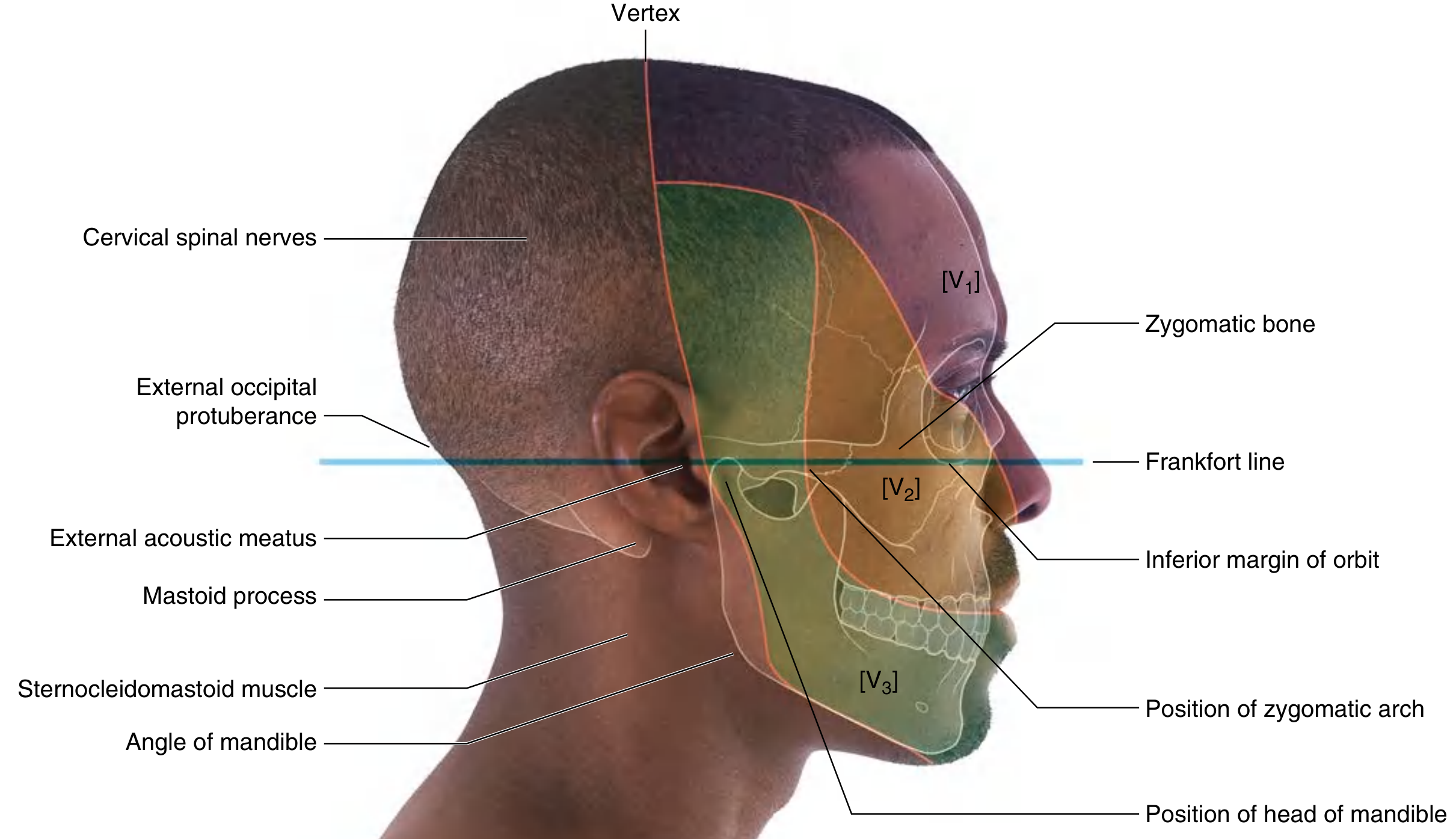
The head is in the anatomical position when the inferior margins of the bony orbits and the superior margins of the external acoustic meatuses lie on the same horizontal plane (the Frankfort plane).
Key palpable landmarks of the head and neck include:
- Mastoid process - posterior to the external acoustic meatus; the sternocleidomastoid (SCM) attaches here
- Zygomatic arch - from the temporomandibular joint to the zygomatic bone
- External occipital protuberance - marks the junction of the neck and head posteriorly
- Vertex - highest point of the head; marks the transition from trigeminal (anterior) to cervical (posterior) scalp innervation
2. Key Vertebral Levels
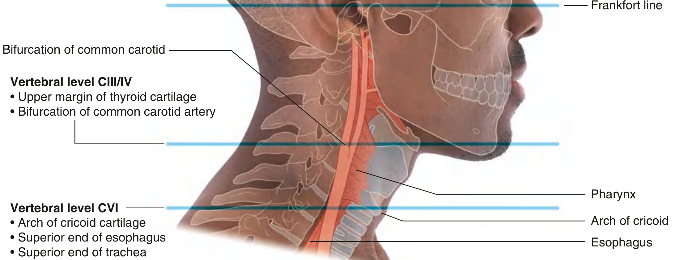
Two cervical levels serve as major anatomical reference points:
| Level | Landmark |
|---|---|
| CIII/CIV | Upper margin of thyroid cartilage; bifurcation of common carotid artery |
| CVI | Inferior margin of cricoid cartilage; junction of pharynx-esophagus and larynx-trachea |
3. Fascia of the Neck
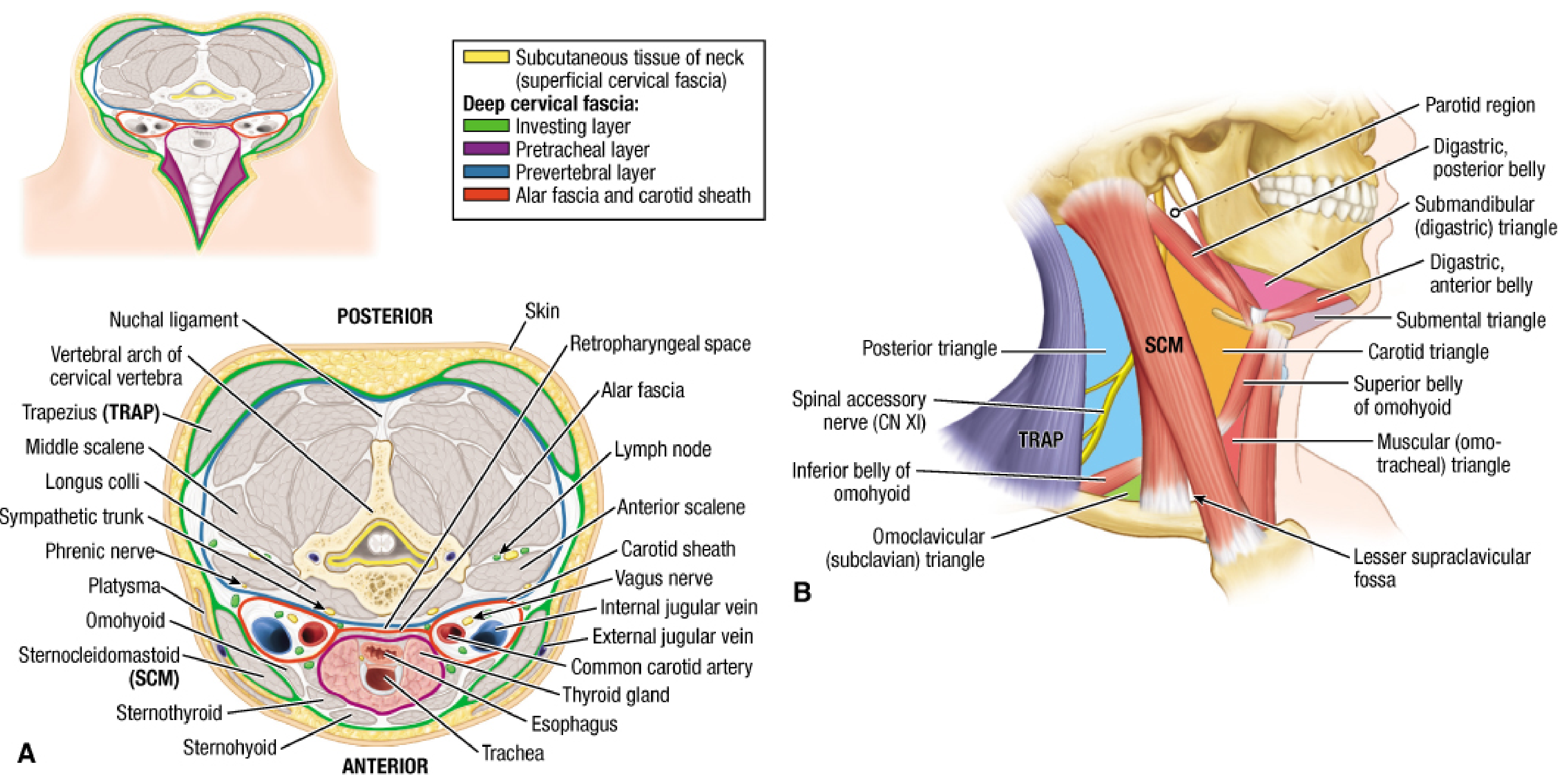
Superficial Fascia
- Loose areolar connective tissue containing the platysma muscle (a muscle of facial expression, innervated by the cervical branch of CN VII)
- Contains: external and anterior jugular veins, branches of the superficial cervical plexus, and superficial lymph nodes
Deep Cervical Fascia - Three Layers
- Investing layer - deepest layer of subcutaneous tissue; encircles the entire neck; splits to envelop the SCM and trapezius
- Pretracheal layer - muscular part envelops the infrahyoid (strap) muscles; visceral part envelops thyroid gland, trachea, and esophagus
- Prevertebral layer - surrounds the vertebral column and its musculature; contains the cervical sympathetic trunk
Carotid Sheath
A tubular fascial compartment extending from the cranial base to the root of the neck. It contains:
- Common and internal carotid arteries
- Internal jugular vein
- Vagus nerve (CN X)
- Sympathetic nerve fibers
- Carotid sinus nerve and lymph nodes
Retropharyngeal Space
Lies between the prevertebral layer and the buccopharyngeal fascia - a clinically important potential space for spread of infection.
4. Triangles of the Neck
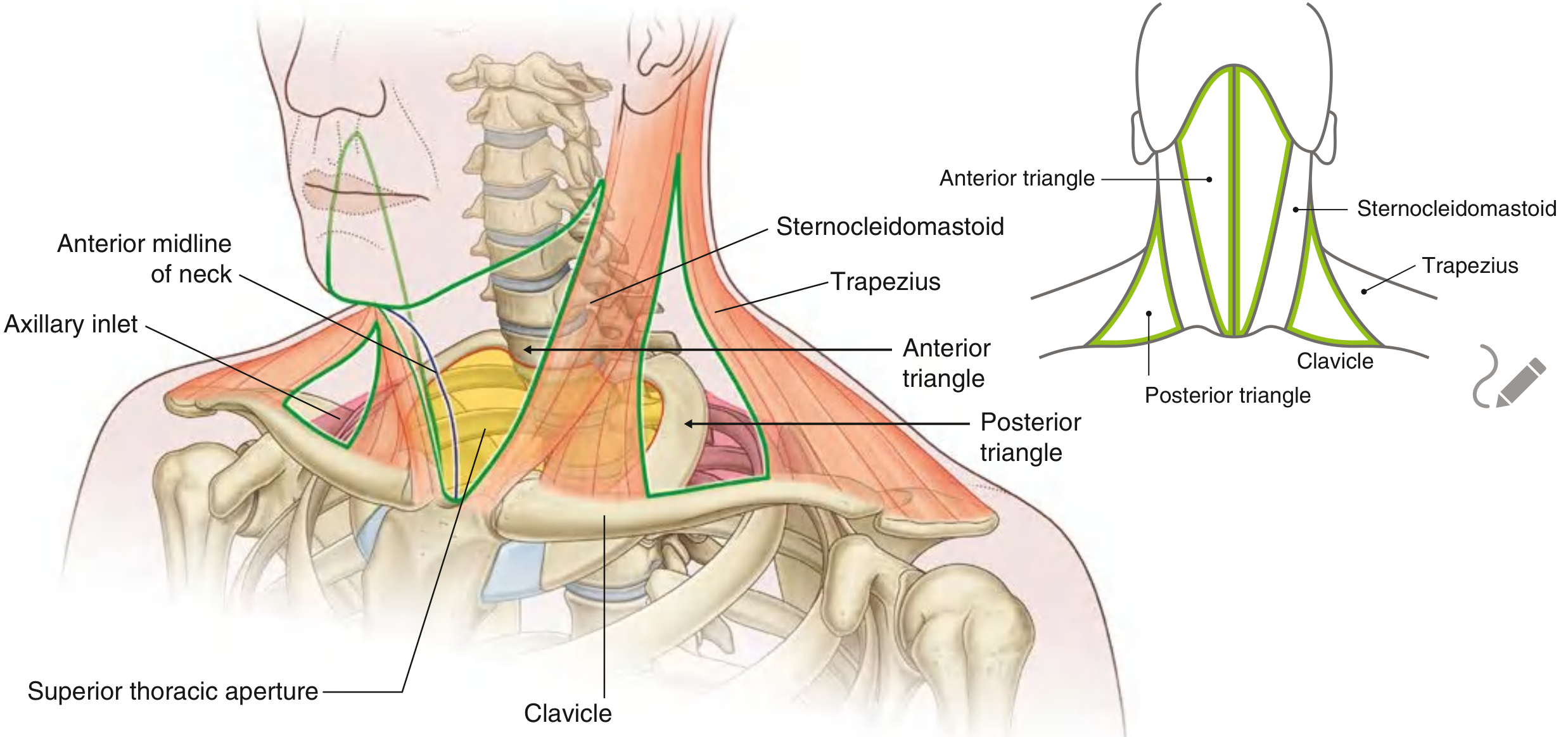
The SCM and trapezius divide each side of the neck into anterior and posterior triangles.
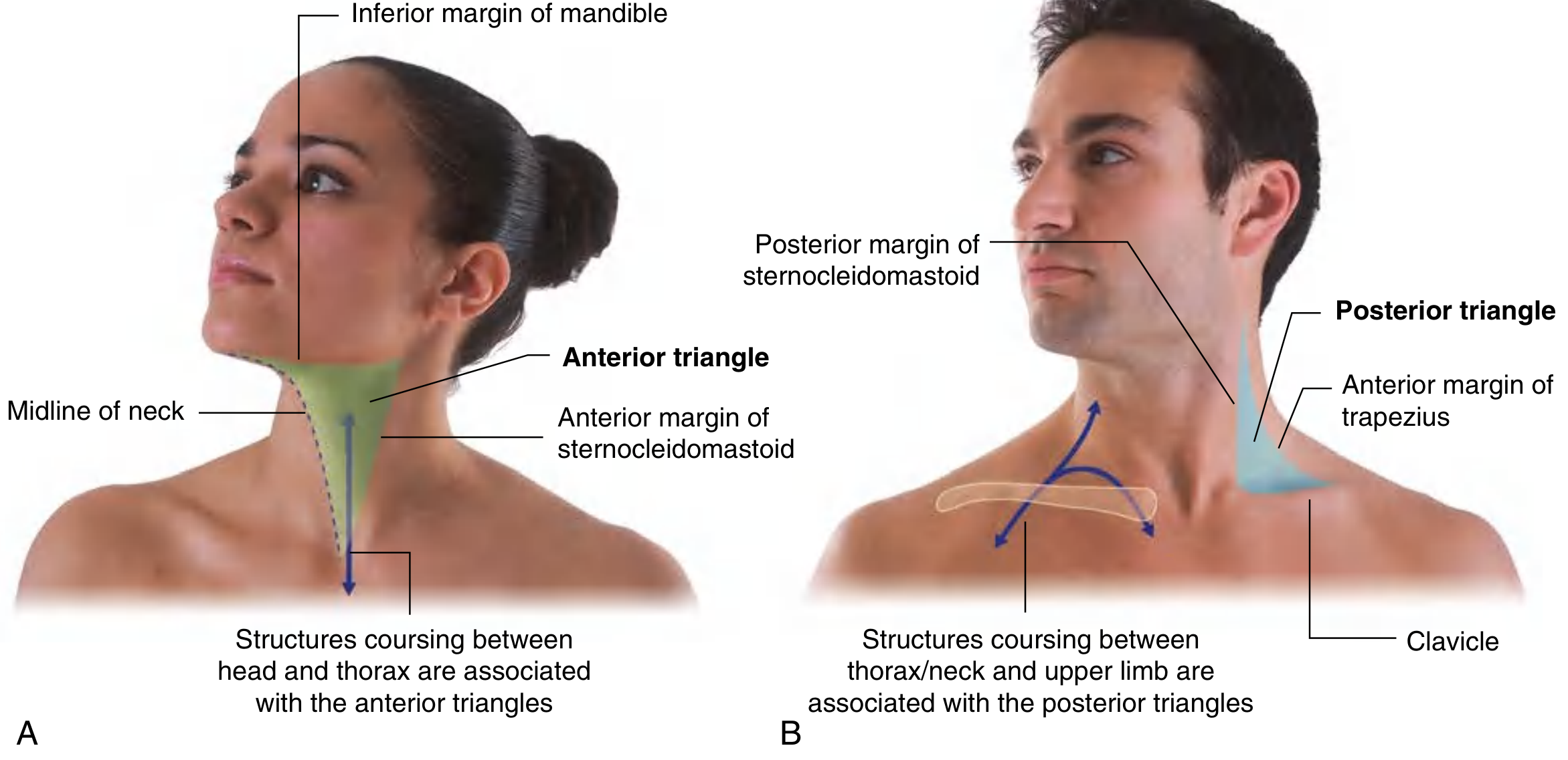
Anterior Triangle
Boundaries:
- Medial: midline of neck
- Superior: inferior border of mandible
- Lateral/posterior: anterior border of SCM
- Apex: inferiorly at the suprasternal notch
Function: Contains airway, digestive tract, thyroid/parathyroid glands, and neurovascular structures passing between head and thorax.
Subdivisions (by the digastric and omohyoid muscles):
| Sub-triangle | Boundaries | Key Contents |
|---|---|---|
| Submental (unpaired) | Hyoid, anterior bellies of both digastrics | Small lymph nodes, small veins |
| Submandibular | Inferior mandible, anterior and posterior bellies of digastric | Submandibular gland, facial artery/vein, CN XII (hypoglossal) |
| Carotid | Posterior belly of digastric (sup.), omohyoid (inf.), SCM (post.) | Common/internal/external carotid arteries, internal jugular vein, CN X (vagus), CN XI, CN XII, ansa cervicalis |
| Muscular (omotracheal) | Midline, omohyoid, SCM | Infrahyoid muscles, thyroid gland, trachea, esophagus |
Posterior Triangle (Lateral Cervical Triangle)
Boundaries:
- Anterior: posterior border of SCM
- Posterior: anterior border of trapezius
- Inferior: middle third of clavicle
- Apex: posteroinferior to the mastoid process
Subdivisions (by the inferior belly of omohyoid):
- Occipital triangle (larger, superior)
- Omoclavicular / subclavian triangle (smaller, inferior)
Floor muscles (anterior to posterior):
- Anterior scalene (C3-C6 → 1st rib)
- Middle scalene (C5-C7 → 1st rib)
- Posterior scalene (C5-C7 → 2nd rib)
- Levator scapulae
- Splenius capitis / semispinalis capitis (at apex)
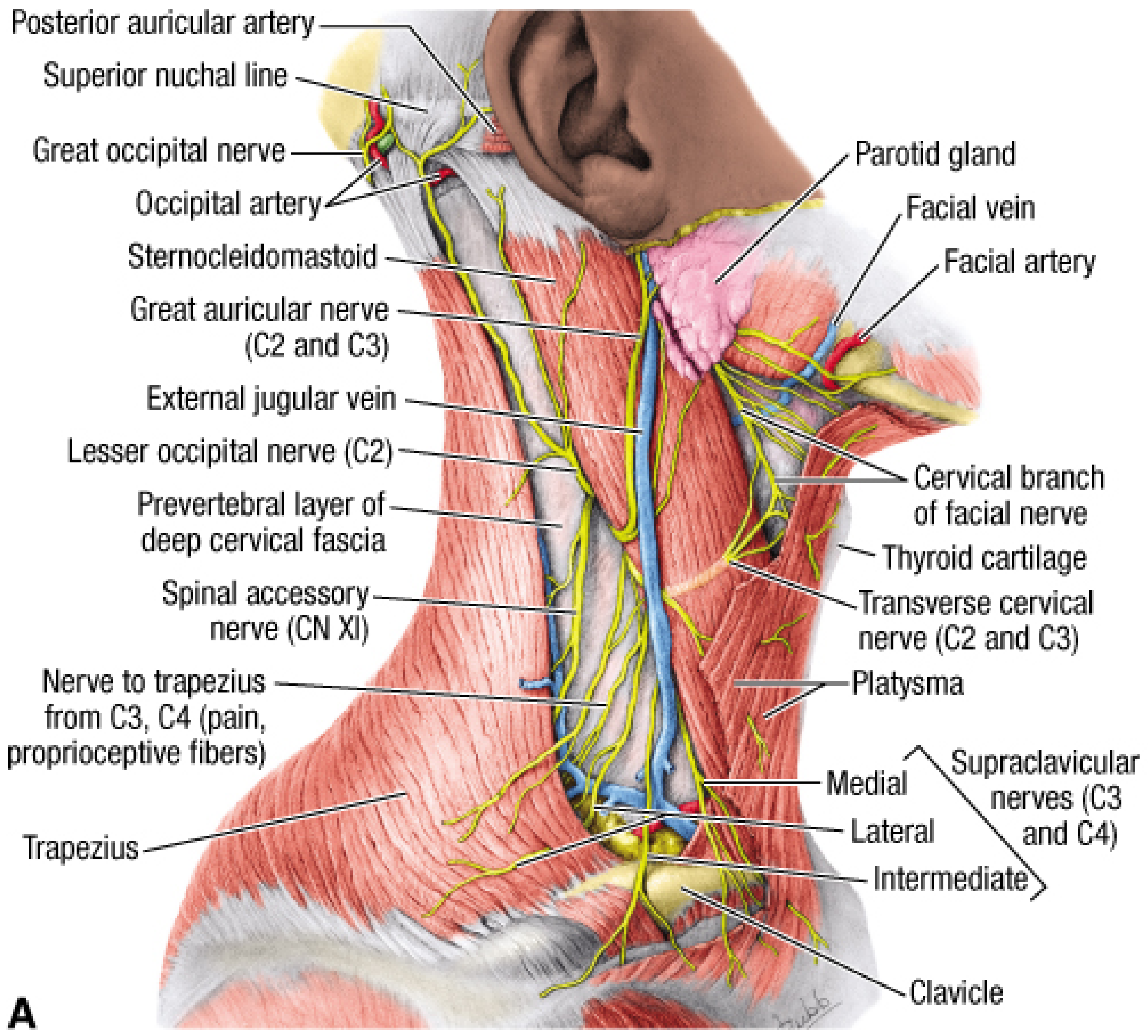
5. Nerves of the Neck
Superficial Cervical Plexus (Sensory)
All branches emerge at the nerve point (posterior border of SCM, midpoint). They include:
| Nerve | Root | Area Supplied |
|---|---|---|
| Lesser occipital nerve | C2 | Scalp posterior to ear |
| Greater auricular nerve | C2-C3 | Skin over parotid, mastoid, ear |
| Transverse cervical nerve | C2-C3 | Anterior neck skin |
| Supraclavicular nerves (medial, intermediate, lateral) | C3-C4 | Clavicular and deltoid region |
Deep Motor Branches
- Ansa cervicalis (C1-C3) - innervates infrahyoid strap muscles (except thyrohyoid which is C1 via CN XII)
- Phrenic nerve (C3,4,5) - sole motor supply to the diaphragm; descends on the anterior scalene muscle deep to prevertebral fascia
Cranial Nerve XI (Spinal Accessory)
- Emerges at the junction of middle and superior thirds of the lateral SCM border ("nerve point")
- Courses superficial to prevertebral fascia, deep to investing fascia, to reach trapezius
- Most superior nerve in the posterior triangle - important surgical landmark
Brachial Plexus (in posterior triangle)
Roots emerge between anterior and middle scalene muscles:
- C5 + C6 → Superior trunk
- C7 → Middle trunk
- C8 + T1 → Inferior trunk
6. Vascular Anatomy
Arteries
- Common carotid artery: Right originates from brachiocephalic; left from aortic arch. Both travel in the carotid sheath. No branches in the neck.
- Bifurcation at the superior margin of thyroid cartilage (CIII/CIV) into:
- Internal carotid artery - no neck branches; enters carotid canal
- External carotid artery - multiple neck branches:
- Anterior: Superior thyroid, lingual, facial arteries
- Posterior: Sternocleidomastoid, occipital, posterior auricular arteries
- Medial: Ascending pharyngeal artery
- Terminal: Superficial temporal + maxillary (at neck of mandible)
- Carotid sinus - dilation at proximal internal carotid; baroreceptor
- Carotid body - ovoid mass at bifurcation; chemoreceptor
Veins
- Internal jugular vein - begins at jugular foramen; runs in carotid sheath lateral to carotid arteries; joins subclavian to form brachiocephalic vein
- External jugular vein - crosses the superficial surface of SCM; drains into subclavian vein lateral to clavicular head of SCM
- Anterior jugular vein - runs in anterior neck
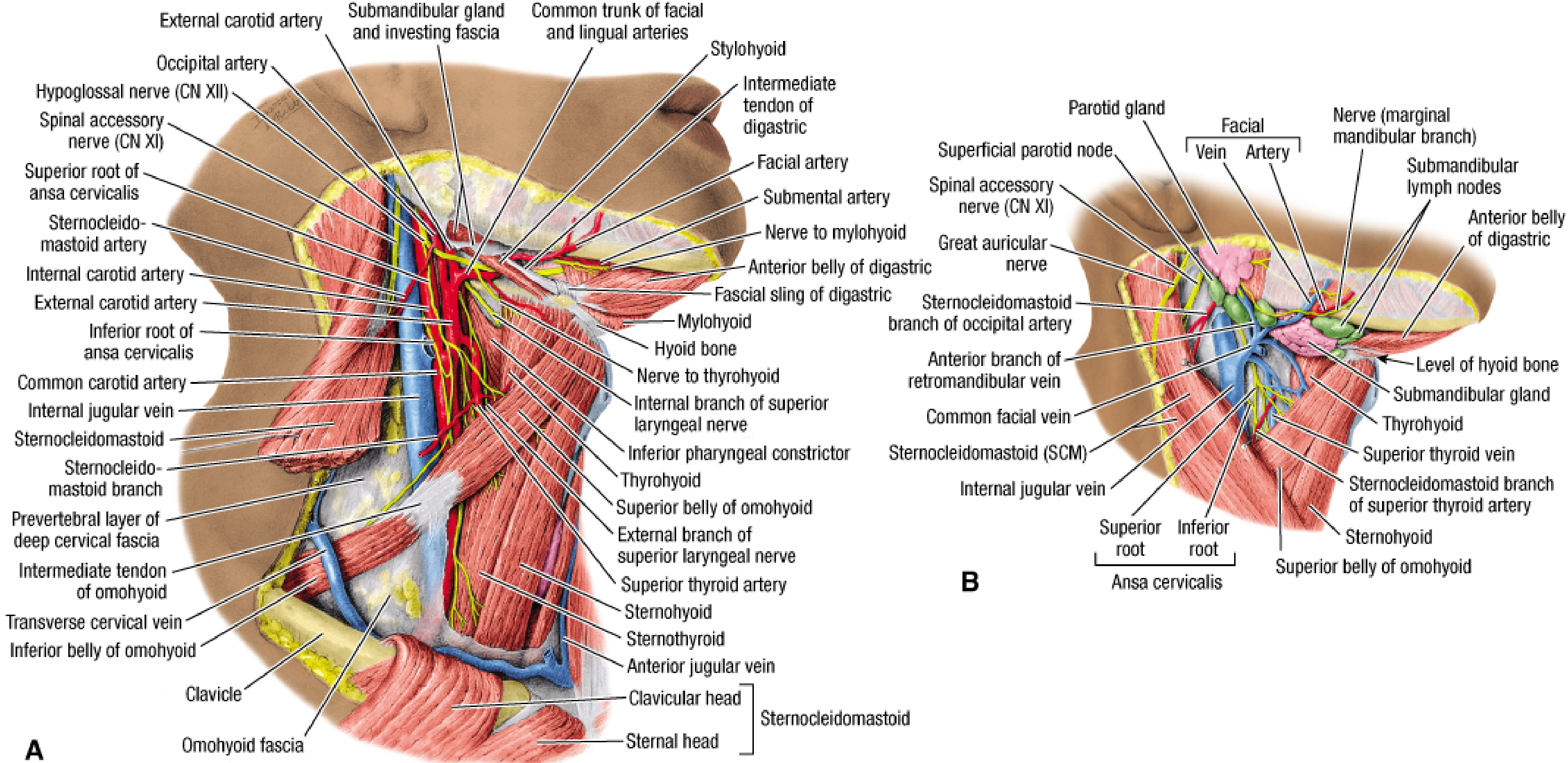
7. Carotid Triangle - Deep Dissection
The carotid triangle contains the major neurovascular bundle of the neck. Key relationships within it:
8. Lymph Nodes of the Neck
The cervical lymph nodes are classified into 7 levels (surgical levels):
| Level | Location |
|---|---|
| I | Submental (Ia) and submandibular (Ib) |
| II | Upper jugular (above hyoid) |
| III | Middle jugular (hyoid to cricoid) |
| IV | Lower jugular (cricoid to clavicle) |
| V | Posterior triangle nodes |
| VI | Central compartment (between carotid sheaths) |
| VII | Superior mediastinum (suprasternal notch to brachiocephalic artery) |
All surgically relevant nodes lie deep to the platysma, along nerves, vessels, and viscera.
9. Clinical Summary
| Structure | Clinical Relevance |
|---|---|
| Carotid sinus | Carotid sinus hypersensitivity; palpation can cause reflex bradycardia |
| Carotid body | Carotid body tumor (paraganglioma) at bifurcation |
| Retropharyngeal space | Spread of deep neck infections; retropharyngeal abscess |
| Phrenic nerve on scalenus anterior | At risk during central venous catheter insertion and thoracic procedures |
| Marginal mandibular nerve | Can lie 2.5 cm below mandible; at risk in submandibular dissection |
| Spinal accessory nerve (CN XI) | Injury causes trapezius palsy and dropped shoulder |
| Ansa cervicalis | Used for nerve grafting/reinnervation procedures |
| Median cricothyroid ligament | Emergency surgical airway access point (cricothyrotomy) |
Sources: Gray's Anatomy for Students (9780323934237); Fischer's Mastery of Surgery, 8th ed. (9781975176433)
This is a shared conversation. Sign in to Orris to start your own chat.