
P63 marker in duct papilloma
Reading File
P63 is a myoepithelial/basal cell marker. In intraductal papilloma, it highlights the presence of myoepithelial cells along the fibrovascular cores, which helps distinguish benign papilloma from malignant papillary lesions.
Searching sources...
Finding Sources
Finding Sources
Searching PubMed
p63 intraductal papilloma breast myoepithelial
Good results from Robbins. Let me read the relevant sections and fetch the images.
Reading File
Reading File
Finding Sources
Now let me search more specifically for p63 in the context of differentiating papillary lesions:
Finding Sources
Finding Sources
Good lead from Schwartz. Let me read that section and also look for more context on the Robbins papillary carcinoma section:
Reading File
Reading File
Now let me search specifically for p63 papillary carcinoma distinction and fetch the intraductal papilloma image:
Finding Sources
Loading Image
Now let me do a web search to supplement with the specific IHC role of p63 in papilloma vs papillary carcinoma, and search for the encapsulated papillary carcinoma context too:
Searching the Web
p63 intraductal papilloma breast myoepithelial fibrovascular cores benign vs malignant papillary lesion
Finding Sources
Excellent - very rich results. Let me extract the PMC article for more detail:
Reading Web Page
https://pmc.ncbi.nlm.nih.gov/articles/PMC8983543
I now have comprehensive information. Here is a complete answer:
P63 in Intraductal (Duct) Papilloma
What P63 Marks
P63 is a nuclear transcription factor that serves as a reliable immunohistochemical (IHC) marker for myoepithelial cells (also called basal cells) in the breast. It stains the nuclei of myoepithelial cells exclusively. Other myoepithelial markers used alongside it include smooth muscle myosin heavy chain (SMMHC), calponin, CD10, and CK5/6 - though p63 is considered among the most specific.
Role of P63 in Intraductal Papilloma (Benign)
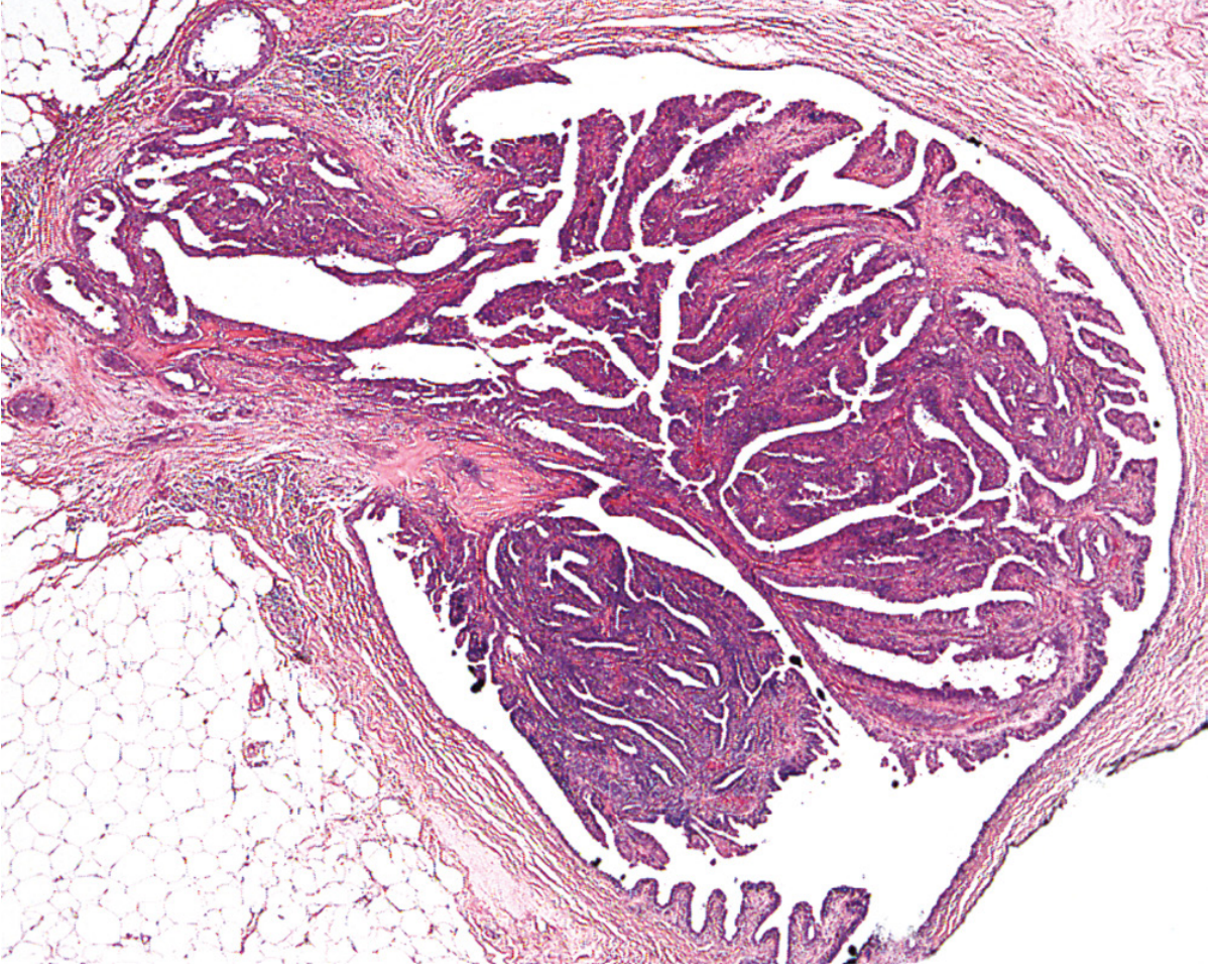
In a benign intraductal papilloma, the architecture consists of branching fibrovascular cores lined by two cell types:
- Luminal (epithelial) cells - the inner layer
- Myoepithelial cells - the outer layer
P63 staining highlights myoepithelial cells in two distinct locations:
| Location | Significance |
|---|---|
| Along the fibrovascular cores (within the papillae) | Hallmark of a benign papillary lesion |
| At the peripheral wall of the surrounding duct | Confirms intact ductal architecture |
"Myoepithelial cells can be demonstrated by p63 immunoreactivity both at the peripheral wall and along the fibrovascular cores." - Kulka et al., 2021, Papillary Lesions of the Breast, PMC8983543
This two-cell-type pattern - evidenced by p63 positivity along the cores - is the defining hallmark of benign papillary lesions.
Here is the histology of a benign intraductal papilloma (H&E):
P63 as a Differentiating Tool: Benign vs. Malignant Papillary Lesions
This is the most clinically important use of p63 in this context:
| Lesion | P63 Along Fibrovascular Cores | P63 at Periphery |
|---|---|---|
| Benign intraductal papilloma | Present (positive) | Present |
| Papilloma with atypia (ADH/DCIS within papilloma) | Present in non-atypical areas; absent where atypia expands | Present |
| Papillary DCIS | Absent along cores | Only at duct periphery |
| Encapsulated papillary carcinoma | Absent (no myoepithelial cells in cores OR periphery) | Typically absent |
| Invasive papillary carcinoma | Absent | Absent |
Key principle: Loss of p63 staining along fibrovascular cores = loss of myoepithelial layer = malignant papillary lesion.
In papillary DCIS, neoplastic cells line the cores but lack myoepithelial cells - p63 will only be seen at the outer duct wall, not along the fronds themselves.
In encapsulated papillary carcinoma, p63 is notably absent both within the cores and at the outer wall (the fibrous capsule substitutes for the myoepithelial layer), which has led to debate about whether it represents an in situ or invasive lesion.
Practical Pitfall
P63 must be interpreted in context. A key caveat from the literature: if absence of p63 alone is used to call a lesion malignant, approximately 50% of papillary carcinomas would be misclassified as benign (since some low-grade papillary carcinomas can focally retain myoepithelial-like cells). Therefore, p63 must be interpreted alongside morphology and a panel of markers (CK5/6, SMMHC).
Summary in One Line
P63 is POSITIVE along fibrovascular cores in benign intraductal papilloma (confirming myoepithelial cell presence) and ABSENT/lost in malignant papillary lesions, making it a key marker for distinguishing benign from malignant papillary breast lesions on core biopsy or excision.
Sources: Robbins & Cotran Pathologic Basis of Disease (9th ed.), p. 707; Kulka J et al., Pathology & Oncology Research, 2021 (PMC8983543); StatPearls: Intraductal Papilloma.
This is a shared conversation. Sign in to Orris to start your own chat.