Now I have everything I need. Let me write a thorough, student-friendly explanation of the entire presentation.
Medical Biology - Detailed Explanation for First Year MBBS
This PowerPoint covers two major topics: Molecular Biology (DNA, RNA, Gene Expression, Replication, Repair) and Microscopy. Below is a detailed, plain-language explanation of each slide group.
PART 1: MOLECULAR BIOLOGY
1. Central Dogma of Molecular Biology (Slide 2)
Think of the central dogma as the "master rule" of how genetic information flows in a living cell:
DNA → RNA → Protein
- DNA is like a blueprint kept safe in the nucleus - it stores all instructions.
- Transcription: A copy of a specific gene is made from DNA in the form of messenger RNA (mRNA). This is done by an enzyme called RNA Polymerase II.
- Translation: The mRNA travels to the ribosome in the cytoplasm, where its code is read and converted into a protein.
Important exceptions (examiners love these!):
- Retroviruses (HIV) work backwards - they use an enzyme called reverse transcriptase to convert RNA → DNA. This is why HIV is so difficult to eliminate.
- Telomerase also uses reverse transcriptase to protect chromosome ends.
- Many RNA molecules (rRNA, tRNA, miRNA, lncRNA) are functional without being translated into proteins.
Why does this matter clinically? Mutations in DNA alter proteins, causing inherited diseases and cancer.
2. Discovery of DNA as Genetic Material (Slide 3)
This is a favourite exam history topic. Know the key experiments:
| Year | Scientist(s) | Experiment | Finding |
|---|
| 1928 | Griffith | Mixed dead virulent + live harmless bacteria | Live bacteria became virulent - "transforming principle" exists |
| 1944 | Avery, MacLeod, McCarty | Destroyed DNA, RNA, or protein separately | Only destroying DNA stopped transformation - DNA is the genetic material |
| 1952 | Hershey & Chase | Labelled DNA with ³²P, protein with ³⁵S in bacteriophage | Only ³²P (DNA) entered bacteria - confirmed DNA is genetic material |
| 1953 | Watson & Crick | Used Franklin's X-ray data | Proposed the double helix model |
Rosalind Franklin's contribution: Her X-ray diffraction images (particularly Photo 51) revealed the helical structure of DNA and the spacing between base pairs - critical data used by Watson and Crick.
3. Structure of DNA (Slide 4)
DNA is a long, ladder-like twisted molecule. Here is how to picture it:
The rungs of the ladder = Nitrogenous bases paired together:
- A (Adenine) pairs with T (Thymine) - 2 hydrogen bonds
- G (Guanine) pairs with C (Cytosine) - 3 hydrogen bonds
Mnemonic: "Apples in Trees, Cars on Grass" = A-T, C-G
The sides of the ladder = Sugar-phosphate backbone:
- Each unit = deoxyribose sugar + phosphate group + one base = nucleotide
- Nucleotides are joined by phosphodiester bonds (3' of one sugar to 5' of the next)
Key features:
- The two strands run antiparallel - one goes 5'→3' while the other goes 3'→5'
- The helix is right-handed, approximately 2 nm in diameter
- Major and minor grooves allow proteins (like transcription factors) to recognize and bind specific sequences
GC-rich DNA is more thermally stable because 3 hydrogen bonds vs 2 in AT.
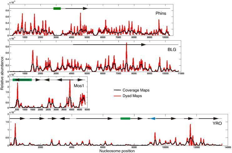
4. DNA Packaging - Histones and Chromatin (Slides 5-6)
The problem: Human DNA, if stretched out, is ~2 metres long. The nucleus is only ~6 micrometres in diameter. DNA must be folded ~250,000 times to fit!
The solution - multiple levels of packaging:
-
Nucleosome - the basic unit of chromatin
- 147 base pairs of DNA wrapped around a histone octamer (2× each of H2A, H2B, H3, H4)
- Histone H1 links adjacent nucleosomes (linker histone)
- Looks like "beads on a string"
-
30 nm fiber - nucleosomes coil into a thicker fiber
-
Loops and further coiling - during cell division, DNA condenses into visible chromosomes
Two forms of chromatin:
| Feature | Euchromatin | Heterochromatin |
|---|
| Compaction | Loosely packed | Tightly packed |
| Staining | Light (pale) | Dark |
| Transcription | Active (genes "ON") | Inactive (genes "OFF") |
| Replication timing | Early S phase | Late S phase |
| Location | Center of nucleus | Near nuclear envelope |
Types of heterochromatin:
- Constitutive = permanently condensed (centromeres, telomeres)
- Facultative = can switch ON/OFF. Classic example: Barr body (inactivated X chromosome in females)
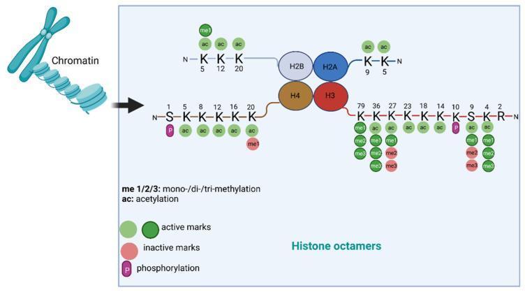
Histone modifications (epigenetic marks):
- Acetylation (by HATs) → opens chromatin → activates genes
- Deacetylation (by HDACs) → closes chromatin → silences genes
- Methylation, phosphorylation, ubiquitination = other regulatory marks
These are called epigenetic changes because they alter gene expression WITHOUT changing the DNA sequence.
5. Chromosomes (Slide 7)
Human somatic cells: 46 chromosomes = 23 pairs
- 22 pairs autosomes + 1 pair sex chromosomes (XX in females, XY in males)
Key chromosome structures:
- Centromere: Where the two sister chromatids are joined; also where the kinetochore forms - this is the attachment site for spindle fibers during cell division. Errors here → aneuploidy (e.g., Trisomy 21 = Down Syndrome)
- Telomeres: Protective caps at chromosome ends made of repetitive TTAGGG sequences bound by shelterin proteins. They shorten with each cell division.
- Sister chromatids: Identical copies of a chromosome after DNA replication; joined at the centromere
Classification by centromere position:
- Metacentric - centromere in middle
- Submetacentric - centromere slightly off-center
- Acrocentric - centromere near one end (humans have these; e.g., chromosomes 13, 14, 15, 21, 22)
- Telocentric - centromere at very end (humans do NOT have these)
Clinical tool: Karyotyping = arranging chromosomes by size/shape to detect abnormalities (deletions, duplications, translocations)
6. Genes: Structure (Slide 8)
A gene is a specific DNA sequence that codes for a functional product (protein or RNA).
Human genome facts:
- ~3.2 billion base pairs
- ~20,000 protein-coding genes
- Protein-coding genes = only 1-2% of total genome!
Parts of a gene:
[Promoter] → [5' UTR] → [Exon 1] → [Intron] → [Exon 2] → [3' UTR] → [Poly A signal]
- Promoter: Binding site for RNA Polymerase and transcription factors; located upstream
- Exons: Expressed sequences - appear in mature mRNA and are translated
- Introns: Intervening sequences - transcribed but then SPLICED OUT; not in final mRNA
- Enhancers/Silencers: Regulatory elements that can be far away from the gene; they increase or decrease transcription
- 5' UTR and 3' UTR: Not translated but regulate mRNA stability and translation efficiency
Why introns are important: They allow alternative splicing - one gene can produce many different proteins!
7. Types of RNA (Slide 9)
| RNA Type | Function |
|---|
| mRNA (messenger) | Carries genetic code from DNA to ribosome |
| tRNA (transfer) | Brings the correct amino acid to the ribosome; has anticodon |
| rRNA (ribosomal) | Forms the ribosome structure; catalyzes peptide bonds (it's a ribozyme!) |
| snRNA (small nuclear) | Part of spliceosome; removes introns |
| miRNA (microRNA) | ~22 nucleotides; suppresses gene expression |
| siRNA (small interfering) | ~21 nucleotides; degrades target mRNA (RNA interference) |
| lncRNA (long non-coding) | Regulates chromatin, transcription; >200 nt; e.g., XIST |
| piRNA | Protects germ cell genome from transposons |
Clinical pearl: siRNA technology is now used as medicine (e.g., inclisiran for cholesterol). mRNA technology underlies COVID-19 vaccines!
8. Genome Content (Slide 10)
| Component | % of Genome | Examples |
|---|
| Protein-coding exons | ~1-2% | Structural genes |
| Introns | ~25% | Removed during splicing |
| Regulatory DNA | Variable | Promoters, enhancers |
| Repetitive DNA | ~50% | LINEs, SINEs, Alu elements |
| Transposable elements | Many are inactive | "Jumping genes" |
Microsatellites (short tandem repeats) are highly variable between individuals → used in forensic DNA profiling and paternity testing
9. 3D Genome Organization (Slide 11)
The nucleus is not just a "bag of DNA" - it is highly organized:
- Each chromosome occupies a discrete chromosome territory
- Gene-rich chromosomes → center of nucleus (more transcription)
- Gene-poor chromosomes → near nuclear envelope (more condensed)
- DNA forms loops that bring enhancers close to promoters
- TADs (Topologically Associating Domains): Chromatin domains organized by CTCF and cohesin proteins; ensure enhancers only talk to the right genes
- Nuclear lamina: Made of lamins; anchors heterochromatin at the nuclear periphery. Mutations → laminopathies (e.g., Hutchinson-Gilford Progeria - premature aging, Emery-Dreifuss muscular dystrophy)
10. DNA Replication (Slides 12-18)
When: S (synthesis) phase of the cell cycle, before mitosis/meiosis
The Semiconservative Model (Meselson-Stahl experiment, 1958):
Each new DNA molecule has one old (parental) strand + one new strand. They proved this using heavy nitrogen (¹⁵N) and density-gradient centrifugation.
Steps at the Replication Fork:
- Origin of Replication (Ori): Where replication starts. Humans have tens of thousands, allowing the whole genome to be copied in 6-8 hours
- ORC (Origin Recognition Complex) + Cdc6, Cdt1 → forms pre-replication complex
- MCM helicase unwinds the double helix → creates the replication fork (Y-shaped)
- SSBPs/RPA stabilize single-stranded DNA
- Topoisomerases relieve the coiling tension ahead of the fork (I = single-strand cuts, II = double-strand cuts)
- Primase makes a short RNA primer (DNA polymerase cannot start from scratch - it needs a free 3' OH)
- DNA polymerases extend DNA only in 5'→3' direction
Leading vs. Lagging Strand:
| Leading Strand | Lagging Strand |
|---|
| Direction | Toward the fork | Away from the fork |
| Synthesis | Continuous | Discontinuous |
| Fragments | One long piece | Okazaki fragments (~100-200 nt in eukaryotes) |
Processing of Okazaki fragments:
- RNA primers removed by RNase H + FEN1
- Gaps filled by DNA Pol δ
- Nicks sealed by DNA Ligase I
DNA Polymerases in Eukaryotes:
| Polymerase | Job |
|---|
| Pol α | Initiates synthesis; extends primer |
| Pol δ | Lagging strand synthesis; proofreading |
| Pol ε | Leading strand synthesis |
| Pol γ | Mitochondrial DNA replication |
Proofreading: DNA polymerases have 3'→5' exonuclease activity - they remove wrongly added nucleotides immediately → error rate ~1 in 10⁹-10¹⁰ nucleotides!
PCNA (Proliferating Cell Nuclear Antigen): A sliding clamp that keeps DNA polymerase attached to DNA (increases processivity).
11. Telomeres and Telomerase (Slide 18)
The End-Replication Problem: When the last RNA primer on the lagging strand is removed, the very tip of the chromosome cannot be filled in (no upstream OH group available). Result: chromosomes get slightly shorter with every cell division.
Consequences of shortening:
- Eventually activates p53/Rb → replicative senescence (cell stops dividing) or apoptosis
- This is a tumor suppressor mechanism
Telomerase: An enzyme with its own RNA template that adds back TTAGGG repeats. Active in:
- Germ cells
- Embryonic stem cells
- 85-90% of cancer cells (which is why they can divide indefinitely!)
Clinical diseases of telomeres:
- Dyskeratosis congenita (mutated telomerase gene)
- Bone marrow failure syndromes
- Pulmonary fibrosis
12. DNA Damage and Repair (Slide 19)
Each cell gets ~10,000-100,000 DNA lesions per day!
| Repair Mechanism | What it fixes | Key proteins | Clinical significance |
|---|
| Mismatch Repair (MMR) | Replication errors (mismatches, small indels) | MSH2, MLH1, MSH6, PMS2 | Defect → Lynch Syndrome (hereditary colon cancer) |
| Base Excision Repair (BER) | Small damaged bases (oxidized, deaminated) | DNA glycosylases | Common everyday repair |
| Nucleotide Excision Repair (NER) | Bulky lesions (UV-induced thymine dimers) | XPA, XPC, etc. | Defect → Xeroderma Pigmentosum (extreme UV sensitivity, skin cancer) |
| Homologous Recombination (HR) | Double-strand breaks (accurate) | BRCA1, BRCA2, RAD51 | BRCA mutations → breast/ovarian cancer |
| Non-Homologous End Joining (NHEJ) | Double-strand breaks (less accurate, error-prone) | Ku70, Ku80 | Used in G1 when no sister chromatid available |
13. Transcription in Detail (Slides 20-27)
Key players:
- RNA Polymerase I - in nucleolus - makes rRNA (28S, 18S, 5.8S)
- RNA Polymerase II - makes mRNA + some miRNA + snRNA
- RNA Polymerase III - makes tRNA, 5S rRNA
Clinically important: α-amanitin (toxin in Amanita phalloides = death cap mushroom) inhibits RNA Pol II → stops mRNA synthesis → fatal liver failure
Promoter elements:
- TATA box (~25-30 bp upstream of start site) → binding site for TBP (TATA-Binding Protein), part of TFIID
- General Transcription Factors (GTFs): TFIID, TFIIA, TFIIB, TFIIF, TFIIE, TFIIH
- TFIIH - has helicase (opens DNA) + kinase (phosphorylates CTD of RNA Pol II to start elongation)
Stages of Transcription:
- Initiation - RNA Pol II + GTFs assemble at promoter → pre-initiation complex → DNA unwinding
- Elongation - RNA Pol II moves along template (3'→5' direction), making RNA (5'→3'). No primer needed!
- Termination - RNA Pol II encounters termination signal; RNA released
Prokaryotes vs. Eukaryotes (High-yield comparison):
| Feature | Prokaryotes | Eukaryotes |
|---|
| Location | Cytoplasm | Nucleus |
| RNA Polymerases | One (sigma factors) | Three (I, II, III) |
| Coupling | Transcription + translation simultaneous | Separated (nucleus vs. cytoplasm) |
| mRNA | Polycistronic | Monocistronic |
| mRNA processing | Minimal | Extensive (capping, splicing, polyadenylation) |
| Operons | Yes | No |
Antibiotic example: Rifampin blocks bacterial RNA polymerase → used in tuberculosis treatment
14. RNA Processing (Slides 28-33)
Pre-mRNA must be processed into mature mRNA before leaving the nucleus. Three major modifications:
A. 5' Capping
- Happens when only ~20-30 nt of RNA are made
- A 7-methylguanosine (m⁷G) cap is added via an unusual 5'-5' triphosphate bond
- Functions:
- Protects mRNA from exonucleases
- Helps ribosome (eIF4E) recognize mRNA
- Promotes export from nucleus
- Required for splicing of the first intron
Viral trick: Influenza performs "cap snatching" - it steals the 5' cap from host mRNAs to make its own proteins!
B. Polyadenylation (3' Poly-A Tail)
- Signal sequence: AAUAAA at 3' end of transcript
- Recognized by CPSF and CstF → mRNA is cleaved
- Poly(A) Polymerase (PAP) adds ~200-250 adenine residues
- Functions:
- Protects from 3' exonucleases
- Enhances stability
- Promotes nuclear export
- Interacts with 5' cap to form circular mRNA (enhances translation)
C. Splicing
- Introns removed; exons joined
- Done by the spliceosome - made of 5 snRNPs: U1, U2, U4, U5, U6
- Key recognition sequences:
- 5' splice site: GU (donor)
- Branch point: adenosine
- 3' splice site: AG (acceptor)
- Mechanism: Branch-point A attacks the 5' splice site → lariat intermediate formed → intron released as lariat → exons joined
Splice site mutations cause diseases:
- β-thalassemia
- Cystic fibrosis
- Retinitis pigmentosa
D. Alternative Splicing
One gene → multiple different proteins by including/excluding different exons
Classic example: Calcitonin/CGRP gene:
- In thyroid C cells → Calcitonin (calcium regulation)
- In neurons → CGRP (pain transmission, vasodilation)
Clinical application: Nusinersen (Spinraza) is an antisense oligonucleotide that modifies splicing of the SMN2 gene → treats Spinal Muscular Atrophy (SMA)
E. RNA Editing
Post-transcriptional nucleotide changes:
- A→I editing (ADAR enzymes): Adenosine → Inosine; important in brain (modifies neurotransmitter receptors)
- C→U editing (APOBEC enzymes): Classic example = ApoB gene
- In liver: no editing → ApoB-100 → needed for LDL
- In intestine: editing creates premature stop codon → ApoB-48 → needed for chylomicrons
15. Translation (Slides 34-40)
Where: Cytoplasm, on ribosomes
What is needed:
- mRNA - the template
- tRNA - the adaptor molecule
- Ribosomes - the molecular machine
Ribosome structure (eukaryotes):
- 80S total = 40S (small subunit) + 60S (large subunit)
- The 40S reads mRNA codons
- The 60S has the Peptidyl Transferase Center (actually rRNA - making it a ribozyme)
- Three sites: A site (incoming tRNA), P site (growing chain), E site (exit)
Antibiotic difference: Bacteria have 70S ribosomes (30S + 50S). Many antibiotics exploit this:
- Tetracyclines → block 30S A site
- Aminoglycosides → 30S misreading
- Chloramphenicol → 50S peptidyl transferase
- Macrolides → 50S translocation
tRNA and Charging:
- tRNA = ~70-90 nt with cloverleaf shape
- Anticodon loop - recognizes the mRNA codon
- Acceptor stem - ends in CCA, where amino acid attaches
- Aminoacyl-tRNA synthetases - charge tRNA with correct amino acid (2-step reaction using ATP); have proofreading activity
- Wobble hypothesis (Crick): flexible pairing at 3rd codon position → one tRNA can recognize several synonymous codons
The Genetic Code:
- Triplet codons: 4³ = 64 possible codons
- 61 sense codons (specify amino acids)
- 3 stop codons: UAA, UAG, UGA ("UAA = U Are Away", "UAG = U Are Gone", "UGA = U Go Away")
- Start codon: AUG (methionine)
- Degenerate: multiple codons can specify the same amino acid (e.g., leucine has 6 codons)
- Unambiguous: one codon = only one amino acid
- Universal: same code in almost all organisms
Mutation types:
- Silent/synonymous: codon change but same amino acid
- Missense: codon change → different amino acid (e.g., sickle cell: GAG→GUG → Glu→Val)
- Nonsense: codon → stop codon → truncated protein (e.g., Duchenne MD, cystic fibrosis)
- Frameshift: insertion/deletion → shifts reading frame → usually nonsense or garbage protein
Stages of Translation:
1. Initiation:
- 40S + eIFs + Met-tRNAi → 43S complex
- eIF4E recognizes 5' cap; poly(A) binding protein at 3' end → mRNA circularizes
- Scanning for AUG in Kozak consensus sequence
- 60S joins → complete 80S ribosome
- Regulated by mTOR pathway (nutrients, growth factors)
2. Elongation:
- Codon recognition: aminoacyl-tRNA + eEF1A + GTP enters A site
- Peptide bond formation: rRNA (peptidyl transferase) transfers growing chain from P site to A site
- Translocation: eEF2 + GTP moves ribosome one codon (A→P→E)
- ~5-10 amino acids/second in eukaryotes
3. Termination:
- Ribosome reaches UAA, UAG, or UGA
- Release factors (eRF1, eRF3) mimic tRNA, enter A site
- Peptidyl transferase hydrolyzes the bond → protein released
- Ribosome dissociates; components recycled
Polysomes: Multiple ribosomes on one mRNA simultaneously → efficient protein production
16. Post-translational Modifications (Slide 41)
After translation, proteins are usually not yet functional. They need:
- Folding - helped by chaperones (Hsp70, Hsp90)
- Proteolytic cleavage - Proinsulin → insulin; Trypsinogen → trypsin
- Phosphorylation - regulates enzyme activity, cell signaling
- Glycosylation - adds sugar chains; important for secreted/membrane proteins; done in ER and Golgi
- Acetylation - histone regulation, gene expression
- Hydroxylation - collagen maturation; requires Vitamin C (deficiency → Scurvy)
- Lipidation - anchors proteins to membranes
- Ubiquitination → targets protein for degradation by proteasome
Disease examples:
- Vitamin C deficiency → poor collagen → Scurvy
- Misfolded proteins → Alzheimer's, Parkinson's, Huntington's, Prion diseases
17. Gene Regulation Summary (Slides 42-44)
Gene expression is regulated at 5 main levels:
| Level | Mechanism |
|---|
| 1. Epigenetic | DNA methylation (silences genes), histone modifications, chromatin remodeling |
| 2. Transcriptional | Transcription factors, promoters, enhancers, RNA Pol II |
| 3. Post-transcriptional | Splicing, RNA editing, mRNA stability, miRNA/siRNA |
| 4. Translational | mTOR pathway, eIF regulation, nutrient availability |
| 5. Post-translational | Phosphorylation, ubiquitination, protein degradation |
Epigenetic drugs in clinical use:
- Azacitidine - DNMT inhibitor (cancer treatment)
- Vorinostat - HDAC inhibitor (cancer treatment)
Regulatory RNAs:
- miRNA (~22 nt): processed by Drosha (nucleus) → Dicer (cytoplasm) → into RISC complex → silences target mRNA
- siRNA (~21 nt): triggers direct mRNA cleavage via RISC; used in RNAi technology
- lncRNA (>200 nt): e.g., XIST mediates X-chromosome inactivation
- piRNA: protects germ cell genome from jumping genes (transposons)
PART 2: MICROSCOPY
18. Introduction & History (Slides 45-46)
Why microscopy? Unaided human eye can only see objects >0.1-0.2 mm. Most cells, bacteria, and organelles are much smaller.
Key historical milestones:
| Year | Scientist | Contribution |
|---|
| ~1590 | Zacharias & Hans Janssen | First compound microscope |
| 1665 | Robert Hooke | Observed cork cells → coined "cell" (from Micrographia) |
| ~1670s | Antonie van Leeuwenhoek | First to see living microorganisms; called "Father of Microbiology" |
| 19th century | Ernst Abbe | Principles of optical resolution, numerical aperture |
19. Optical Principles (Slide 47)
| Property | Definition | Key point |
|---|
| Magnification | How much larger the image appears | Total = Objective × Eyepiece (e.g., 40× × 10× = 400×) |
| Resolution | Ability to distinguish two close points as separate | Light microscope limit = 0.2 μm (limited by wavelength of light) |
| Contrast | Brightness difference between specimen and background | Most tissues need staining |
| Numerical Aperture (NA) | Light-gathering ability of objective | Higher NA = better resolution |
| Working distance | Space between objective and slide when focused | High-power objectives have SHORTER working distance |
20. Structure of Compound Light Microscope (Slides 48-50)
Mechanical parts: base, arm, stage, stage controls (X-Y), coarse adjustment knob, fine adjustment knob, revolving nosepiece
Optical parts: eyepiece (10×), objective lenses (4×, 10×, 40×, 100×), condenser, iris diaphragm, illuminator (LED/halogen)
Objective lenses:
| Objective | Total Magnification (with 10× eyepiece) | Use |
|---|
| 4× (scanning) | 40× | Overview, locate specimen |
| 10× (low power) | 100× | Tissue organization |
| 40× (high dry) | 400× | Individual cell detail |
| 100× (oil immersion) | 1000× | Bacteria, blood smears |
Oil immersion: Immersion oil has the same refractive index as glass → reduces light scatter → increases resolution
Golden rules:
- Always start with 4× objective
- Use coarse focus only at 4× and 10×; use fine focus only at 40× and 100×
- Clean objectives only with lens paper, not tissue
- After use: clean oil off with lens paper, return to 4×, lower stage, cover microscope
21. Histological Sample Preparation (Slides 52-54)
Steps to prepare a tissue for light microscopy:
- Fixation - preserves tissue; most common: 10% neutral buffered formalin (cross-links proteins)
- Dehydration - removes water using increasing ethanol concentrations (70% → 80% → 95% → 100%)
- Clearing - xylene replaces ethanol; makes tissue transparent; prepares for paraffin
- Embedding - molten paraffin infiltrates and hardens around tissue (provides support for cutting)
- Sectioning (Microtomy) - microtome cuts sections 4-6 μm thick; sections floated on warm water bath then mounted on slides
- Deparaffinization and Staining - xylene removes paraffin; sections stained
Types of microtomes:
- Rotary - routine paraffin sections
- Cryostat - frozen sections for rapid intraoperative diagnosis (patient on operating table, surgeon needs quick result!)
- Ultramicrotome - 50-100 nm sections for electron microscopy
Common staining techniques:
| Stain | What it stains | Color | Use |
|---|
| Hematoxylin | Nuclei, chromatin, RER | Blue/purple (basophilic) | Routine H&E |
| Eosin | Cytoplasm, collagen, muscle | Pink/red (eosinophilic) | Routine H&E |
| PAS | Glycogen, basement membranes, mucus, fungi | Magenta | Glycogen storage, fungal infection |
| Masson's Trichrome | Collagen vs. muscle | Blue/green vs. red | Fibrosis |
| Silver stains | Reticular fibers | Black/brown | Reticulin |
| Ziehl-Neelsen | Acid-fast bacteria (TB) | Red on blue | Tuberculosis |
| Giemsa | Blood cells, malaria | Variable | Hematology, microbiology |
Memory tip: Basophilic structures (DNA, RNA, RER) are stained by hematoxylin because they carry a negative charge, attracted to the positively charged dye.
22. Specialized Microscopy Techniques (Slides 55-61)
Stereo (Dissecting) Microscope
- 3D image using reflected light
- Magnification: 5-80×
- Large working distance
- For: dissection, gross specimen examination, microsurgery, embryology
Phase Contrast Microscopy (Frits Zernike, Nobel 1953)
- Converts phase differences → brightness differences
- For: living, unstained cells; cell culture; semen analysis
- Disadvantage: halo artifacts
Dark Field Microscopy
- Bright specimen on dark background (scattered light only)
- For: thin bacteria, Treponema pallidum (syphilis), live organisms
Polarizing Microscopy
- Examines birefringent (double-refracting) materials
- Has polarizer + analyzer
- Key clinical use: Crystal analysis in joint fluid
- Monosodium urate crystals (Gout) = negatively birefringent (yellow when parallel)
- Calcium pyrophosphate crystals (Pseudogout) = positively birefringent
- Also detects: amyloid, asbestos, silica, foreign bodies
Fluorescence Microscopy
- Uses fluorescent dyes (fluorophores) excited by specific wavelengths
- Common dyes: DAPI (blue, stains DNA), FITC (green), TRITC (red)
- Immunofluorescence:
- Direct: fluorescent dye on primary antibody
- Indirect: fluorescent secondary antibody (amplifies signal)
- Uses: autoimmune diseases (lupus, pemphigus), kidney biopsy, cancer markers, infectious organisms
Confocal Laser Scanning Microscopy (CLSM)
- Laser scans one point at a time
- Pinhole blocks out-of-focus light → very sharp optical sections
- Can reconstruct 3D images
- Uses: cell biology, neuroscience, live-cell imaging, corneal imaging
Electron Microscopy (EM)
- Uses electrons instead of light → wavelength much shorter → resolution in nanometers
| Type | How it works | What you see | Uses |
|---|
| TEM (Transmission EM) | Electrons pass through ultrathin sections (50-100 nm) | Internal ultrastructure: ribosomes, membranes, viruses, organelles | Ultrastructural pathology, virology, glomerular diseases |
| SEM (Scanning EM) | Electrons scan surface | 3D surface morphology | Cell surfaces, bacteria morphology, implants |
- Requires vacuum, fixation with glutaraldehyde, heavy metal staining (osmium, uranium, lead)
- Cannot examine living specimens
Advanced Modern Techniques
- Cryo-EM: Rapid freezing in vitreous ice (no staining), then 3D reconstruction → near-atomic resolution. 2017 Nobel Prize in Chemistry. Revealed structures of ribosomes, ion channels, viral particles.
- Super-resolution microscopy (STED, PALM, STORM): Overcomes 200 nm light diffraction limit → 20-50 nm resolution
- Live-cell imaging: Fluorescence + environmental chambers → watch cell division, organelle transport, signaling in real time
23. Clinical Applications of Microscopy (Slide 62)
| Specialty | What microscopy provides |
|---|
| Histopathology | Diagnose tumors, inflammation, fibrosis (H&E stains) |
| Cytology | Pap smear (cervical cancer screening), FNA, body fluids |
| Hematology | Blood smears (malaria, leukemia, anemia, platelet disorders) |
| Microbiology | Gram stain, Ziehl-Neelsen (TB), dark field (syphilis), Giemsa (malaria) |
| Renal pathology | Immunofluorescence (glomerulonephritis patterns) + EM (podocyte/basement membrane) |
| Neuromuscular | EM for muscle diseases, ciliary disorders |
| Digital Pathology | Whole-slide scanning + AI for diagnosis, telepathology, biomarker quantification |
Quick Revision Summary
Central Dogma: DNA → (Transcription) → RNA → (Translation) → Protein
DNA structure: Antiparallel double helix; A-T (2 H-bonds), G-C (3 H-bonds); phosphodiester backbone
Replication: Semiconservative; 5'→3' only; leading (continuous) vs. lagging (Okazaki fragments); proofreading by DNA polymerases
Transcription: RNA Pol II makes mRNA; Pol I makes rRNA; Pol III makes tRNA; rifampin blocks bacterial RNA Pol
Processing: 5' cap (m7G), 3' poly-A tail (~200-250 A), splicing (GU...AG rule, spliceosome)
Translation: mRNA codon → tRNA anticodon → amino acid; 80S ribosome; A-P-E sites; initiation-elongation-termination
Microscopy: Compound (4×,10×,40×,100×); H&E staining; phase contrast for living cells; dark field for syphilis; polarizing for crystals; fluorescence for immune markers; EM for ultrastructure
Good luck in your studies! These topics form the foundation for Biochemistry, Pathology, Genetics, and Pharmacology throughout your MBBS course.