
Neuromodulation in chronic pain management 10 mark answer
neuromodulation chronic pain management
spinal cord stimulation neuromodulation chronic pain dorsal column
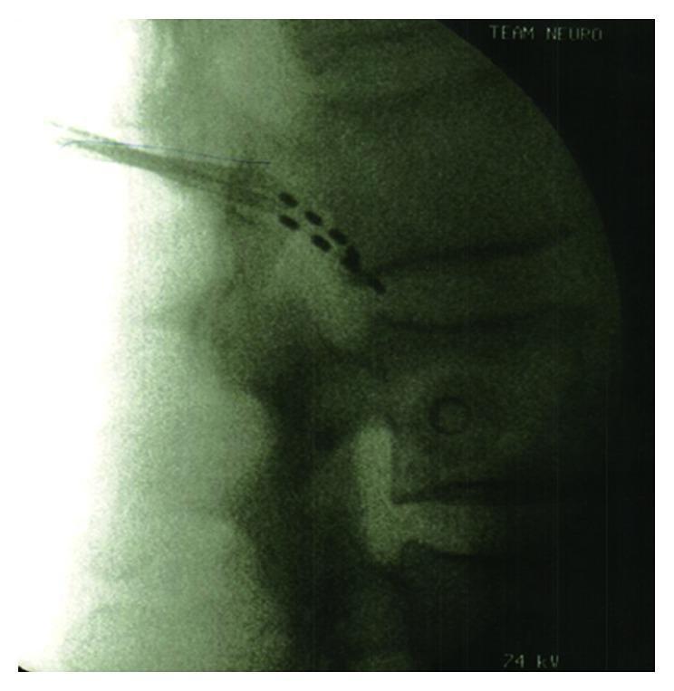
This diagnostic fluoroscopic image provides a lateral radiographic view of the lumbar spine, focusing on the vertebral column and neural foramina. The visual content demonstrates the placement of two radio-opaque neurostimulation electrode arrays near the L2 vertebral level. Each lead appears as a linear series of four distinct, rectangular, high-density metallic contacts. The arrays are positioned in the dorsal epidural space or near the dorsal root ganglion (DRG) region for chronic pain management. Anatomical landmarks include the rectangular lumbar vertebral bodies, the intervertebral disc spaces, and the posteriorly projecting spinous processes. The thin, radio-opaque lead wires are seen extending from the electrode contacts towards a cephalad direction, indicating a translaminar or percutaneous approach. This imaging is typically used intraoperatively to confirm the precise lead position and ensure appropriate target coverage for neuromodulation therapies, such as spinal cord stimulation (SCS) or dorsal root ganglion stimulation.
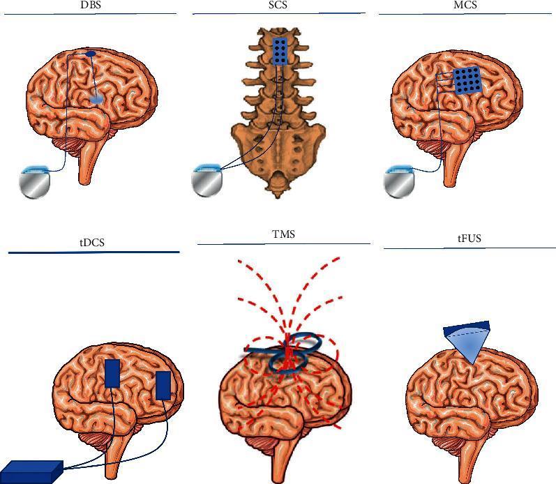
Educational infographic illustrating six neurostimulation modalities for chronic pain management. The content is organized into invasive and non-invasive categories. The top row depicts invasive surgical interventions: Deep Brain Stimulation (DBS) with intraparenchymal electrodes; Spinal Cord Stimulation (SCS) showing an epidural lead array over the dorsal columns; and Motor Cortex Stimulation (MCS) featuring a paddle electrode array on the cortical surface. All three are connected via subcutaneous leads to an Implantable Pulse Generator (IPG). The bottom row displays non-invasive transcranial techniques: Transcranial Direct Current Stimulation (tDCS) using scalp-mounted anode and cathode electrodes; Transcranial Magnetic Stimulation (TMS) utilizing an external figure-of-eight coil to generate magnetic fields (indicated by dashed lines); and Transcranial Focused Ultrasound Stimulation (tFUS) demonstrating acoustic energy delivery through a transducer. The diagrams emphasize anatomical targeting differences, ranging from deep subcortical nuclei and the spinal cord to superficial cortical regions, highlighting various mechanisms of neuromodulation used in clinical neurology and pain medicine.
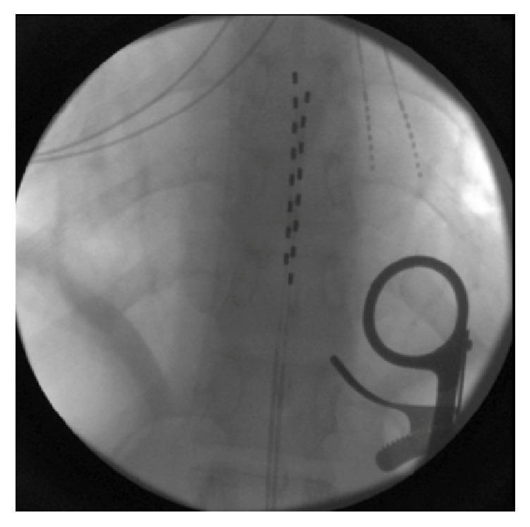
An intraoperative fluoroscopic image (X-ray) showing the mid-thoracic spine in an anteroposterior view. Centrally located within the spinal canal are two parallel epidural stimulator leads. Each lead contains eight distinct, radiopaque rectangular contacts arranged linearly. The leads are positioned in the dorsal epidural space for spinal cord stimulation (SCS). To the right of the spinal column, a radiopaque surgical retractor or handle is visible. Background anatomical structures include the shadows of the vertebral bodies and the articulation of the ribs. This diagnostic image illustrates the successful placement of bilateral multi-contact electrodes in a patient undergoing neuromodulation for chronic pain management.
Neuromodulation in Chronic Pain Management
Definition
- Firestein & Kelley's Textbook of Rheumatology, p. 697
Pathophysiological Basis
| Level | Target | Intervention |
|---|---|---|
| Peripheral | Nociceptors, A-beta fibres | TENS, PNS |
| Spinal cord | Dorsal horn, dorsal columns | SCS, DRG stimulation |
| Supraspinal | Thalamus, PAG, motor cortex | DBS, TMS, MCS |
- Morgan & Mikhail's Clinical Anesthesiology, p. 2081
Modalities of Neuromodulation
1. Transcutaneous Electrical Nerve Stimulation (TENS)
- Mechanism: Stimulates large A-beta fibres (gate control), and at low frequency activates endogenous opioid release (reversed by naloxone)
- Parameters: Conventional TENS uses 80-100 Hz, 10-30 mA, pulse width 50-80 μs; acupuncture-like TENS uses <10 Hz, pulse width >200 μs
- Indications: Mild-to-moderate chronic pain, low back pain, arthritis, neuropathic pain
- Limitations: Evidence is mixed; limited short-term benefit for neuropathic pain, not well supported for back pain
- Goldman-Cecil Medicine, p. 276; Morgan & Mikhail, p. 2081
2. Spinal Cord Stimulation (SCS)
- Activation of inhibitory interneurons in the dorsal horn
- Suppression of wide dynamic range neurons
- Enhancement of descending inhibition via serotonergic and GABAergic pathways
- Modulation of sympathetic activity (relevant in ischemic pain and CRPS)
- Temporary trial with percutaneous leads for 5-7 days to confirm efficacy (minimum 50% pain reduction is considered success)
- Permanent implantable pulse generator if trial is successful
- Conventional/tonic SCS (40-60 Hz): Paresthesia-based, primarily for radicular pain
- High-frequency SCS (10 kHz, HF10): Paresthesia-free; the SENZA-RCT showed superiority over conventional SCS for chronic back and leg pain
- Burst SCS: Mimics natural neural firing patterns; effective for both axial and radicular pain
- Dorsal root ganglion (DRG) stimulation: Targets the sensory ganglion at a specific spinal level; particularly useful for focal neuropathic pain (CRPS, post-surgical pain)
- Failed back surgery syndrome (persistent radicular pain after lumbar surgery) - strongest indication
- Complex Regional Pain Syndrome (CRPS) Type 1 - RCT showed 2.4 cm improvement on VAS vs. 0.2 cm with physiotherapy alone
- Ischemic limb pain (peripheral vascular disease, refractory angina)
- Post-herpetic neuralgia, phantom limb pain, peripheral neuropathies
- Visceral abdominal and pelvic pain
- Morgan & Mikhail's Clinical Anesthesiology, pp. 2081-2085; Rheumatology 2-Volume Set, p. 697
3. Peripheral Nerve Stimulation (PNS)
- Leads placed in close proximity to an identified peripheral nerve (percutaneously, with or without ultrasound guidance, or surgically)
- Indications: Neuropathic pain in the distribution of one or two peripheral nerves; occipital neuralgia; migraine
- Recent advances include less invasive implantable leads with external pulse generators
- Relief can persist for more than 1 year in neuropathic pain; evidence for migraine is mixed
- Occipital nerve stimulation is a specific form used for chronic occipital neuralgia and refractory migraine
- Goldman-Cecil Medicine, p. 276; Morgan & Mikhail, p. 2084
4. Dorsal Root Ganglion (DRG) Stimulation
- Electrodes placed adjacent to the DRG within the lateral epidural space at the appropriate spinal level
- The DRG acts as a sensory gateway; stimulation reduces ectopic discharge from injured neurons
- Advantages: Precise dermatomal targeting; lower energy requirements; reduced positional paresthesia
- Particularly effective for CRPS, groin/inguinal pain, foot pain, post-amputation pain
- Morgan & Mikhail's Clinical Anesthesiology, p. 2083
5. Deep Brain Stimulation (DBS)
- Electrodes implanted stereotactically into deep brain targets
- Targets for nociceptive pain (e.g., cancer, chronic low back pain): periaqueductal grey (PAG) and periventricular grey (PVG)
- Targets for neuropathic pain: ventral posterolateral (VPL) and ventral posteromedial (VPM) thalamic nuclei
- Also used for thalamic stroke pain (central post-stroke pain), cluster headache (posterior hypothalamus)
- Complications: Intracranial haemorrhage (most serious), infection; several haemorrhages including one death have been reported in hypothalamic DBS for cluster headache
- Morgan & Mikhail, p. 2085; Bailey & Love's Surgery, p. 5434
6. Motor Cortex Stimulation (MCS)
- Paddle electrode placed extradurally over the motor cortex
- Acts via activation of corticospinal and corticothalamic pathways to modulate thalamo-cortical pain circuits
- Used for central post-stroke pain, trigeminal deafferentation pain, phantom limb pain
- Less invasive than DBS with a lower complication profile
7. Repetitive Transcranial Magnetic Stimulation (rTMS)
- Non-invasive; uses a rapidly changing magnetic field to induce cortical electrical currents
- High-frequency rTMS over the motor cortex or dorsolateral prefrontal cortex can reduce chronic pain and improve mood (relevant in comorbid depression)
- Being evaluated for spinal conditions (transcranial magnetic stimulation of motor cortex for back pain)
- Rheumatology 2-Volume Set, p. 697
8. Intrathecal Drug Delivery (ITDD)
- Implantable pump delivers drugs (opioids, ziconotide, baclofen, local anaesthetics) directly into the intrathecal space
- Achieves therapeutic concentrations at the spinal cord with 1/300th of the systemic dose, minimising side effects
- Indications: Cancer pain, failed back surgery syndrome, CRPS, spasticity
- Also used in CRPS where it has been employed alongside other interventions
Patient Selection and Trial Criteria
- Reversible procedures (SCS trial) must precede permanent implantation
- Psychological screening is mandatory - untreated depression, secondary gain, or psychosocial issues predict poor outcomes
- Patients with predominantly central sensitisation benefit less from peripheral/spinal stimulation
- Multidisciplinary pain team assessment is required
Indications Summary Table
| Condition | Preferred Modality |
|---|---|
| Failed back surgery syndrome | SCS (strongest evidence) |
| CRPS Type 1 | SCS, DRG stimulation |
| Ischemic limb/angina | SCS |
| Neuropathic pain (focal) | PNS, DRG stimulation |
| Occipital neuralgia | Occipital PNS |
| Central post-stroke pain | DBS (VPL/VPM), MCS |
| Cancer pain | DBS (PAG/PVG), ITDD |
| Refractory chronic back pain | High-frequency/burst SCS, rTMS |
Limitations and Future Directions
- Long-term efficacy decreases in some SCS patients (habituation)
- Most trials are industry-funded; independent long-term data are limited
- Closed-loop (adaptive) SCS systems that respond in real time to neural activity are emerging
- Transcranial focused ultrasound stimulation (tFUS) is a newer non-invasive modality under investigation
- Combination of neuromodulation with pharmacotherapy, physiotherapy, and cognitive-behavioural therapy gives best outcomes in a multidisciplinary pain programme
Key Evidence
- SENZA-RCT: 10 kHz high-frequency SCS superior to conventional SCS for chronic back and leg pain
- SCS vs. physiotherapy in CRPS: VAS improvement 2.4 cm vs. 0.2 cm (SCS group)
- Meta-analysis by Kong et al., 2024 on brain stimulation targets for chronic pain (PMID 38237403) confirms VPL thalamus and motor cortex as optimal targets for DBS and MCS respectively
- Systematic review - Gish et al., 2024 (PMID 37726930): Neuromodulation shows benefit for chronic pelvic pain syndromes