I now have rich, comprehensive content from Smith & Tanagho's General Urology (19th Ed), Brenner & Rector's The Kidney, and Campbell-Walsh Urology - the three definitive urology texts. Let me now compose the complete, highly memorable clinical anatomy guide:
🫘 Complete Clinical Anatomy of Urology
From Smith & Tanagho's General Urology (19th Ed), Campbell-Walsh-Wein Urology, and Brenner & Rector's The Kidney
🗺️ THE BIG PICTURE - MASTER FLOWCHART
UROLOGICAL SYSTEM
│
├── UPPER URINARY TRACT (Urologic function ONLY)
│ ├── Adrenal Glands (capping each kidney)
│ ├── Kidneys (paired, retroperitoneal)
│ ├── Renal Calyces (8-12 minor → 2-3 major)
│ └── Renal Pelvis → tapers into Ureter (30 cm)
│
├── MIDDLE TRACT (Urologic function ONLY)
│ └── Ureters (30 cm, 3 narrowings = stone traps!)
│
└── LOWER TRACT (Both URINARY + GENITAL functions)
├── Bladder
├── Prostate (males) + Urethra
├── Seminal Vesicles / Vas Deferens (males)
├── Testes / Epididymis / Scrotum (males)
├── Penis (males)
└── Female urethra / Vagina (females)
Smith & Tanagho's General Urology, 19th Ed, Chapter 1
1. ADRENAL GLANDS - "The Hat on the Kidney"
🎯 Memory Hook: "Right is Triangle, Left is Crescent"
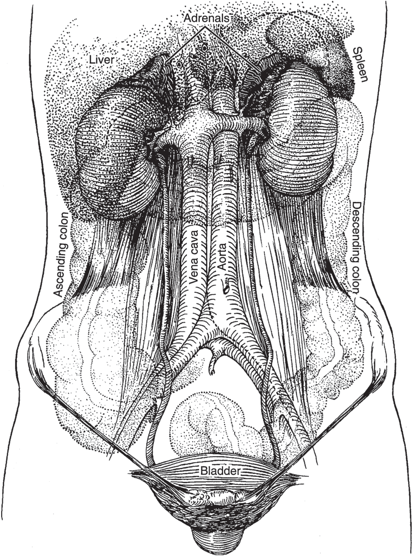
Fig. 1-2: Relations between the kidneys, ureters, and bladder (Smith & Tanagho)
| Feature | Details |
|---|
| Weight | 4-5 g each |
| Right shape | Triangular |
| Left shape | Rounded/Crescentic |
| Dimensions | 3 cm wide × 5 cm long × 1 cm thick |
| Enclosed in | Gerota's fascia (= perirenal fascia) |
Blood Supply - "Right is SHORT, Left is LONG"
- Arteries (3 per gland): Inferior phrenic + Aorta + Renal artery
- Right vein → drains DIRECTLY into vena cava (very short)
- Left vein → drains into left renal vein (longer path)
Histology (Cortex = 85% of mass)
CORTEX (85%) — outer to inner:
├── Zona Glomerulosa → Mineralocorticoids (Aldosterone)
├── Zona Fasciculata → Glucocorticoids (Cortisol)
└── Zona Reticularis → Sex steroids (DHEA)
MEDULLA (15%) — central:
└── Chromaffin cells (polyhedral) → Catecholamines
+ Sympathetic ganglion cells
Mnemonic: GFR = "Go Find Receptors" (Glomerulosa, Fasciculata, Reticularis - outer to inner)
Lymphatics
Accompany the suprarenal vein → lumbar lymph nodes
2. KIDNEYS - "The Bean-Shaped Workhorse"
🎯 Memory Hook: "Right is Lower, Left is Higher" (liver pushes right down)
| Measurement | Male | Female |
|---|
| Weight | 125-170 g | 115-155 g |
| Length | 10-12 cm | 10-12 cm |
| Width | 5-7 cm | 5-7 cm |
| Thickness | 3-5 cm | 3-5 cm |
Position
- Between T12 and L3 vertebrae, retroperitoneal
- Lie along the psoas muscle borders (obliquely placed)
- Average descent on inspiration or standing = 4-5 cm
- Enclosed in perirenal fat within Gerota's fascia
3D Relationships (Crucial for Surgery!)
RIGHT KIDNEY relations:
Anterior: Liver, hepatic flexure of colon, duodenum (2nd part)
Posterior: Diaphragm, psoas, quadratus lumborum, 12th rib
LEFT KIDNEY relations:
Anterior: Stomach, spleen, pancreas (tail), jejunum, descending colon
Posterior: Same muscles + 11th and 12th ribs
Internal Structure Flowchart
KIDNEY (cut section)
│
├── CORTEX (outer, homogeneous)
│ ├── Columns of Bertin (project inward between pyramids)
│ ├── Nephrons (1-1.5 million per kidney!)
│ └── Bowman's capsule + Glomerulus
│
└── MEDULLA (inner, striated)
├── 8-18 Renal Pyramids (conical)
│ └── Base at corticomedullary junction
│ └── Apex = Papilla → opens into Minor Calyx
├── Collecting ducts (Ducts of Bellini) → Area Cribrosa
└── Loop of Henle (thin limbs)
The Nephron - "The Real Kidney Unit"
Renal Corpuscle:
Glomerulus → Bowman's capsule
↓
Proximal Convoluted Tubule (PCT)
↓
Loop of Henle (descending + thin + ascending limbs)
↓
Distal Convoluted Tubule (DCT)
↓
Collecting Duct (Cortical → Medullary → Papillary)
↓
Area Cribrosa at Papilla tip
↓
Minor Calyx → Major Calyx → Renal Pelvis → Ureter
Blood Supply - "Segmental = No Collaterals!"
Aorta
└── Renal Artery → Anterior + Posterior divisions
├── Anterior → 3 segmental arteries (upper, middle, lower thirds)
│ + Apical branch (most commonly from anterior)
└── Posterior → >50% of posterior surface
└── NO ANASTOMOSES between segments!
(infarction = true wedge-shaped ischemia)
Venous: Renal Vein → Inferior Vena Cava
Left renal vein is LONGER and receives:
- Left gonadal vein
- Left adrenal vein
- Left inferior phrenic vein
Nutcracker syndrome = Left renal vein compressed between Aorta and SMA - causes hematuria/flank pain. Why? Because the left gonadal vein drains into the left renal vein!
Lymphatics
Drain to lumbar (para-aortic) lymph nodes - same drainage as testes! This is why testicular cancer spreads to para-aortic nodes, NOT inguinal nodes.
3. CALYCES, RENAL PELVIS & URETER
The Collecting System
8-12 Minor Calyces
↓ (cup each papilla)
2-3 Major Calyces
↓
Renal Pelvis (intra- or extra-renal)
↓ (tapers inferomedially)
URETER (30 cm long)
↓
Urinary Bladder
⚠️ THE 3 URETERIC NARROWINGS - "Where Stones Get STUCK"
STONE TRAP #1: Ureteropelvic Junction (UPJ)
└── Where pelvis meets ureter
STONE TRAP #2: Crossing of Iliac Vessels
└── At the pelvic brim (ureter crosses anterior to vessels)
STONE TRAP #3: Ureterovesical Junction (UVJ)
└── Where ureter enters the bladder obliquely
└── NARROWEST point! (~2mm)
Mnemonic: "PIP" - Pelvis, Iliac vessels, Pelvic entry
Ureteric Relations (Clinically Critical!)
Fig. 1-6: Anatomy of the bladder, ureters, and posterior male pelvic structures (Smith & Tanagho)
ABDOMINAL URETER:
On psoas muscle, covered by posterior peritoneum
Right ureter: lateral to vena cava
Left ureter: lateral to aorta
PELVIC URETER:
Passes medial to sacroiliac joints
Swings lateral near ischial spines
Then turns medial to enter bladder base
SURGICAL DANGER ZONE (Females!):
Uterine artery crosses OVER the ureter at the juxtavesical level
"WATER UNDER THE BRIDGE" = Ureter goes under uterine artery
→ Risk of ureteric ligation in hysterectomy!
MALES:
Vas deferens sweeps over ureter from lateral → medial
just before entering bladder base
Histology of Ureter/Pelvis/Calyces
- Mucosa: Transitional cell epithelium (urothelium)
- Lamina propria: Loose connective tissue
- Muscle: Helical + longitudinal smooth muscle (NOT distinct layers)
- Adventitia: Fibrous connective tissue outermost
Ureteric Blood Supply (Rule of Thirds!)
Upper ureter/pelvis → Renal artery branches
Middle ureter → Gonadal arteries (internal spermatic/ovarian)
Lower ureter → Superior/inferior vesical arteries
+ Uterine artery (females)
Clinical pearl: Blood supply enters the ureter from its MEDIAL side in the abdominal portion and LATERAL side in the pelvis. Therefore, during surgery, dissect along the LATERAL side of the abdominal ureter and MEDIAL side in the pelvis to preserve blood supply!
4. URINARY BLADDER
🎯 Memory Hook: "Detrusor = Random mesh; Neck = 3 layers"
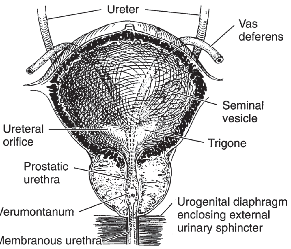
Fig. 1-5: Bladder opened anteriorly (Smith & Tanagho)
Position
- Empty bladder: behind pubic symphysis, largely pelvic
- Full bladder: rises above symphysis, can be palpated/percussed
- In infants/children: positioned higher than adults
- Median umbilical ligament = obliterated urachus (dome → umbilicus)
Trigone - "The Control Zone"
TRIGONE boundaries:
├── Superior (base): Interureteric ridge (Bell's muscle)
├── Sides: from each ureteral orifice
└── Inferior point: Bladder neck (internal urethral meatus)
Ureteral orifices:
- 5 cm apart when bladder full
- 2.5 cm apart at rest
Bladder neck = NOT a true sphincter
= Thickening from interlaced detrusor muscle fibers
Bladder Wall Layers (Outside → In)
DETRUSOR MUSCLE (smooth, random orientation except at neck)
↓ At bladder neck, 3 DEFINITE LAYERS appear:
├── Outer longitudinal
├── Middle CIRCULAR (most important!)
└── Inner longitudinal
SUBMUCOSA (connective + elastic tissue)
TRANSITIONAL EPITHELIUM (Urothelium, 7 layers thick!)
└── Apical "umbrella cells" = contact with urine
└── Express UROPLAKINS (unique marker)
Bladder Parts (McNeal-inspired classification)
BODY (dome + lateral walls above ureteral orifices)
→ Parasympathetic control → Micturition
BASE = Trigone + Bladder Neck
→ Different neuromorphology! Alpha-adrenergic rich
→ Males: rich noradrenergic innervation for ejaculation
→ Females: less adrenergic = no "internal sphincter" equivalent
Relations
| Sex | Posterior Relations |
|---|
| Male | Seminal vesicles, vasa deferentia, ureters, rectum |
| Female | Uterus + vagina interposed between bladder & rectum |
Both sexes: dome and posterior wall covered by peritoneum (related to small intestine and sigmoid colon)
Blood Supply
- Arteries: Superior, middle, inferior vesical arteries (from internal iliac / hypogastric artery)
- Venous: Rich perivesical plexus → internal iliac veins
- Lymphatics: Vesical → external iliac → internal iliac → common iliac lymph nodes
Innervation (Summary)
PARASYMPATHETIC (S2-S4, pelvic splanchnic nerves):
→ Contracts DETRUSOR (M3 receptors)
→ Micturition
SYMPATHETIC (T10-L2, hypogastric nerve):
→ Relaxes detrusor (β3 receptors)
→ Contracts bladder NECK (α1 receptors)
→ Storage phase
SOMATIC (S2-S4, pudendal nerve):
→ External urethral sphincter (rhabdosphincter)
→ Voluntary continence
5. PROSTATE GLAND - "The Chestnut"
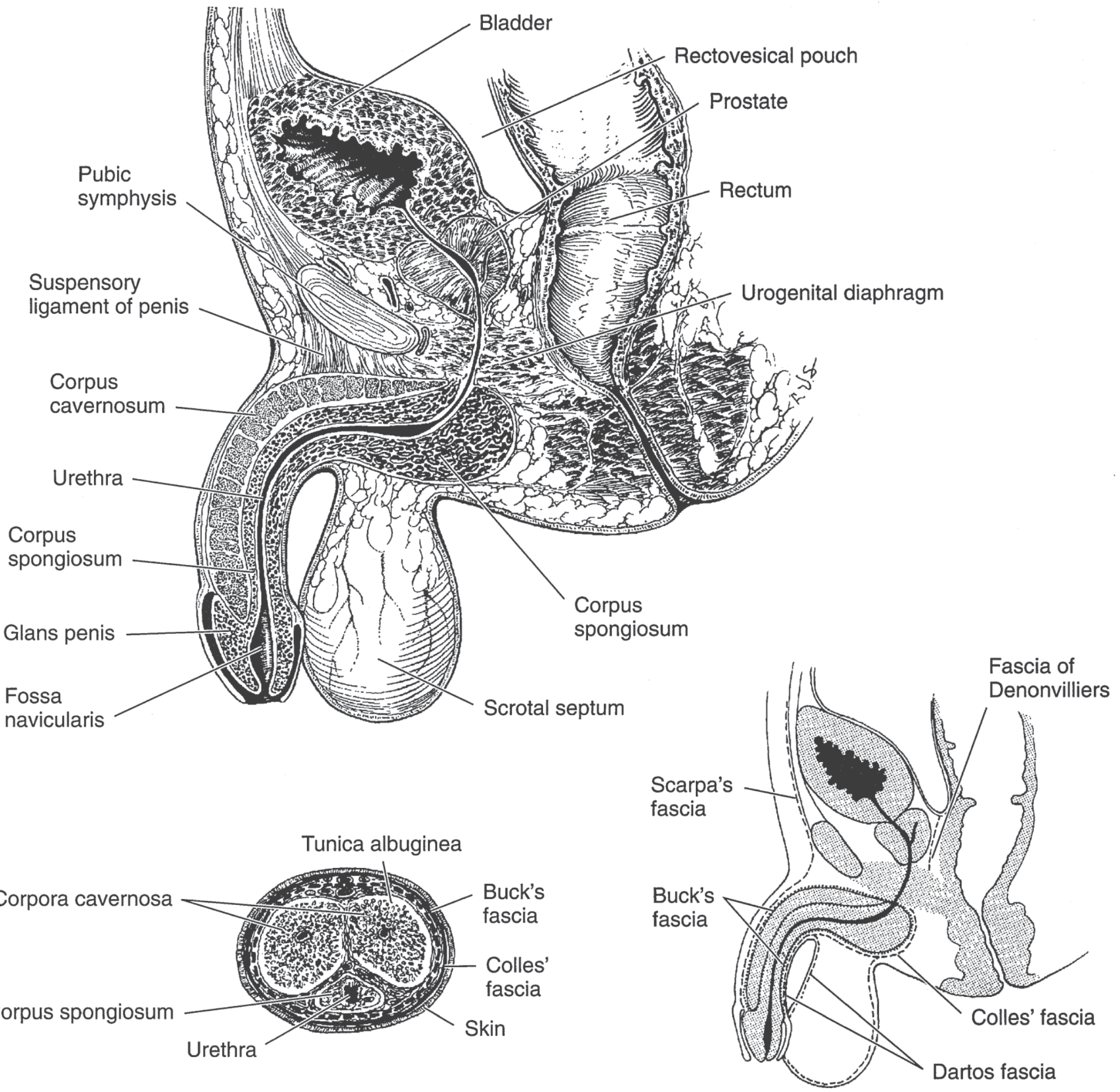
Fig. 1-8: Male pelvic anatomy - sagittal and cross-sectional views (Smith & Tanagho)
Basic Facts
| Feature | Detail |
|---|
| Weight | ~20 g (normal) |
| Prostatic urethra | 2.5 cm long (within the gland) |
| Support (anterior) | Puboprostatic ligaments |
| Support (inferior) | Urogenital diaphragm |
| Posterior covering | Denonvilliers' fascia (separates from rectum) |
| DRE landmark | Posterior surface palpable rectally |
Zones of the Prostate - "McNeal's Zones" (Pathology uses this)
PERIPHERAL ZONE (70% of glandular tissue)
└── Posterior and lateral
└── SITE OF MOST PROSTATE CANCERS (70-80%)
└── Palpable on DRE
CENTRAL ZONE (25%)
└── Surrounds ejaculatory ducts
└── Resistant to cancer
TRANSITIONAL ZONE (5% at baseline, but grows with BPH!)
└── Surrounds proximal prostatic urethra
└── SITE OF BPH (Benign Prostatic Hyperplasia)
ANTERIOR FIBROMUSCULAR ZONE
└── No glands, pure stroma
Mnemonic: "PCT - Prostate Cancer is Peripheral, Central is cool, Transition grows BPH"
Lobar Classification (Used clinically/cystoscopically)
5 lobes: Anterior, Posterior, Median, Right Lateral, Left Lateral
- Median lobe enlargement in BPH causes bladder outlet obstruction
Urethra Within the Prostate
VERUMONTANUM (colliculus seminalis):
├── On FLOOR of prostatic urethra
├── Ejaculatory ducts open HERE
└── Landmark for TUR surgery (stop here to protect sphincter!)
Above verumontanum → bladder neck (involuntary smooth sphincter)
Below verumontanum → membranous urethra → external sphincter
Blood Supply
- Branches of inferior vesical artery (from internal iliac)
- Venous: Periprostatic plexus of Santorini → internal iliac veins
- Lymphatics: Internal iliac + obturator lymph nodes
6. MALE URETHRA - "The 4-Part Tube"
PROSTATIC URETHRA (3-4 cm)
└── Runs through prostate
└── Verumontanum on posterior wall
└── Ejaculatory ducts open at verumontanum
└── Lined: Transitional epithelium → Pseudostratified columnar
MEMBRANOUS URETHRA (1-2 cm) ← SHORTEST, LEAST DISTENSIBLE
└── Traverses urogenital diaphragm
└── External urethral sphincter here (voluntary, striated)
└── MOST vulnerable in pelvic fractures!
BULBAR URETHRA (part of spongy/penile)
└── Surrounded by corpus spongiosum bulb
└── Cowper's (bulbourethral) gland ducts enter here
PENILE (SPONGY) URETHRA (15 cm)
└── In corpus spongiosum
└── Fossa navicularis at glans (dilated portion)
└── External urethral meatus = NARROWEST normal point
└── Glands of Littre (mucus glands in submucosa)
Mnemonic: "Pretty Men Buy Penises" - Prostatic, Membranous, Bulbar, Penile
Clinical: Sites of Stricture
#1 Bulbar urethra (most common stricture site - straddle injury)
#2 Penile urethra (instrumentation, gonorrhea)
#3 Membranous urethra (pelvic fracture)
7. PENIS - "The 3-Cylinder Engine"
Gross Anatomy
CROSS-SECTION of PENIS (dorsal to ventral):
[Dorsal]
Dorsal arteries & deep dorsal vein
┌────────────────────────────────┐
│ Corpus Cavernosum │ Corpus │ ← paired, dorsolateral
│ (tunica albuginea)│ Cavernosum│
└────────────────────────────────┘
← Buck's fascia (deep) →
┌─────────────────────────────────┐
│ Corpus Spongiosum │ ← single, ventral
│ (contains urethra) │
└─────────────────────────────────┘
← Colles' fascia (superficial) →
Skin (no subcutaneous fat!)
[Ventral]
Fascial Layers (Know for surgery & trauma!)
| Layer | Location | Continuous With |
|---|
| Tunica albuginea | Around each corpus | Unique to penis |
| Buck's fascia | Around all 3 corpora together | Colles' fascia proximally |
| Colles' fascia | Under penile skin | Scarpa's fascia (abdomen), Dartos (scrotum) |
Why this matters clinically: In urethral rupture below Buck's fascia, urine/blood tracks along Colles' fascia - into scrotum, perineum, and up anterior abdominal wall (but NOT into thighs because Colles' attaches to fascia lata)!
Root of the Penis
Proximal attachments:
├── Corpora cavernosa → attached to ischial tuberosities as CRURA
│ └── Covered by ischiocavernosus muscles
└── Corpus spongiosum → connected to urogenital diaphragm as BULB
└── Covered by bulbospongiosus muscle
Suspensory ligament of penis:
Arises from linea alba + pubic symphysis
Inserts into fascial covering of corpora cavernosa
Blood Supply
Internal Pudendal Artery (from internal iliac)
└── Perineal artery
└── Cavernous artery of penis → supplies CORPORA CAVERNOSA
└── Dorsal artery of penis → glans and prepuce
└── Bulbourethral artery → corpus spongiosum + glans + urethra
VENOUS DRAINAGE:
Superficial dorsal vein → external to Buck's fascia → saphenous vein
Deep dorsal vein → under Buck's fascia, between dorsal arteries
→ drains into periprostatic venous plexus (Santorini)
Cavernous veins → internal pudendal vein
Nerve Supply (Erection vs. Ejaculation!)
ERECTION: Parasympathetic (S2-S4) via pelvic plexus and cavernous nerve
+ NANC (VIP, NO pathways) → major mediator of corporal smooth muscle relaxation
EMISSION: Sympathetic (T10-L2) → smooth muscle contraction → emission
EJACULATION: Somatic (pudendal nerve S2-S4) → bulbospongiosus contraction
SENSATION: Dorsal nerve of penis → pudendal nerve → S2-S4
8. SEMINAL VESICLES, VAS DEFERENS & EJACULATORY DUCTS
SEMINAL VESICLES:
Location: Cephalic to prostate, under bladder base
Size: ~6 cm long, very soft
Histology: Pseudostratified mucosa, dense connective tissue, thin muscle
Function: 60-70% of seminal fluid (fructose, prostaglandins)
Relations: Ureters medial, rectum posterior, bladder superior
VAS DEFERENS:
├── Originates from tail of epididymis
├── Travels in spermatic cord through inguinal canal
├── Loops over ureter (anterior) at internal inguinal ring
│ → "Vas crosses OVER ureter then sweeps medial"
├── Joins seminal vesicle to form:
└── EJACULATORY DUCT → enters prostate → opens at VERUMONTANUM
EJACULATORY DUCTS:
Length: ~2 cm
Course: Posterior → oblique → floor of prostatic urethra
Open at: Verumontanum (colliculus seminalis)
9. TESTIS & EPIDIDYMIS
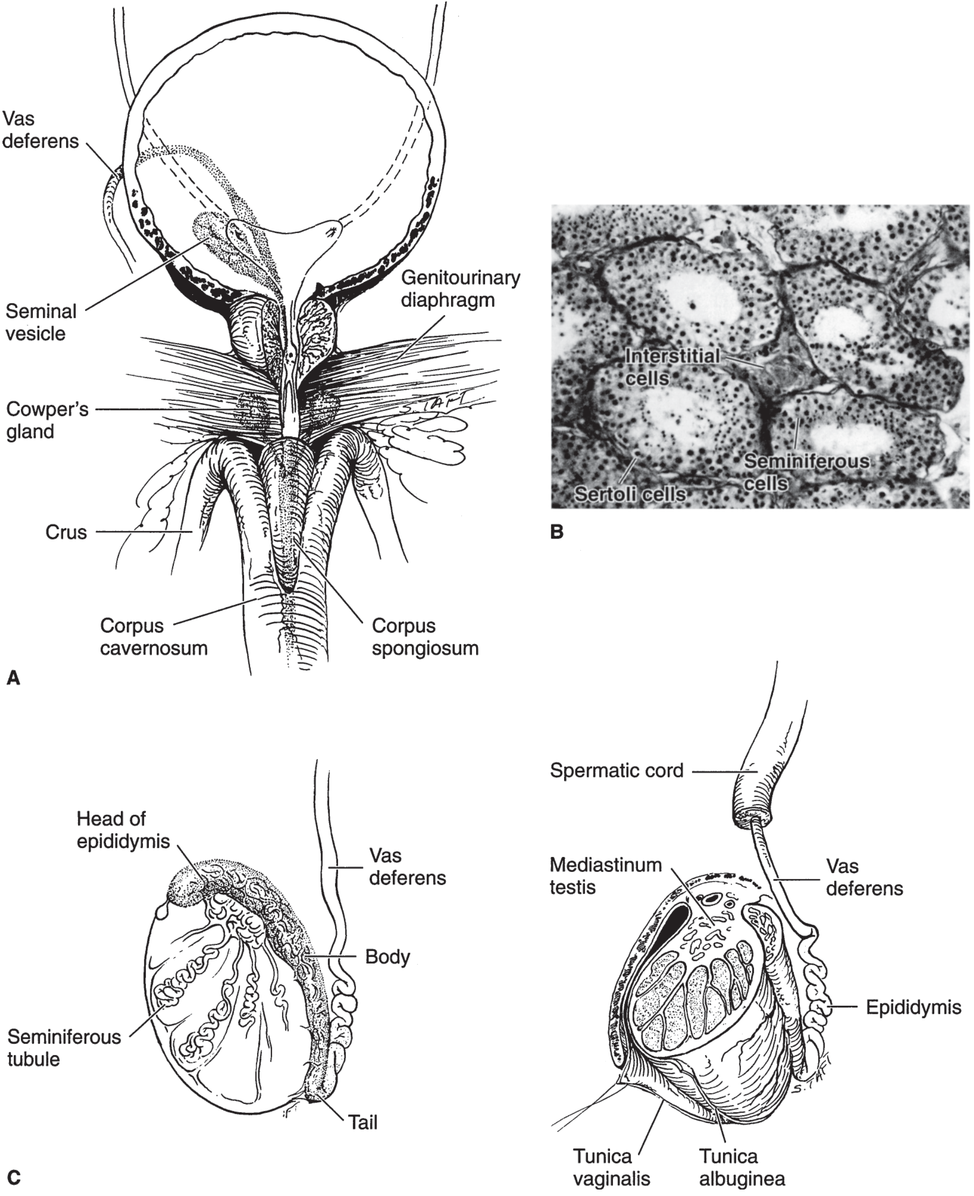
Fig. 1-7: Bladder/prostate relations (A), testis histology (B), testis and epididymis anatomy (C) - Smith & Tanagho
Testis
| Feature | Measurement |
|---|
| Dimensions | 4 × 3 × 2.5 cm |
| Volume formula | L × W × H × 0.71 |
| Average volume | 18 mL (range 12-30 mL) |
Coverings (Outside → In)
Skin
Dartos fascia (smooth muscle)
External spermatic fascia (from external oblique aponeurosis)
Cremasteric fascia + muscle (from internal oblique)
Internal spermatic fascia (from transversalis fascia)
Parietal layer of tunica vaginalis
← small fluid-filled space (hydrocele here when too much) →
Visceral layer of tunica vaginalis
TUNICA ALBUGINEA (dense fibrous capsule)
└── Mediastinum testis (posterior invagination)
└── Fibrous septa → 250 lobules
Mnemonic: "Some Damn Englishmen Call It The Testes"
= Skin, Dartos, External spermatic, Cremaster, Internal spermatic, Tunica vaginalis (parietal), Tunica vaginalis (visceral), Tunica albuginea
Histology
Each lobule: 1-4 seminiferous tubules (each ~60 cm long!)
└── Basement membrane
├── SERTOLI cells (supporting = "Nurse cells")
└── SPERMATOGENIC cells (various stages)
Between tubules:
└── Interstitium with LEYDIG cells (testosterone production)
Blood Supply - "Same origin as kidneys - embryologic memory"
ARTERIES: Testicular arteries (internal spermatic)
→ arise from AORTA just BELOW renal arteries
(mirrors embryologic origin from mesonephros = same level as kidneys!)
→ anastomose with deferential artery (from internal iliac)
VENOUS: Pampiniform plexus → Spermatic vein
RIGHT spermatic vein → IVC (just below right renal vein)
LEFT spermatic vein → LEFT RENAL VEIN (90° angle = more back pressure!)
└── This is why VARICOCELE is 90% LEFT-SIDED!
LYMPHATICS: Para-aortic/lumbar nodes (NOT inguinal!)
└── Except scrotal skin → inguinal nodes
Key surgical insight: If you violate the scrotal skin and then do orchiectomy (as in testicular cancer), the lymphatic drainage shifts to inguinal nodes - you must now address both para-aortic AND inguinal nodal fields!
Epididymis
HEAD (Globus Major) - upper pole
→ Efferent ductules from testis enter here (~12-15 ducts)
BODY
→ Single highly coiled duct (~6 meters when uncoiled!)
→ Sperm maturation occurs here
TAIL (Globus Minor) - lower pole
→ Becomes VAS DEFERENS
Appendix epididymis = vestigial remnant of mesonephric duct
(can TORT and present like torsion!)
10. SPERMATIC CORD
Contents - "VAST COLLECTION"
V - Vas deferens
A - Arteries (internal spermatic, external spermatic, deferential)
S - Spermatic veins (pampiniform plexus)
T - Testicular lymphatics
C - Cremasteric muscle fibers
O - ? (nerve: Genital branch of genitofemoral + sympathetics)
L - Lymph vessels
L - ?
E - ?
C - ?
T - Tunica vaginalis (processus)
I - ?
O - ?
N - Nerves (ilioinguinal, genitofemoral genital branch)
Real contents summary: Vas deferens + 3 arteries (internal spermatic/testicular, external spermatic, deferential) + pampiniform plexus + lymphatics + nerves (genitofemoral genital branch, sympathetic fibers) + remnant of processus vaginalis
Course: Internal inguinal ring → inguinal canal → external inguinal ring → scrotum
11. SCROTUM
LAYERS (outside → in):
1. Skin (rugose/corrugated, pigmented)
2. Dartos muscle (smooth muscle - no fat; responds to temperature!)
3. External spermatic fascia
4. Cremasteric fascia + muscle
5. Internal spermatic fascia
6. Tunica vaginalis (parietal layer)
--- fluid space ---
7. Tunica vaginalis (visceral layer on testis)
BLOOD SUPPLY:
Arteries: Femoral + internal pudendal + inferior epigastric branches
Veins: Paired with arteries
Lymphatics: SUPERFICIAL INGUINAL nodes (NOT para-aortic!)
SEPTUM: Divides scrotum into 2 compartments
└── Reflected as raphe on skin surface
12. FEMALE UROLOGY ANATOMY
Female Urethra - "Short & Straight = More UTIs"
Length: 4 cm (vs 20 cm in males)
Course: Under pubic symphysis, anterior to vagina
Meatus: Between clitoris and vaginal introitus
LAYERS:
Outer striated muscle (rhabdosphincter)
Inner smooth muscle
Mucosa: Squamous → transitional epithelium
SPHINCTER:
NO anatomic internal sphincter equivalent in females!
(unlike males)
External sphincter = rhabdosphincter around urethra
+ Pelvic floor muscles
RELATIONS:
Anterior: Pubic symphysis
Posterior: Anterior vaginal wall (closely adherent!)
→ Urethrocele if supports fail → stress incontinence
Female Pelvic Supports - Crucial for Urologic Function
LEVEL I: Cardinal + Uterosacral ligaments (apex support)
LEVEL II: Arcus tendineus fascia pelvis (lateral bladder/vaginal wall)
LEVEL III: Perineal body + external sphincter (outlet)
Failure of Level II → Cystocele (bladder prolapse into vagina)
→ Anterior vaginal wall bulge
→ Can cause stress urinary incontinence
Failure of Level I → Vault/uterine prolapse
Failure of Level III → Gaping introitus, perineal descent
13. COMPLETE LYMPHATIC DRAINAGE SUMMARY
ORGAN → LYMPH NODE DRAINAGE
─────────────────────────────────────────────────────
Kidney + Ureter (upper) → Para-aortic (lumbar) nodes
Ureter (middle/lower) → Iliac nodes
Bladder → Vesical → External/Internal iliac → Common iliac
Prostate → Internal iliac + Obturator nodes
Seminal vesicles → Same as prostate
Testes/Ovaries → Para-aortic (lumbar) nodes [T10 level]
Epididymis → External iliac nodes
Penis (skin) → Superficial inguinal → deep inguinal
Penis (glans) → Deep inguinal → external iliac
Scrotum (skin) → Superficial inguinal nodes
CRITICAL EXAM POINT: Testicular cancer spreads to para-aortic nodes (because embryologic origin = retroperitoneum). But if the scrotum is involved or previous scrotal surgery has been performed, inguinal node dissection is also required!
14. COMPLETE NERVE SUPPLY SUMMARY
ORGAN SYMPATHETIC (T) PARASYMPATHETIC (S) SOMATIC (pudendal)
───────────────────────────────────────────────────────────────────────────────
Kidneys/Ureter T10-L1 S2-S4 (minor) -
Bladder body T11-L2 (relaxes) S2-S4 (contracts) -
Bladder neck T10-L2 (contracts) - -
Urethra EUS - - S2-S4
Prostate T10-L2 S2-S4 -
Penis (erect.) - S2-S4 -
Penis (ejacu.) T10-L2 - S2-S4
Testes T10 (pain) - -
Why testicular pain refers to the T10 dermatome (umbilicus)? Because testes originated retroperitoneally at the level of the kidneys and descended - they kept their T10 nerve supply. So a stone in the ureter or torsion of the testis can both cause periumbilical/flank pain!
15. FASCIAL PLANES - The Surgeon's GPS
GEROTA'S FASCIA (Perirenal fascia):
→ Encloses kidney + adrenal + perirenal fat
→ Open in partial nephrectomy, renal surgery
DENONVILLIERS' FASCIA:
→ Between prostate/seminal vesicles and rectum
→ Serosal remnant of peritoneal cul-de-sac
→ Dissect ANTERIOR to it in radical prostatectomy to protect rectum
BUCK'S FASCIA:
→ Deep fascia of penis enclosing all 3 corpora
→ Rupture of corpus cavernosum = blood contained within Buck's → "eggplant deformity"
→ Urethral rupture below Buck's → blood/urine into Colles' fascia territory
SCARPA'S FASCIA:
→ Abdominal membranous fascia
→ Continuous with Colles' (perineum) and Dartos (scrotum)
ENDOPELVIC FASCIA:
→ Covers pelvic organs, important in radical prostatectomy
16. MASTER MEMORY TABLES
"The 3s of Urology Anatomy"
| What | The "3" |
|---|
| Arteries to adrenal | 3 (phrenic, aorta, renal) |
| Ureteric narrowings | 3 (UPJ, iliac vessels, UVJ) |
| Ureteral blood supply zones | 3 (renal, gonadal, vesical) |
| Urethral segments in males | 4 (but "3 surgical" = prostatic, membranous, spongy) |
| Prostate zones (McNeal) | 4 (peripheral, central, transitional, fibromuscular) |
| Corpora of penis | 3 (2 cavernosa + 1 spongiosum) |
| Arteries to penis (from pudendal) | 3 (cavernous, dorsal, bulbourethral) |
Key Measurements to Memorize
| Structure | Measurement |
|---|
| Kidney length | 10-12 cm |
| Kidney weight (M) | 125-170 g |
| Ureter length | 30 cm |
| Ureteral descent per inspiration | 4-5 cm |
| Prostatic urethra | 2.5 cm |
| Membranous urethra | 1-2 cm (shortest!) |
| Penile urethra | ~15 cm |
| Male total urethra | ~20 cm |
| Female urethra | ~4 cm |
| Seminal vesicle length | ~6 cm |
| Testis volume | 18 mL (12-30 mL) |
| Adrenal weight | 4-5 g |
| Prostate weight | ~20 g |
🧠 FINAL MEGA-MNEMONIC FLOWCHART
URINE JOURNEY: "Kidneys Make Urine, Ureters Move It, Bladder Stores It, Urethra Expels It"
AORTA
↓
RENAL ARTERIES (at L1-L2)
↓
KIDNEYS (T12-L3, retroperitoneal, psoas border)
↓ glomerular filtration → tubular processing
URINE → Minor Calyces (8-12) → Major Calyces (2-3) → Renal Pelvis
↓
URETERS (30 cm, 3 narrowings = stone traps: UPJ, Iliac, UVJ)
↓ ureteral jets every 2-3 seconds
BLADDER (detrusor + trigone, capacity 400-500 mL)
↓ micturition (S2-S4 parasympathetics)
URETHRA: Male = 20 cm (Prostatic→Membranous→Bulbar→Penile)
Female = 4 cm (short, straight, simple)
↓
EXTERNAL MEATUS
Sources:
- Smith & Tanagho's General Urology, 19th Edition - Chapter 1: Anatomy of the Genitourinary Tract (Tanagho & Lue)
- Campbell-Walsh-Wein Urology, 3-Volume Set - Chapter 110: Lower Urinary Tract Anatomy
- Brenner & Rector's The Kidney, 2-Volume Set - Chapter 2: Anatomy of the Kidney (Verlander & Clapp)