Veins of head and neck
Reading File
Finding Sources
Finding Sources
Reading File
Reading File
Finding Sources
Reading File
Reading File
Reading File
Finding Sources
Loading Image
Loading Image
Reading File
Loading Image
Reading File
I have comprehensive content to compile a thorough answer. Let me now present it.
Veins of the Head and Neck
The venous drainage of the head and neck is organized into three main systems: superficial veins of the neck, the deep internal jugular system, and the intracranial dural venous sinuses.
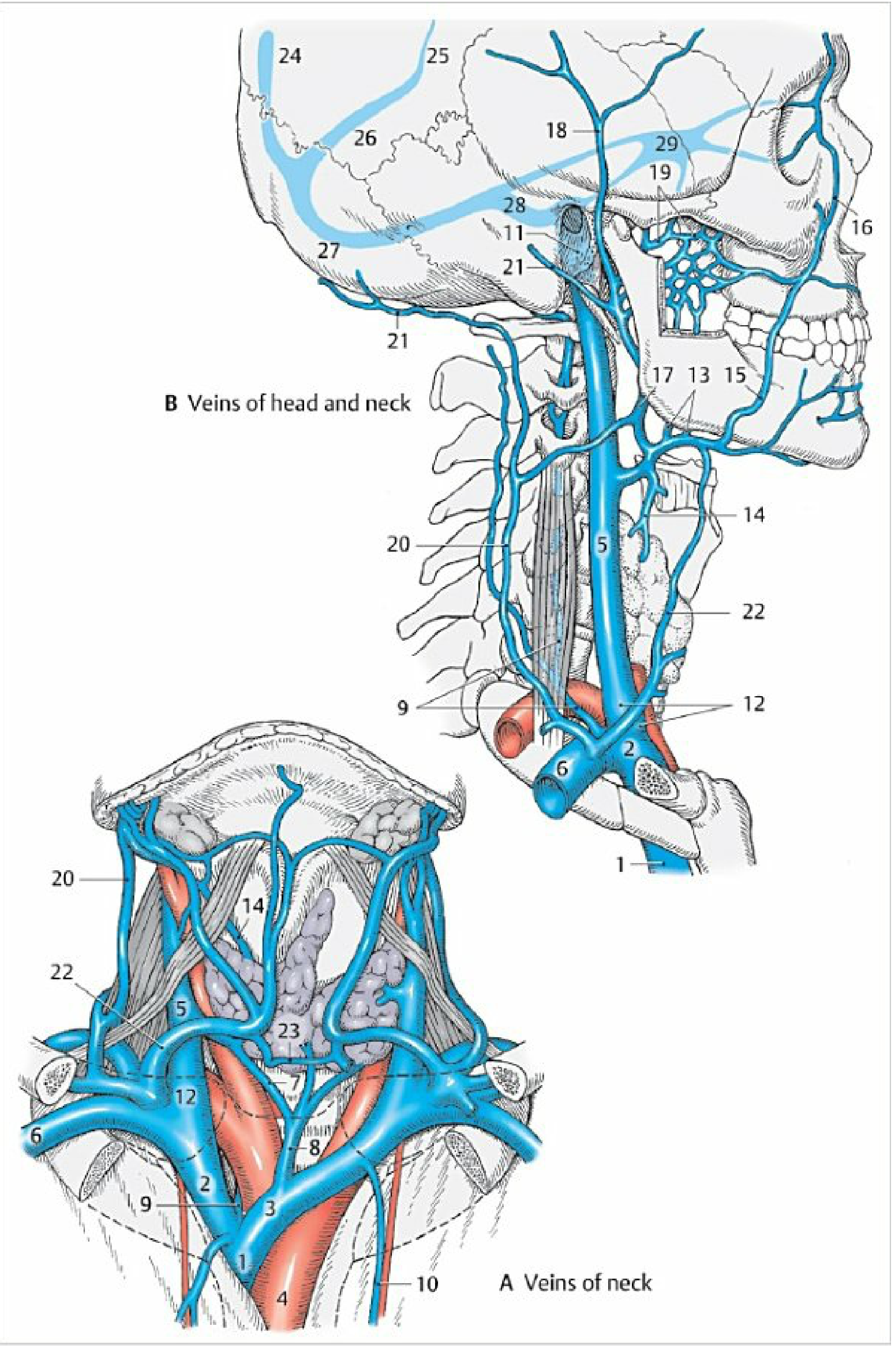
1. Superficial Venous Drainage of the Neck
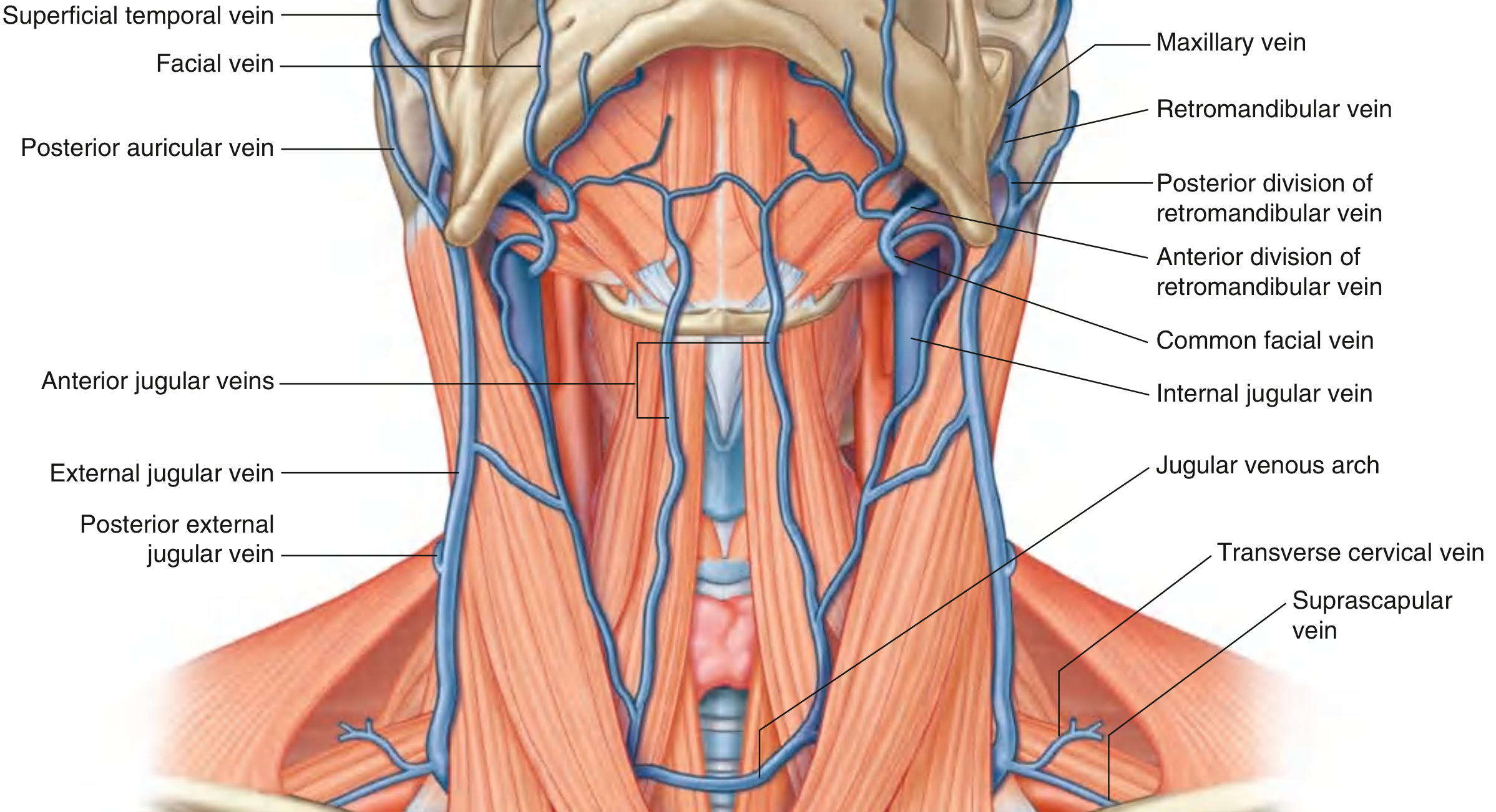
External Jugular Vein (EJV)
The EJV is formed posterior to the angle of the mandible by the union of:
- Posterior auricular vein — drains the scalp behind and above the ear
- Posterior division of the retromandibular vein
Course: Passes straight down the neck in the superficial fascia, lying superficial to the sternocleidomastoid (SCM) muscle and crossing it diagonally. Inferiorly, just above the clavicle and posterior to the SCM, it pierces the investing layer of cervical fascia and enters the subclavian vein.
Tributaries of the EJV:
- Posterior external jugular vein (superficial back of neck)
- Transverse cervical vein
- Suprascapular vein
- Anterior jugular vein (occasionally)
Retromandibular Vein
Formed in the parotid gland by union of the superficial temporal and maxillary veins. Descends to the angle of the mandible, then divides:
- Posterior division → joins posterior auricular vein → forms External Jugular Vein
- Anterior division → joins facial vein → forms Common Facial Vein → drains into Internal Jugular Vein
Anterior Jugular Vein (AJV)
Variable and inconsistent. Usually forms near or just superior to the hyoid bone from small anterior neck veins. Each vein descends on either side of the midline, then pierces the investing fascia near the medial SCM attachment to enter the subclavian vein (or occasionally the EJV).
The two AJVs frequently communicate via the jugular venous arch in the suprasternal notch region.
2. Internal Jugular Vein (IJV)
The IJV is the chief vein draining the cranial cavity, head, and neck.
Origin: Begins at the jugular foramen as a dilated continuation of the sigmoid sinus — this dilatation is the superior bulb of the jugular vein. It exits the skull with the glossopharyngeal [IX], vagus [X], and accessory [XI] nerves.
Course: Travels within the carotid sheath, initially posterior to the internal carotid artery, then lateral to the common carotid artery, with the vagus nerve [X] lying posterior between them.
Termination: Joins the subclavian vein posterior to the sternal end of the clavicle to form the brachiocephalic vein. Just before this junction it expands as the inferior bulb of the jugular vein.
Tributaries of the IJV
| Tributary | Region Drained |
|---|---|
| Inferior petrosal sinus | Intracranial (cavernous sinus) |
| Facial vein (via common facial) | Face, scalp |
| Lingual vein | Tongue |
| Pharyngeal veins | Pharyngeal plexus |
| Superior thyroid vein (+ superior laryngeal vein) | Thyroid, larynx |
| Middle thyroid vein | Thyroid |
| Occipital vein | Occipital scalp |
| Sternocleidomastoid vein | SCM muscle |
| Meningeal veins | Dura mater |
The facial vein begins at the medial angle of the eye as the angular vein, which anastomoses with the ophthalmic vein (providing a communication between the face and the cavernous sinus). It receives tributaries from the face and the pterygoid plexus (via the retromandibular vein).
Clinical note — Jugular venous pulse (JVP): The IJV provides an important clinical window into right heart function. The venous pressure and waveform reflected in the IJV allow assessment of right atrial and ventricular pressure.
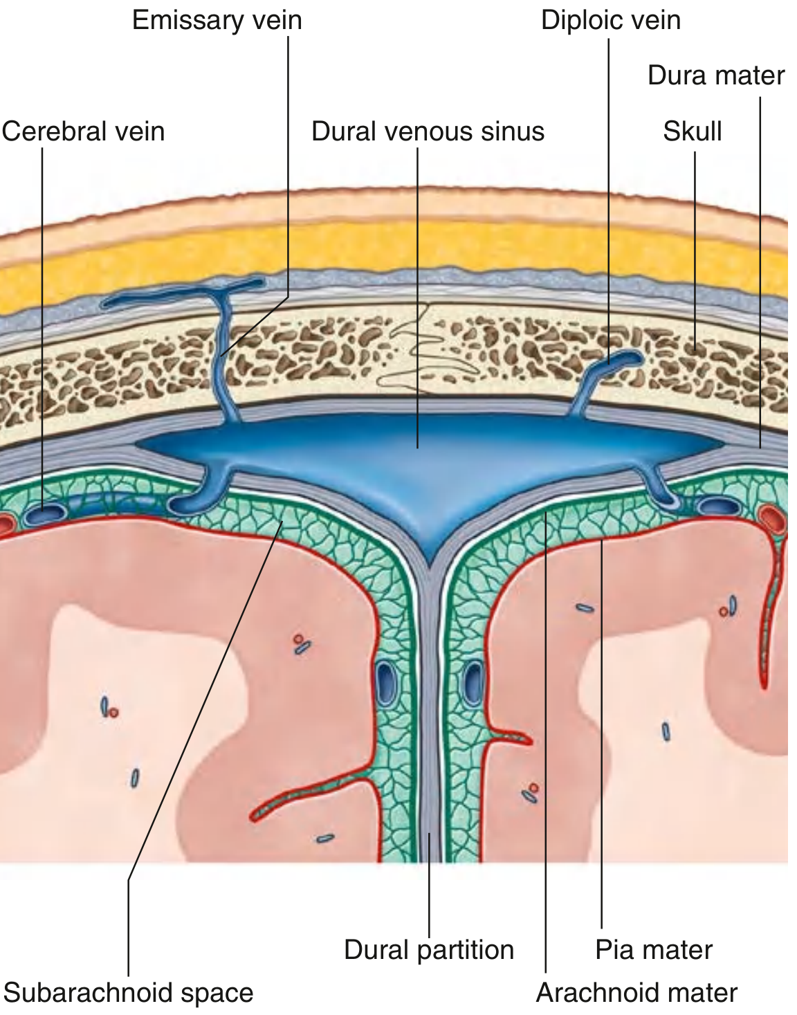
3. Dural Venous Sinuses
These are endothelium-lined, valveless channels with rigid walls formed by cranial periosteum and dura mater. They drain the brain into the IJV.
Major Dural Sinuses
| Sinus | Location | Drains Into |
|---|---|---|
| Superior sagittal sinus | Superior border of falx cerebri | Confluence of sinuses |
| Inferior sagittal sinus | Inferior margin of falx cerebri | Straight sinus |
| Straight sinus | Junction of falx cerebri and tentorium cerebelli | Confluence of sinuses; receives great cerebral vein |
| Occipital sinus | Falx cerebelli against occipital bone | Confluence of sinuses |
| Confluence of sinuses | Internal occipital protuberance | Transverse sinuses (bilateral) |
| Transverse sinus | Posterior attachment of tentorium cerebelli | Sigmoid sinus |
| Sigmoid sinus | Groove of parietal, temporal, occipital bones | Internal jugular vein (at jugular foramen) |
| Cavernous sinus (paired) | Lateral to sella turcica/pituitary | Inferior petrosal sinus → IJV; Superior petrosal sinus → sigmoid sinus |
| Sphenoparietal sinus (paired) | Inferior surface of lesser wing of sphenoid | Cavernous sinus |
| Superior petrosal sinus | Superior margin of petrous temporal | Cavernous sinus → sigmoid sinus |
| Inferior petrosal sinus | Groove between petrous temporal and occipital | Cavernous sinus → IJV directly |
| Basilar plexus | Clivus | Connects cavernous sinus to marginal sinus |
| Marginal sinus | Encircles foramen magnum | Connects dural sinuses to vertebral venous plexuses |
Cavernous Sinus — Key Relations
Structures within the cavernous sinus: internal carotid artery, abducent nerve [VI].
Structures in the lateral wall: oculomotor [III], trochlear [IV], ophthalmic [V1], maxillary [V2] nerves.
Communicates with:
- Angular vein (face) via the superior ophthalmic vein — a clinically important route for spread of facial infections intracranially
- Superior sagittal sinus via sphenoparietal sinus
- Contralateral cavernous sinus via intercavernous sinuses
- IJV via inferior petrosal sinus
- Sigmoid sinus via superior petrosal sinus
4. Additional Drainage Pathways
Diploic Veins
Lie within the spongy diploe of cranial bones. Communicate with both dural venous sinuses and superficial scalp veins. Four groups:
- Frontal, anterior temporal, posterior temporal, occipital diploic veins
Emissary Veins
Pass through preformed cranial openings, providing direct connections between dural sinuses and extracranial veins (valveless — flow can reverse):
| Emissary Vein | Connects |
|---|---|
| Parietal emissary | Superior sagittal sinus ↔ superficial temporal vein |
| Mastoid emissary | Sigmoid sinus ↔ occipital vein |
| Condylar emissary | Sigmoid sinus ↔ external vertebral venous plexus |
| Occipital emissary | Confluence of sinuses ↔ occipital vein |
| Venous plexus of foramen ovale | Cavernous sinus ↔ pterygoid plexus |
Cerebral Veins
- Superficial cerebral veins — drain cortex directly into dural sinuses
- Deep cerebral veins — drain deep structures via the great cerebral vein (of Galen) into the straight sinus
Overview Diagram
Summary of Drainage Flow
Scalp/Brain
↓
Dural Venous Sinuses (Superior Sagittal → Straight/Transverse → Sigmoid)
↓
Internal Jugular Vein (at Jugular Foramen)
↓ ← also receives: facial, lingual, pharyngeal, thyroid veins
Brachiocephalic Vein (IJV + Subclavian)
Face/Scalp (superficial)
↓
Retromandibular Vein → External Jugular Vein → Subclavian Vein
Sources: Gray's Anatomy for Students (9780323934237), pp. 1143–1144, 1152–1153, 1024; Color Atlas of Human Anatomy Vol. 2 (9783132424487), pp. 131–137.
This is a shared conversation. Sign in to Orris to start your own chat.