
A 14 year old male child presented in OPD with complaints of with recurrent unprovoked nasal bleed. He also had unilateral cheek swelling and proptosis. Anterior rhinoscopy shows lobulated, pink to purplish, smooth surfaced mass. 1. What is the diagnosis? (1) 2. Name two radiological signs associated with the same.(1)
juvenile nasopharyngeal angiofibroma CT scan Holman Miller sign
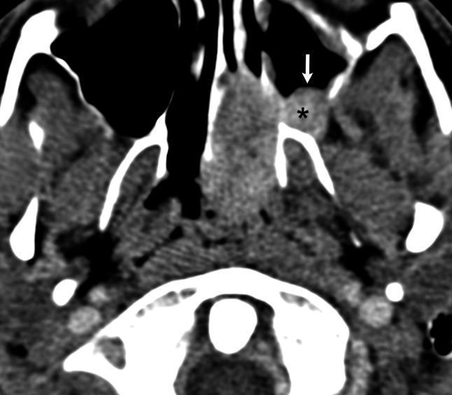
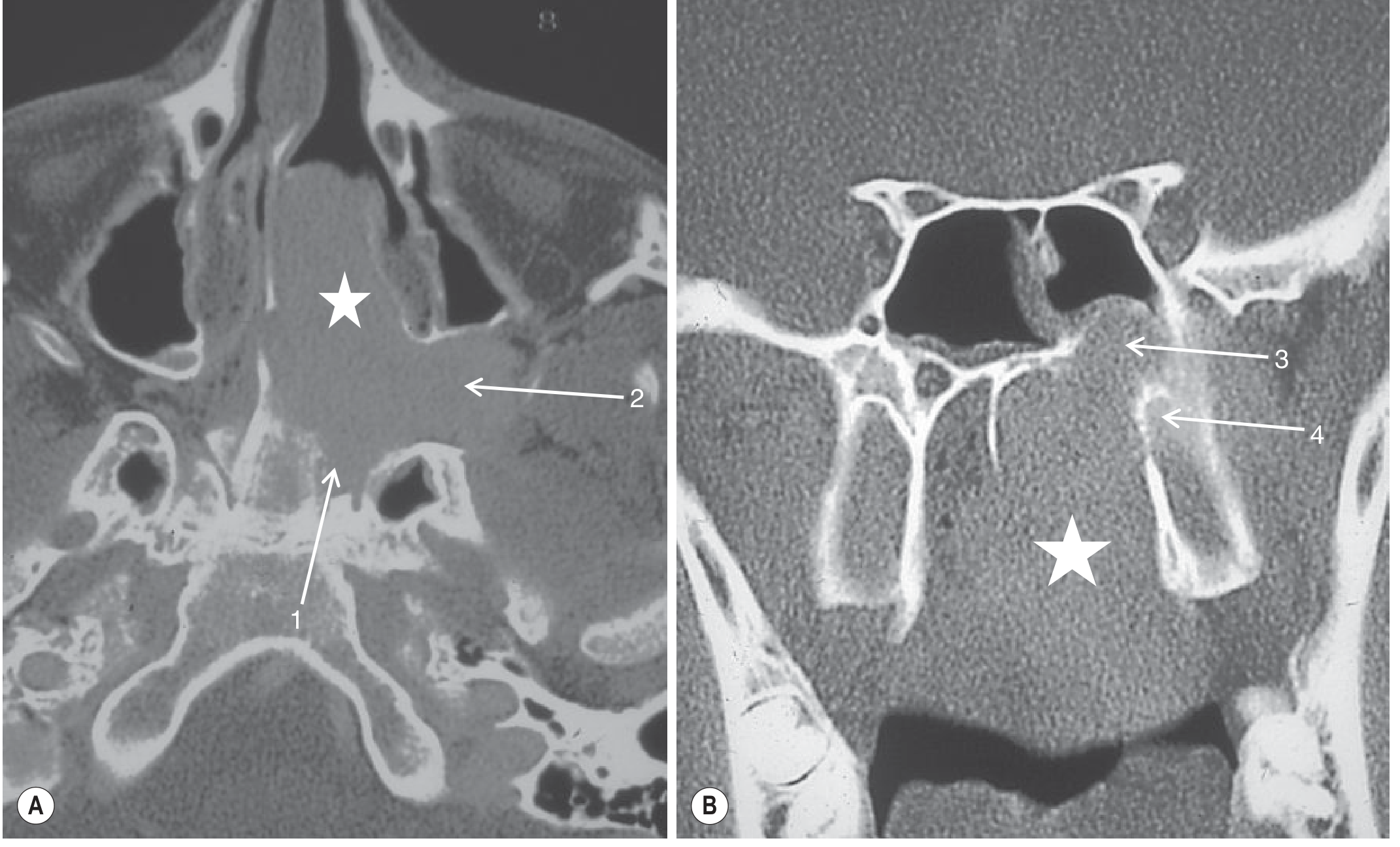
This diagnostic image is an axial contrast-enhanced CT scan of the maxillofacial region and skull base. The scan demonstrates characteristic features of a juvenile nasopharyngeal angiofibroma (JNA) with significant local extension. A prominent soft tissue mass of intermediate density is visible, originating in the nasopharynx and extending laterally. A key diagnostic feature shown is the 'Holman-Miller sign,' characterized by the anterior (forward) displacement and bowing of the posterior wall of the maxillary sinus, indicated by a white arrow. The tumor has also invaded the pterygopalatine fossa, marked by an asterisk, appearing as an area of abnormal soft tissue density with contrast enhancement. This lateral spread pattern often precedes further extension into the infratemporal fossa. The imaging modality highlights the relationship between the hypervascular tumor and surrounding bony structures, illustrating bone remodeling and displacement rather than aggressive destruction. This material is suitable for teaching radiological signs of benign but locally invasive sinonasal tumors.
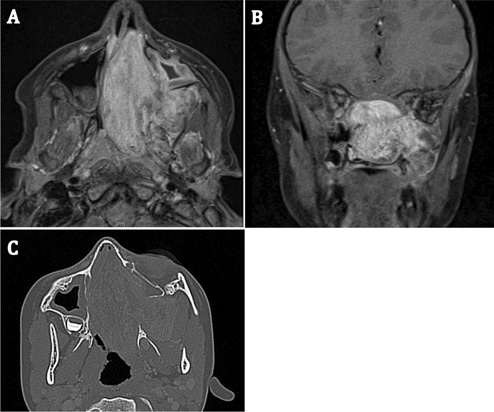
This composite diagnostic image displays a Juvenile Nasopharyngeal Angiofibroma (JNA) in a 7-year-old patient using three radiological views. Panels A (axial) and B (coronal) are MRI scans showing a large, heterogeneously enhancing mass that occupies the nasal cavity and extends into the left pterygopalatine fossa. The mass exhibits irregular boundaries and mixed signal intensity, characteristic of this hypervascular tumor. Panel C provides an axial CT scan (bone window), illustrating the tumor's impact on local anatomy, including bone remodeling, significant widening of the pterygopalatine fossa (Holman-Miller sign), and erosion of adjacent bony structures. The images demonstrate the typical local aggressiveness and spread patterns of JNA into the paranasal sinuses and skull base regions. These findings are critical for preoperative staging and surgical planning, particularly for endoscopic trans-pterygoid approaches.
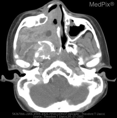
**Imaging Modality:** Axial Computed Tomography (CT) scan with soft tissue window settings. **Anatomical Region:** Skull base and paranasal sinuses, specifically focusing on the nasopharynx and the pterygopalatine fossa. **Observed Pathology:** Large, heterogeneous soft tissue mass consistent with Juvenile Nasopharyngeal Angiofibroma (JNA). The lesion is centered in the right nasopharyngeal region with significant aggressive extension. **Characteristic Visual Features:** * **Expansion and Destruction:** The mass demonstrates aggressive local behavior, characterized by the destruction of the posterior wall of the right maxillary sinus and the pterygoid plates. * **Infiltration:** The tumor fills the right nasal cavity and extends laterally into the right infratemporal fossa and pterygopalatine fossa. * **Displacement:** There is marked opacification of the right maxillary sinus and deviation of the nasal septum to the left. * **Bony Hallmarks:** The image displays a widening of the sphenopalatine foramen, a classic radiologic indicator for JNA. **Key Diagnostic Features:** The combination of a highly vascular-appearing soft tissue mass in the nasopharynx of a young patient, widening of the sphenopalatine foramen, and anterior bowing or destruction of the posterior maxillary sinus wall (Holman-Miller sign) are definitive features for this entity.
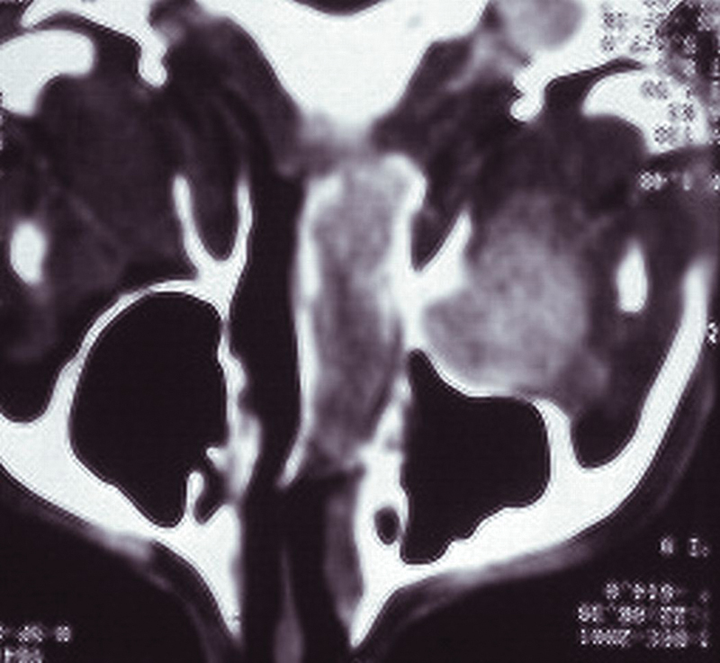
**Imaging Modality:** Axial computed tomography (CT) scan of the midface and paranasal sinuses. **Anatomical Region:** Maxillofacial region, specifically the right retromaxillary space, pterygopalatine fossa, and nasal cavity. **Observed Pathology:** An expansive, soft-tissue density mass is localized to the right retromaxillary and infratemporal regions. The lesion demonstrates significant mass effect, resulting in the characteristic anterior bowing of the posterior wall of the right maxillary sinus (Holman-Miller sign). There is total obliteration and opacification of the right nasal cavity with a marked leftward deviation of the nasal septum. **Characteristic Visual Features:** * **Bone Remodeling:** Smooth, pressure-induced anterior displacement of the posterior maxillary cortex rather than aggressive destruction. * **Expansion:** The mass extends medially through the sphenopalatine foramen area into the nasopharynx and nasal fossa. * **Density:** Heterogeneous soft-tissue attenuation. **Key Diagnostic Features:** The combination of a retromaxillary mass with the Holman-Miller sign and nasal cavity extension is highly characteristic of a juvenile nasopharyngeal angiofibroma (JNA) or similar slow-growing, benign but locally aggressive neoplasm.
1. Diagnosis: Juvenile Nasopharyngeal Angiofibroma (JNA)
- Adolescent male (14 years old)
- Recurrent unprovoked epistaxis (often profuse)
- Unilateral nasal mass - lobulated, pink-to-purplish, smooth surface on anterior rhinoscopy
- Unilateral cheek swelling - from lateral extension through the pterygomaxillary fissure into the infratemporal fossa, pushing the posterior wall of the maxillary sinus forward
- Proptosis - from orbital extension
2. Two Radiological Signs
Sign 1: Holman-Miller Sign (Antral Sign)
- Anterior bowing / forward displacement of the posterior wall of the maxillary antrum (sinus)
- Seen on CT (axial view) and plain X-ray (lateral view)
- Caused by the tumor expanding anteriorly from the pterygopalatine fossa, pushing the posterior maxillary wall forward
- This is the most classic and eponymous radiological sign of JNA
Sign 2: Widening of the Pterygopalatine Fossa (PPF)
- Seen on CT (axial and coronal views)
- The tumor's epicenter in the PPF causes erosion and widening of the pterygopalatine fossa and the sphenopalatine foramen
- On coronal CT, this appears as an expanded soft-tissue density mass at the PPF with erosion of the medial pterygoid plate and the vidian canal aperture
| Sign | Radiological Finding | Modality |
|---|---|---|
| Holman-Miller sign | Anterior bowing of posterior wall of maxillary sinus | CT/X-ray lateral view |
| Widening of pterygopalatine fossa | Expansion and erosion of PPF / sphenopalatine foramen | CT axial/coronal |
- Cummings Otolaryngology Head and Neck Surgery, Chapter 50 (Juvenile Angiofibroma)
- Grainger & Allison's Diagnostic Radiology, Fig. 61.45
Child with ear pain, fever, irritability; otoscopy shows congested, bulging tympanic membrane with reduced Mobillity what diagnosis
acute otitis media bulging tympanic membrane otoscopy
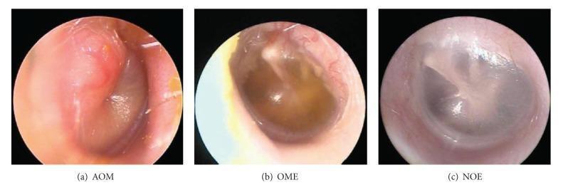
This clinical comparison image, captured via otoscopy/endoscopy, presents three distinct diagnostic states of the tympanic membrane (TM) to illustrate otitis media classification. Image (a) depicts Acute Otitis Media (AOM), characterized by a significantly erythematous, opaque, and bulging TM, which obscures the malleus and other anatomical landmarks. Image (b) shows Otitis Media with Effusion (OME), demonstrating a semi-opaque TM with an amber-colored fluid level or sterile effusion behind it; the membrane is less inflamed than in AOM but lacks normal translucency. Image (c) represents No Effusion (NOE), displaying a healthy, translucent, pearly-gray TM with clearly visible anatomical landmarks, including the lateral process and handle of the malleus. This visual aid serves as a diagnostic tool for distinguishing bacterial infection (AOM) from sterile fluid collection (OME) and normal middle ear status (NOE) based on variations in color, position (bulging vs. neutral), and opacification. Educational focus includes otolaryngology physical examination findings and the clinical signs of middle ear inflammation.
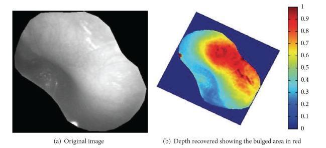
This medical figure illustrates the computational analysis of a tympanic membrane for diagnosing acute otitis media (AOM). (a) A grayscale clinical photograph of a tympanic membrane obtained via otoscopy shows a smooth, opaque surface with subtle brightness variations and a specular highlight from the otoscope's light source. (b) A corresponding depth map generated using shape-from-shading techniques. Depth information is visualized via a pseudocolor gradient (ranging from blue for low depth to red for high depth/bulging). A prominent red region in the upper and central portions of the membrane indicates significant bulging, a primary diagnostic indicator of AOM. This comparative visualization demonstrates how 2D image data can be transformed into 3D topographical information to assist in the automated classification of middle-ear inflammation and the identification of pathological protrusions.
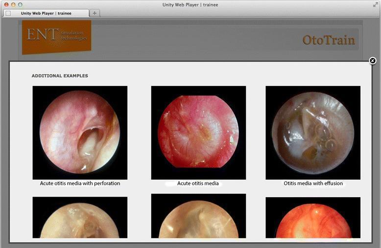
A comparison chart of otoscopic diagnostic images from a medical simulator, focusing on pathologies of the tympanic membrane (TM). The visual contains three labeled primary circular otoscopic views: 1) 'Acute otitis media with perforation,' showing an inflamed, erythematous TM with a large, irregular central perforation through which the middle ear space is visible; 2) 'Acute otitis media,' depicting an intensely hyperemic, opaque, and bulging TM with loss of normal landmarks and light reflex; and 3) 'Otitis media with effusion,' illustrating a translucent, amber-colored TM with visible air-fluid levels and bubbles in the middle ear space. These clinical photographs serve as educational examples for ENT trainees to differentiate between infectious stages (AOM) and non-purulent fluid accumulation (OME). The interface includes secondary supportive images below each category to demonstrate variability in presentation. This content is designed for medical students and residents to master diagnostic otoscopy and middle ear pathology recognition.
Diagnosis: Acute Otitis Media (AOM)
Why This Diagnosis?
| Feature | Significance |
|---|---|
| Ear pain (otalgia) | Ear-specific symptom of middle ear inflammation |
| Fever | Systemic sign of acute infection |
| Irritability | Common nonspecific symptom, especially in children |
| Congested, bulging tympanic membrane | The defining otoscopic sign of AOM - distinct bulging indicates middle ear effusion under pressure |
| Reduced mobility of TM | Assessed by pneumatic otoscopy; reduced mobility confirms middle ear effusion (MEE) - an essential prerequisite for AOM diagnosis |
"AOM is an acute infection characterized by distinct bulging of the tympanic membrane that is often accompanied by rapid onset of signs and symptoms of middle ear inflammation... signs include bulging or fullness of the TM, TM erythema, and acute onset of otorrhoea." - Cummings Otolaryngology
Otoscopic Findings - Visual Reference
Diagnostic Criteria (AAP Guidelines)
- Moderate-to-severe bulging of the tympanic membrane
- Mild bulging + signs of acute inflammation (erythema or otalgia)
- New-onset otorrhoea (not due to otitis externa)
Pathophysiology (Why it Happens in Children)
- Shorter (13 mm vs 35 mm in adults)
- More horizontal (~10° vs 45° in adults)
- Floppier and wider
Common Causative Organisms
- Streptococcus pneumoniae - most common (pre-vaccination era, now decreasing with PCV13)
- Non-typeable Haemophilus influenzae - increasingly common
- Moraxella catarrhalis
Treatment (Summary)
| Age / Severity | First-line Treatment |
|---|---|
| < 2 years or severe symptoms (any age) | High-dose Amoxicillin × 10 days |
| 2-5 years | High-dose Amoxicillin × 7 days |
| ≥ 5 years | High-dose Amoxicillin × 5 days |
| Amoxicillin in past 30 days | Amoxicillin-clavulanate |
| Treatment failure at 48-72 hrs | Amoxicillin-clavulanate × 10 days OR IM Ceftriaxone |
Key Differentials to Exclude
| Condition | Distinguishing Feature |
|---|---|
| Otitis Media with Effusion (OME / Glue ear) | No acute symptoms; TM retracted/dull but NOT bulging; NO fever |
| Otitis Externa | Tragus tenderness; canal swollen/inflamed; TM normal |
| Myringitis | TM erythema without effusion or bulging |
- Cummings Otolaryngology, Chapter 199 (AOM Definition, Symptoms, and Signs)
- Harriet Lane Handbook, 23rd ed. (Pediatric Infections - AOM)