
how to identify gluten intolerance
Gluten intolerance (also called non-celiac gluten sensitivity) can be tricky to identify because it overlaps with celiac disease and irritable bowel syndrome. The main clues are GI symptoms (bloating, diarrhea, abdominal pain) that improve when gluten is removed from the diet.
Searching sources...
Reading File
Searching the Web
how to identify gluten intolerance diagnosis 2025
Finding Sources
Searching PubMed
"non-celiac gluten sensitivity" diagnosis
Finding Sources
Reading File
Reading File
Now let me fetch the diagnostic flowchart image and read more of the clinical section:
Loading Image
Reading File
I now have comprehensive information to give a thorough answer. Here it is:
How to Identify Gluten Intolerance
The term "gluten intolerance" is commonly used to describe two distinct conditions that need to be distinguished from each other:
- Celiac disease - an autoimmune disorder causing intestinal damage
- Non-celiac gluten sensitivity (NCGS) - a sensitivity without autoimmune markers or intestinal damage
Step 1: Recognizing the Symptoms
Both conditions share overlapping symptoms triggered by eating wheat, rye, or barley:
GI symptoms:
- Chronic diarrhea (present in 27-50% of celiac patients)
- Abdominal bloating and pain
- Steatorrhea (fatty stools)
- Weight loss (6-22% of cases)
Non-GI / "non-classical" symptoms (now actually more common than classical GI symptoms):
- Unexplained iron-deficiency anemia
- Fatigue
- Osteoporosis / osteopenia (due to vitamin D deficiency and secondary hyperparathyroidism)
- Aphthous mouth ulcers
- Abnormal liver biochemical tests
- Skin rash (dermatitis herpetiformis - itchy blistering rash, strongly associated with gluten sensitivity)
- Infertility
Symptoms usually appear between ages 10-40, affect women ~1.5x more than men, and resolve within weeks to months on a gluten-free diet.
- Symptom to Diagnosis: An Evidence Based Guide, 4th Ed., p. 598-599
Step 2: Blood Tests (Serology)
These should be done while the patient is still eating gluten - going gluten-free before testing causes false negatives.
| Test | Sensitivity | Specificity | Notes |
|---|---|---|---|
| IgA tTG (tissue transglutaminase) | 90-98% | 95-97% | First-line; LR+ ~24, LR- 0.06 |
| IgA EMA (endomysial antibody) | Lower than tTG | Very high (~100%) | More operator-dependent |
| IgA/IgG DGP (deaminated gliadin peptide) | Moderate | High | Useful alternative |
| Total IgA level | - | - | Must check - IgA deficiency causes false negatives |
Key point: Negative EMA and tTG together make celiac disease very unlikely (negative predictive value ~99%). However, positive results alone are not enough to confirm celiac disease - biopsy is still needed (positive predictive value only 29-76% due to low overall prevalence).
- Symptom to Diagnosis: An Evidence Based Guide, 4th Ed., p. 598
Step 3: Endoscopy and Small Bowel Biopsy (Gold Standard)
A biopsy of the duodenum/proximal small intestine remains the definitive test for celiac disease. It looks for:
- Villus atrophy (flattening of intestinal villi)
- Crypt hyperplasia
- Increased intraepithelial lymphocytes
This is the Marsh classification. Full villus atrophy confirms celiac disease.
Step 4: HLA Genetic Typing (if needed)
- Virtually 100% of celiac disease patients carry HLA-DQ2 or HLA-DQ8 haplotypes
- LR+ = 2.3, LR- = 0 (so negative HLA virtually rules out celiac disease)
- Useful when patients have already started a gluten-free diet before testing (antibodies drop, biopsy may be normal)
- Limitation: 25-30% of the general Caucasian population also carry these genes without having celiac disease
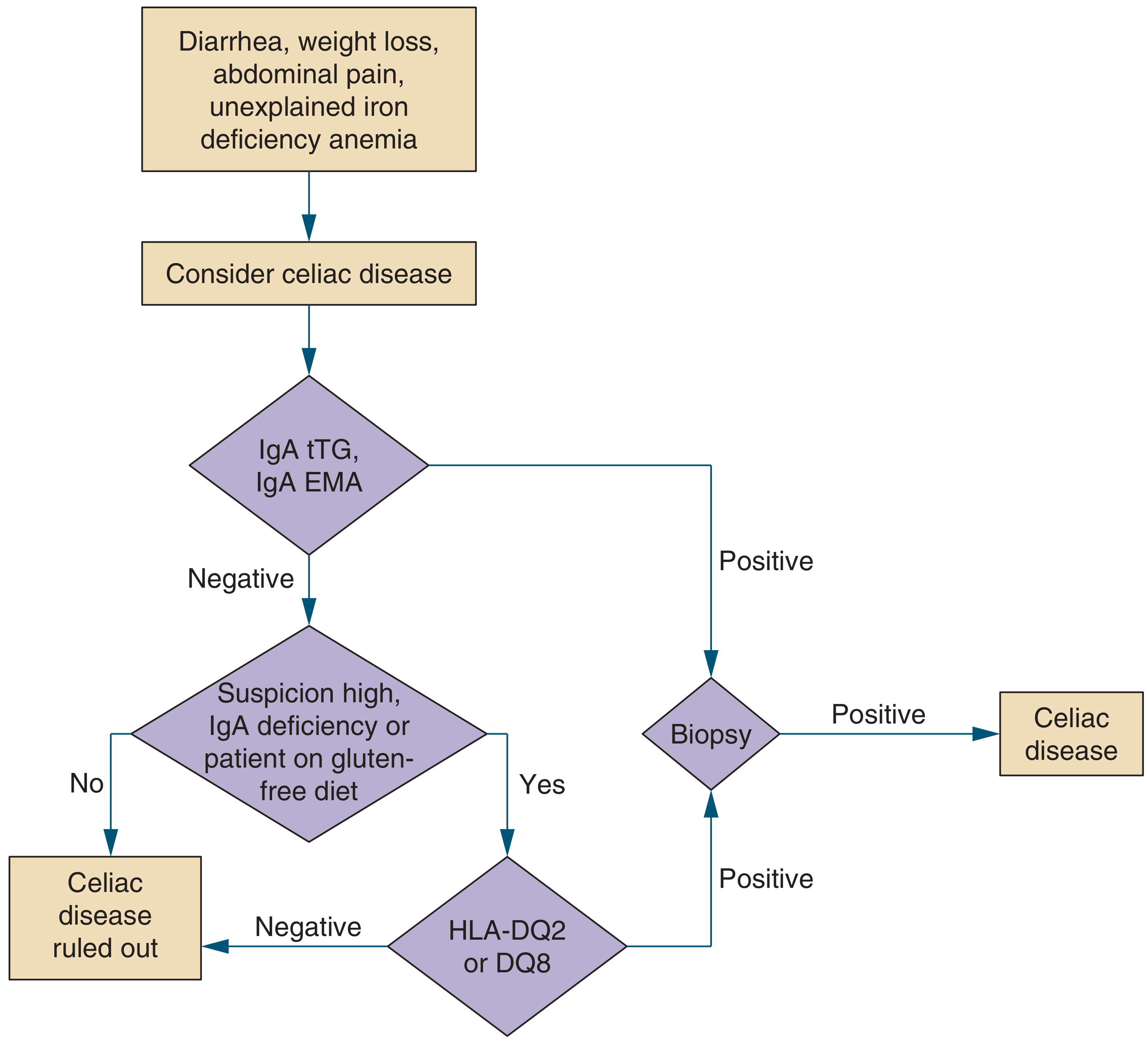
Diagnostic Flowchart
Here is the evidence-based diagnostic pathway:
Step 5: Identifying NCGS (Non-Celiac Gluten Sensitivity)
NCGS is a diagnosis of exclusion. According to Cleveland Clinic guidelines, it requires:
- Negative blood tests for celiac disease (IgA tTG, IgA EMA)
- Negative wheat allergy test (IgE-mediated)
- Negative duodenal biopsy (no villus atrophy)
- Symptomatic improvement on a gluten-free diet
As stated in Sleisenger & Fordtran's GI and Liver Disease: "Celiac disease and nonceliac gluten sensitivity usually cannot be differentiated based on history alone." A gluten challenge (reintroducing gluten after exclusion) is sometimes used to confirm the diagnosis.
Gluten Challenge (Confirmatory Step)
If a patient has already started a gluten-free diet, testing becomes unreliable. A gluten challenge means:
- Eating ~3g of gluten/day for 2-6 weeks before re-testing serology or repeating biopsy
- This helps distinguish celiac disease from NCGS, as symptom improvement on GFD alone cannot reliably differentiate the two
Summary Table: Celiac vs. NCGS
| Feature | Celiac Disease | NCGS |
|---|---|---|
| Autoimmune? | Yes | No |
| Antibodies (tTG, EMA) | Positive | Negative |
| Intestinal villus atrophy | Yes | No |
| HLA-DQ2/DQ8 | Present | May or may not be present |
| Diagnosis method | Serology + biopsy | Rule out celiac + symptom response |
| Risk of malignancy | Elevated | Not established |
Emerging Test (2025)
A new IL-2 blood test developed at the Walter and Eliza Hall Institute (published in Gastroenterology, 2025) can detect celiac disease with ~90% sensitivity and 97% specificity even in people already on a gluten-free diet, by measuring interleukin-2 spike after in-vitro gluten exposure. This may eventually replace the need for gluten challenge testing. (Celiac Disease Foundation)
Bottom line: Always rule out celiac disease with serology (IgA tTG + total IgA) and biopsy before labeling someone as "gluten intolerant." Never go gluten-free before completing testing - it invalidates the results.
treatment
The cornerstone of treatment for both celiac disease and non-celiac gluten sensitivity is a strict, lifelong gluten-free diet. Nutritional deficiencies are corrected, and in refractory cases, immunosuppressants may be needed.
Searching sources...
Finding Sources
Searching PubMed
"celiac disease" treatment management
Searching the Web
gluten intolerance celiac disease treatment guidelines 2025
Reading File
Reading File
Reading File
I now have everything needed for a thorough, evidence-based treatment answer.
Treatment of Gluten Intolerance (Celiac Disease & NCGS)
1. The Gluten-Free Diet (GFD) - The Only Proven Treatment
A strict, lifelong gluten-free diet is the sole effective treatment for both celiac disease and non-celiac gluten sensitivity (NCGS). There are no approved medications that replace it.
What to completely eliminate:
| Grain | Toxic protein | Hidden sources |
|---|---|---|
| Wheat (incl. spelt, kamut, triticale) | Gliadin | Bread, pasta, crackers, cereal, baking mixes, soy sauce, soups, gravies, salad dressings |
| Rye | Secalin | Rye bread, some beers |
| Barley | Hordein | Malt, barley water, many beers, some soups |
Important: "Wheat-free" does NOT mean "gluten-free" - a product can be wheat-free but still contain rye or barley.
What is safe to eat:
-
Rice, corn, quinoa, millet, buckwheat, sorghum, teff, amaranth
-
Potatoes, legumes, fruits, vegetables, meat, fish, eggs, dairy
-
Oats - acceptable only if certified uncontaminated (pure, gluten-free oats); regular oats are frequently cross-contaminated during processing
-
Yamada's Textbook of Gastroenterology, 7th Ed., p. 1206
2. Essential Steps in Management (NIH Consensus Framework)
According to NIH Consensus guidelines cited in Yamada's Textbook of Gastroenterology:
- Consultation with a skilled dietitian (at diagnosis and yearly thereafter)
- Patient education about the disease and label reading
- Lifelong adherence to a gluten-free diet
- Identification and correction of nutritional deficiencies
- Access to an advocacy/support group
- Continuous long-term follow-up by a multidisciplinary team
3. Correcting Nutritional Deficiencies
Celiac disease impairs absorption from the small intestine. At diagnosis, check and correct:
| Nutrient | Why deficient | Supplement/action |
|---|---|---|
| Iron | Poor duodenal absorption | Iron supplementation; recheck |
| Folic acid (B9) | Small bowel malabsorption | Supplement |
| Vitamin B12 | Ileal involvement | Supplement or IM if severe |
| Vitamin D | Fat malabsorption | Supplement; monitor 25-OH VitD |
| Calcium | Vitamin D deficiency + malabsorption | Supplement + dietary calcium |
| Zinc, Magnesium | General malabsorption | Supplement if deficient |
Note: Supplementation should be targeted to documented deficiencies - routine blanket supplementation is no longer universally recommended (2025 updated guidelines).
- Symptom to Diagnosis: Evidence Based Guide, 4th Ed., p. 599
4. Monitoring and Follow-Up
Do not assume improvement equals cure - ongoing monitoring is essential:
- 3-6 months after diagnosis: physician review, symptom check, celiac serology (IgA tTG)
- 1 year: repeat serology, nutritional labs (CBC, iron, thyroid, liver enzymes, 25-OH Vitamin D)
- DEXA bone scan within 1 year of diagnosis (osteoporosis is a common complication)
- Repeat duodenal biopsy at 2-3 years to confirm mucosal healing (serology alone is poorly sensitive to persistent villous atrophy)
- Annual follow-up indefinitely thereafter
Rising antibody titers on follow-up = dietary non-compliance.
- Yamada's Textbook of Gastroenterology, 7th Ed., p. 1207
5. Managing Persistent Symptoms Despite GFD
The most common reason for treatment failure is inadvertent gluten exposure - through:
- Hidden gluten in processed foods, medications, supplements
- Cross-contamination at restaurants or shared kitchens
- Mislabeled products
If symptoms persist after 6-12 months of strict GFD, consider:
| Condition | Action |
|---|---|
| Inadvertent gluten ingestion | Repeat dietitian consultation, review labels, reduce restaurant meals |
| Lactose intolerance | Common co-morbidity; trial lactose-free diet |
| Fructose malabsorption | FODMAPs-restricted diet (only when needed, not routinely) |
| Small intestinal bacterial overgrowth (SIBO) | Test and treat with antibiotics |
| Microscopic colitis | Colonoscopy + biopsy |
| Irritable bowel syndrome (IBS) | Manage per IBS guidelines |
| Refractory celiac disease (RCD) | See below |
6. Refractory Celiac Disease (RCD)
Defined as ongoing villous atrophy despite strict GFD for >6-12 months, after excluding inadvertent gluten ingestion and other causes.
RCD Type 1 (normal intraepithelial lymphocytes):
- Budesonide or prednisolone (corticosteroids) - first-line
- Azathioprine (2-2.5 mg/kg/day) or mercaptopurine as steroid-sparing agents
- Nutritional support (often requires parenteral or enteral nutrition)
RCD Type 2 (abnormal clonal intraepithelial lymphocytes - high malignancy risk):
- More aggressive immunosuppression
- Close monitoring for enteropathy-associated T-cell lymphoma (EATL) - a rare but serious complication
- Specialist referral to a tertiary center
The 2026 British Society of Gastroenterology guidelines recommend azathioprine and mercaptopurine in RCD Type 1 with careful risk-benefit assessment.
7. Treatment for Non-Celiac Gluten Sensitivity (NCGS)
- Same dietary approach: remove gluten from the diet
- Symptoms typically resolve within days to weeks
- No biopsy healing to monitor (no villous atrophy)
- No established long-term complications
- Some patients eventually tolerate low levels of gluten; periodic re-evaluation is reasonable
- Per Harrison's Principles of Internal Medicine (2025): "Upon discontinuation of gluten, they have relief of abdominal pain, diarrhea, headaches/migraines, and other celiac disease-type symptoms."
8. Vaccinations
- Pneumococcal vaccine is recommended by some guidelines (celiac disease is associated with hyposplenism)
- Influenza vaccination is also advised in some guidelines
9. Emerging / Future Therapies (2025-2026)
Several non-dietary treatments are in clinical trials and may become adjuncts to the GFD in the coming years:
- Larazotide acetate - tight junction regulator to reduce intestinal permeability
- ALV003 / TAK-062 - oral proteases that digest gluten in the gut
- Nexvax2 - gluten peptide immunotherapy vaccine
- Anti-IL-15 monoclonal antibodies - for RCD Type 2
- IL-2 blood test (Novoleukin-C) for monitoring treatment response even in those already on GFD
The 2025 ESsCD guidelines (PMID: 40999951) represent the most current European framework for diagnosis and management.
Key takeaway: The GFD is not optional - it is the treatment. With strict adherence, most patients achieve complete symptomatic relief, mucosal healing, normalization of antibodies, and correction of nutritional deficiencies within 1-2 years. Lifelong monitoring is necessary to catch complications early.
This is a shared conversation. Sign in to Orris to start your own chat.