
Properties of receptors from guyton and hall for 1st mbbs 10 marks
Reading File
Now let me search the medical library for receptor properties from Guyton and Hall.
Finding Sources
Finding Sources
Finding Sources
Finding Sources
Reading File
Finding Sources
Reading File
Finding Sources
Reading File
Reading File
I now have all the key content from Guyton and Hall on receptor properties. Let me also fetch the receptor types classification from slightly earlier in the text and the image of types of sensory receptors.
Reading File
Loading Image
I now have all the content needed from Guyton and Hall Chapter 47 to write a comprehensive 10-mark answer on properties of receptors. Let me compile the full answer.
Properties of Receptors
(Guyton and Hall Textbook of Medical Physiology, Chapter 47)
1. Classification of Sensory Receptors
Guyton and Hall classifies receptors into five basic types based on the stimuli they detect:
| Type | Stimulus Detected | Examples |
|---|---|---|
| Mechanoreceptors | Mechanical compression/stretch | Pacinian corpuscle, Meissner's corpuscle, muscle spindle, baroreceptors |
| Thermoreceptors | Temperature changes | Cold receptors, warm receptors |
| Nociceptors | Physical or chemical tissue damage | Free nerve endings (pain) |
| Electromagnetic receptors | Light energy | Rods and cones of the retina |
| Chemoreceptors | Chemical stimuli | Taste buds, olfactory receptors, carotid body O2 receptors |
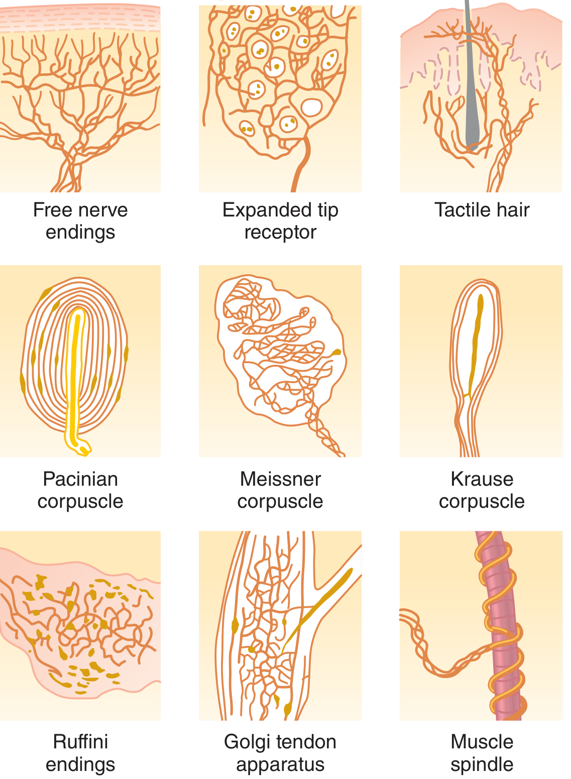
2. Differential Sensitivity (Specificity)
Each receptor is highly sensitive to one type of stimulus (its adequate stimulus) and almost nonresponsive to other stimuli. Examples:
- Rods and cones respond to light but not to heat, cold, or pressure.
- Osmoreceptors in the hypothalamus detect minute changes in osmolality but not sound.
- Pain receptors are not stimulated by normal touch but become active only when stimuli are intense enough to damage tissues.
This property is called differential sensitivity and is the basis for encoding specific sensory modalities.
3. Modality of Sensation and Neural Coding
Labeled Line Coding: Each sensory nerve fiber transmits only one modality of sensation, determined by where it terminates in the CNS. If a pain fiber is stimulated electrically, mechanically, or thermally, the person still perceives pain - because the fiber leads to pain areas of the brain.
Population Coding: In some cases, one cell can encode more than one sensory modality. Combinations of signals from many cells are needed for full sensory perception - for example, perceiving an object requires combining signals about color, size, contour, and texture from many photoreceptors.
4. Transduction of Sensory Stimuli into Nerve Impulses - Receptor Potential
What is a Receptor Potential?
All sensory receptors share one feature in common: whatever stimulus excites the receptor, its immediate effect is to change the membrane electrical potential of the receptor. This change is called the receptor potential.
Mechanisms of Generating a Receptor Potential
Receptors are excited in one of four ways:
- Mechanical deformation - stretches the receptor membrane and opens stretch-sensitive ion channels (e.g., Pacinian corpuscle)
- Chemical application - opens ion channels or binds to membrane receptors and activates second messenger systems
- Temperature change - alters activity of temperature-sensing ion channels and metabotropic receptors
- Electromagnetic radiation - e.g., light on retinal receptors, which changes membrane permeability directly or indirectly
In all cases, the basic mechanism is a change in membrane permeability, allowing ions to diffuse more or less readily and thereby altering the transmembrane potential.
Maximum Amplitude
The maximum amplitude of most sensory receptor potentials is approximately 100 mV, occurring at extremely high stimulus intensity (similar to action potential amplitude, corresponding to maximal sodium permeability).
Relationship to Action Potentials
When the receptor potential rises above the threshold, action potentials are generated in the attached nerve fiber. The greater the receptor potential above threshold, the higher the frequency of action potentials generated (frequency coding of stimulus intensity).
5. Receptor Potential of the Pacinian Corpuscle (Example)
The Pacinian corpuscle has a central unmyelinated nerve fiber core surrounded by concentric capsule layers. Compression of the outside deforms the central fiber, generating a receptor potential. The receptor potential is proportional (not linear) to stimulus strength - it increases in a logarithmic (compressive) fashion, allowing response across an extreme range from very weak to very intense stimuli.
6. Adaptation of Receptors
Definition: When a continuous sensory stimulus is applied, receptors respond at a high impulse rate initially, then progressively slower, until action potentials decrease to very few or none - this is adaptation.
Types Based on Rate of Adaptation
| Type | Also Called | Examples | Function |
|---|---|---|---|
| Rapidly adapting | Phasic receptors / Rate receptors / Movement receptors | Pacinian corpuscles (adapt in hundredths of a second), hair receptors (adapt in ~1 second) | Detect change and rate of change in stimulus; not useful for constant signals |
| Slowly adapting | Tonic receptors | Muscle spindles, joint capsule receptors, pain receptors, arterial baroreceptors, chemoreceptors of carotid/aortic bodies, macula of vestibular apparatus | Detect continuous, sustained stimulus strength |
Mechanisms of Adaptation (Pacinian Corpuscle as Example)
- Structural/viscoelastic mechanism: The corpuscle is a viscoelastic structure. When a distorting force is applied, the viscous component instantly transmits it to the central fiber, generating a receptor potential. Within hundredths of a second, fluid redistributes within the corpuscle, eliminating the deformation and the receptor potential - even though compression continues.
- Accommodation (electrical mechanism): The nerve fiber itself undergoes accommodation - progressive inactivation of sodium channels in the nerve fiber membrane. Sodium current flow through channels causes them to gradually close, reducing excitability.
These two mechanisms (structural readjustment + electrical accommodation) apply broadly to other mechanoreceptors as well.
Clinical Significance of Adaptation Types
- Tonic receptors (slowly adapting) transmit continuous information for hours or days - essential for maintaining awareness of body position, pain, blood pressure, and chemistry.
- Phasic receptors (rapidly adapting) have a predictive function - detecting rates of change allows the nervous system to anticipate future body positions. E.g., joint rate receptors during running allow the nervous system to predict where feet will land in the next fraction of a second, enabling anticipatory motor corrections. Loss of this function makes coordinated running impossible.
7. Intensity Discrimination (Weber-Fechner Relation)
The receptor potential does not increase linearly with stimulus strength. Instead, it follows a logarithmic relationship (consistent with the Weber-Fechner law of psychophysics). This logarithmic response compresses a wide dynamic range of stimulus intensities into a manageable range of receptor potentials, allowing the receptor to respond meaningfully from very weak to extremely strong stimuli.
Summary Table
| Property | Key Feature |
|---|---|
| Specificity / Differential sensitivity | Each receptor responds to its adequate stimulus only |
| Receptor potential | Common transduction event for all receptors; change in membrane permeability |
| Amplitude | Max ~100 mV; proportional (log) to stimulus strength |
| Frequency coding | Action potential frequency codes stimulus intensity |
| Adaptation | Rapid (phasic) vs. slow (tonic); structural + electrical mechanisms |
| Modality coding | Labeled line + population coding |
Source: Guyton and Hall Textbook of Medical Physiology, 14th edition, Chapter 47 - Sensory Receptors, Neuronal Circuits for Processing Information.
This is a shared conversation. Sign in to Orris to start your own chat.