
Intercoastal muscles easy diagram
Generating Image
Simple labeled anatomy diagram of intercostal muscles showing external intercostal, internal intercostal, and innermost intercostal muscle layers between two ribs, with clear arrows and labels, educational medical illustration style, clean white background
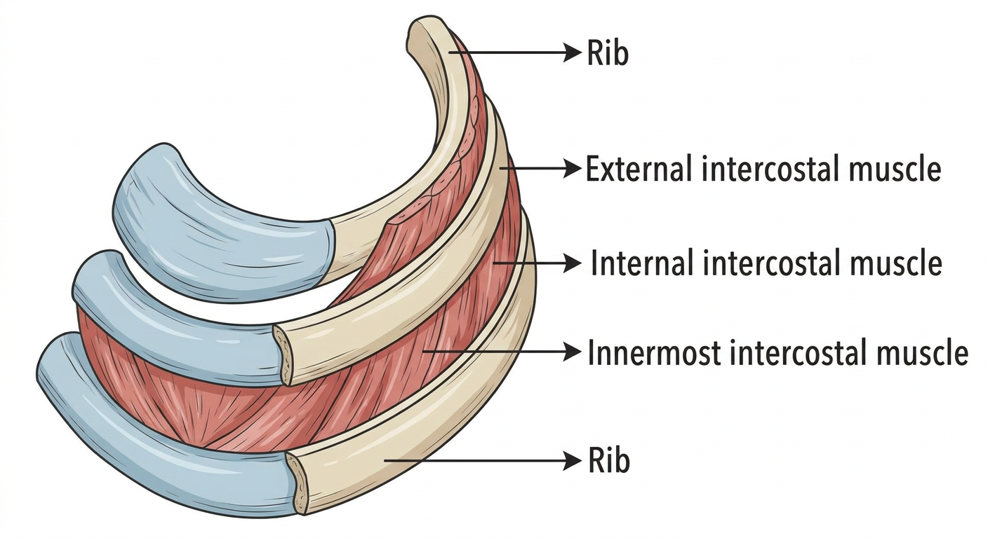
Here's a quick breakdown to go with the diagram:
3 Layers of Intercostal Muscles (outer to inner):
| Layer | Fiber Direction | Main Action |
|---|---|---|
| External intercostal | Downward & forward (like hands in pockets) | Elevates ribs - inspiration |
| Internal intercostal | Downward & backward (opposite to external) | Depresses ribs - forced expiration |
| Innermost intercostal | Same as internal | Assists internal intercostal |
Key points to remember:
- The intercostal neurovascular bundle (vein, artery, nerve - VAN from top to bottom) runs in the groove on the inferior border of each rib, between the internal and innermost layers.
- When inserting a needle (e.g., thoracocentesis), always go just above the upper border of the lower rib to avoid the bundle.
- External fibers are incomplete posteriorly (replaced by posterior intercostal membrane) and anteriorly by the anterior intercostal membrane.
This is a shared conversation. Sign in to Orris to start your own chat.