
What is Huntington disease
Reading File
Now I'll search the medical library and PubMed in parallel for Huntington disease.
Finding Sources
Searching PubMed
"Huntington disease"[MeSH Terms]
Good results. Now let me read the most comprehensive sections in parallel.
Reading File
Reading File
Reading File
Let me get the morphology section and management details.
Reading File
Reading File
Now let me fetch the key brain pathology image and read the Goldman-Cecil management section.
Loading Image
Reading File
Let me get the treatment/management section from Goldman-Cecil.
Reading File
Reading File
I now have comprehensive information to write a detailed answer. Let me compile it.
Huntington Disease (HD)
Definition & Epidemiology
Huntington disease is a fully penetrant autosomal dominant neurodegenerative disorder caused by an expanded CAG trinucleotide repeat in the HTT gene on chromosome 4p16.3, which encodes the protein huntingtin. It was first described by George Huntington in 1872.
- Prevalence: ~2.71–10 per 100,000 worldwide; up to 5.7/100,000 in Europeans, as low as 0.4/100,000 in Asians
- Mean age of onset: 45 years (range: 30–55 years); 5–10% have juvenile onset before age 20
- Course: Relentlessly progressive, leading to death over ~15 years
(Goldman-Cecil Medicine, p. 3972; Tietz Textbook of Laboratory Medicine, p. 2724)
Genetics & Molecular Pathogenesis
CAG Repeat Thresholds
| Allele Type | CAG Repeat Count | Clinical Significance |
|---|---|---|
| Normal | 11–34 | No disease |
| Intermediate | 27–35 | More behavioral abnormalities (apathy, suicidal ideation); unstable, can expand |
| Reduced penetrance | 36–39 | Variable; may cause disease |
| Full penetrance | ≥40 | Invariably causes clinical illness |
- Anticipation: Larger repeats cause earlier onset in successive generations. CAG repeats expand preferentially during spermatogenesis (paternal transmission), so 69% of father-child pairs show expansion vs. 32% of mother-child pairs. Juvenile HD is therefore usually paternally inherited.
- HTT gene has 67 exons and encodes a 3,144 amino acid, ~350 kDa protein. Huntingtin is involved in intracellular trafficking, cytoskeletal organization, endocytosis, and transcription regulation, and is required for neuronal development.
Disease Mechanism (Toxic Gain of Function)
The expanded polyglutamine tract in mutant huntingtin causes toxic gain of function. The mutant protein undergoes ubiquitination and proteolysis, yielding fragments that form intranuclear aggregates. These aggregates:
- Sequester transcription factors
- Disrupt protein degradation (ubiquitin-proteasome) pathways
- Perturb mitochondrial function
Mismatch repair gene variants (e.g., MSH3 p.Pro67Ala) can modify somatic CAG expansion and disease progression.
(Robbins Basic Pathology, p. 855; Tietz, p. 2725)
Neuropathology
On gross examination, there is striking atrophy of the caudate nucleus and putamen (striatum), with secondary globus pallidus atrophy and lateral + third ventricle dilation. Frontal and parietal lobe atrophy also occurs.
Microscopically, there is severe loss of medium-sized spiny neurons that release GABA, enkephalin, dynorphin, and substance P. These neurons are selectively vulnerable and disappear early. Remaining neurons contain intranuclear inclusions of ubiquitinated huntingtin fragments.
- Degree of striatal degeneration correlates with motor symptom severity
- Cortical neuronal loss correlates with dementia
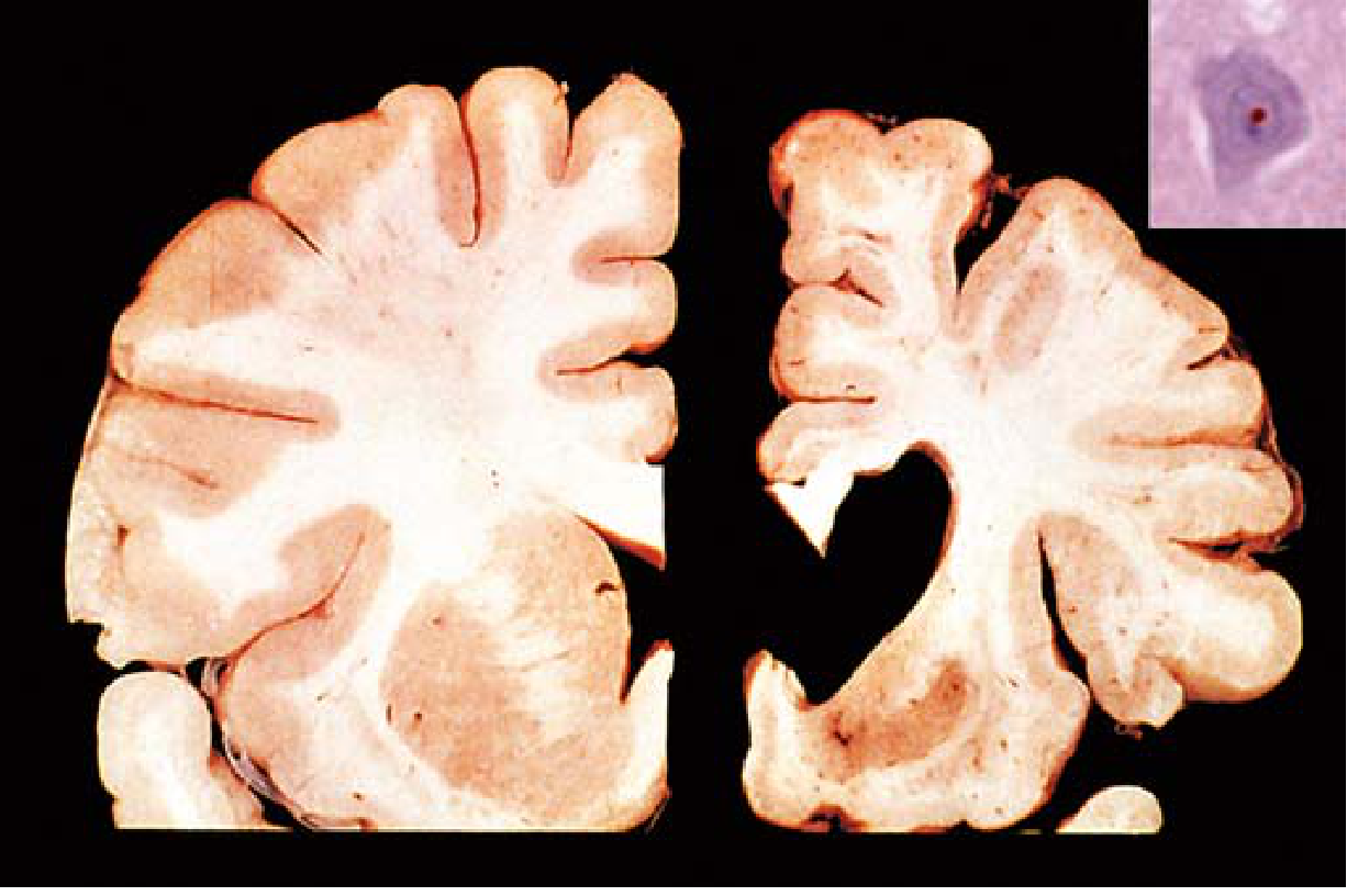
FIG: Huntington disease. Healthy hemisphere (left) compared to HD hemisphere (right) showing atrophy of the striatum and ventricular dilation. Inset: intranuclear ubiquitin-immunoreactive inclusion in a cortical neuron. (Robbins Basic Pathology)
(Robbins Basic Pathology, p. 855)
Clinical Features
HD is characterized by a triad of motor, cognitive, and psychiatric symptoms that are insidious and progressive.
1. Motor Symptoms
- Chorea (involuntary jerky, writhing movements) - predominant early
- Dystonia and parkinsonism develop later
- Westphal variant (juvenile onset): akinetic-rigid/dystonic form rather than choreic
- Oculomotor dysfunction: difficulty with refixating gaze; blinks and head thrusts to compensate
2. Cognitive Symptoms
- Bradyphrenia (slowed thinking), forgetfulness, inattention
- Executive dysfunction, impaired working memory
- Deficits in emotion recognition, smell identification, time discrimination/production
- Progresses to subcortical dementia (different from Alzheimer's cortical pattern - attention, concentration, judgment, and problem-solving are predominantly affected)
- Cognitive decline detectable decades before motor diagnosis on tests like Symbol Digit Modalities Test, Trail Making Test, Stroop
3. Psychiatric Symptoms
- Universal but widely variable
- Apathy, depression, anxiety, aggression, disinhibition
- Impulsiveness, personality changes, paranoid psychosis
- Obsessive and perseverative behaviors
- May precede motor symptoms - can be misattributed to psychotropic drugs
(Bradley & Daroff's Neurology, p. 902; Goldman-Cecil, p. 3972)
Diagnosis
- Clinical: Motor diagnosis requires an unequivocal extrapyramidal movement disorder, though cognitive and behavioral features may dominate
- Genetic testing: Confirmatory - counts CAG repeats in the IT15 gene
- MRI: Gray matter volume loss (caudate especially) detectable in premanifest and early-stage patients; useful for monitoring progression
- Biofluid markers: Mutant huntingtin protein (ultrasensitive single-molecule counting) associates with symptom onset and functional decline; NfL (neurofilament light chain) is an emerging marker
Treatment & Management
There is currently no disease-modifying treatment that slows or reverses progression. Care is multidisciplinary (neurology, psychiatry, genetics, psychology, social work, speech/occupational/physical therapy, nutrition).
Chorea Management
| Drug | Mechanism | Starting Dose |
|---|---|---|
| Tetrabenazine (first-line) | Vesicular monoamine transporter 2 (VMAT2) inhibitor - reduces dopamine | 12.5 mg 2–3x/day, up to 100–200 mg/day |
| Deutetrabenazine (FDA-approved) | VMAT2 inhibitor (longer half-life, better tolerated) | 6 mg/day, increase by 6 mg/week, max 48 mg/day |
| Amantadine | Glutamate antagonist | 300–400 mg/day |
| Riluzole | Glutamate release inhibitor | 200 mg/day |
| Haloperidol / Fluphenazine / Pimozide | Dopamine receptor blockers | Reserved for disabling chorea |
Monitor for: depression, parkinsonism, weight gain (especially with tetrabenazine).
Psychiatric Symptoms
- Treated with standard agents for the respective condition (SSRIs for depression, antipsychotics for psychosis)
Disease-Modifying Strategies (Investigational)
- Intrathecal antisense oligonucleotides targeting mutant huntingtin mRNA - unfortunately, early trials were not helpful
- Fetal cell transplantation - not found to be helpful
- RAN proteins are potential future targets
- Genome editing approaches remain under investigation
Prognosis
- Inexorable decline at a relatively constant rate
- Institutionalization and death over ~15 years from symptom onset
- Prognosis varies with disease burden (CAG repeat length influences age of onset but not rate of decline once symptoms appear)
Differential Diagnosis of Chorea
Other causes of chorea to distinguish from HD include: benign hereditary chorea, neuroacanthocytosis, Wilson disease, Sydenham chorea, SLE, antiphospholipid syndrome, drug-induced chorea (levodopa, antipsychotics, oral contraceptives), hyperthyroidism, and paraneoplastic syndromes.
(Goldman-Cecil Medicine, p. 3972)
Recent literature note: A 2025 meta-analysis (PMID 41081429) identified biofluid markers for HD that may aid in future monitoring and trial design. A 2024 systematic review (PMID 38528808) examined depression treatment in HD specifically. Neither contradicts the standard textbook framework above.
Corpus striatum it receptors and neurons
Reading File
The corpus striatum is a broad topic covering anatomy, neurotransmitter receptors, and neuronal circuits. Let me search the library in parallel across multiple angles.
Searching sources...
Finding Sources
Finding Sources
Finding Sources
Good - found a dedicated corpus striatum section in Gray's Anatomy for Students, and relevant content in Kaplan & Sadock's. Let me now read these in parallel along with more targeted searches.
Reading File
Reading File
Finding Sources
Excellent - found key sources in Kandel's Principles of Neural Science, Ganong's, Bradley & Daroff, and Katzung's. Let me read all of these in parallel.
Reading File
Reading File
Reading File
Excellent content. Let me now get the direct/indirect pathway details from Ganong's (continued) and the Katzung dopamine receptor section, plus the key circuit diagram from Kandel.
Reading File
Reading File
Loading Image
I now have a comprehensive, multi-source picture. Let me also fetch the direct/indirect pathway diagram from Ganong's and get the Katzung section on dopamine in striatum.
Reading File
Loading Image
I now have comprehensive, multi-source material covering anatomy, neurons, receptors, pathways, and neurotransmitters of the corpus striatum. Here is the full answer:
Corpus Striatum: Anatomy, Neurons, and Receptors
1. Anatomy & Subdivisions
The corpus striatum (Latin: "striped body") gets its name from the striated appearance of fiber bands connecting the caudate nucleus and putamen through the anterior limb of the internal capsule.
It is composed of:
| Structure | Component | Notes |
|---|---|---|
| Caudate nucleus | Head, body, tail | C-shaped; follows lateral ventricle; tail ends at amygdala |
| Lentiform nucleus | Putamen + globus pallidus | Lens-shaped on lateral view |
| Putamen | Part of lentiform nucleus | Together with caudate = striatum |
| Globus pallidus (GPe/GPi) | Part of lentiform nucleus | Main output of basal ganglia |
The striatum (caudate + putamen) is the primary input nucleus of the basal ganglia. The internal capsule separates these structures from the thalamus and caudate medially.
(Gray's Anatomy for Students, p. 1337)
2. Neuronal Types in the Striatum
The striatum has a distinctive cellular architecture that is consistent across all its functional territories.
A. Medium Spiny Neurons (MSNs) - ~95% of all striatal neurons
- The dominant cell type - inhibitory, GABAergic
- Receive massive glutamatergic input from the cortex and thalamus
- Send long-range inhibitory projections to globus pallidus and substantia nigra
- Also send local axon collaterals to adjacent MSNs (mutual inhibition)
- Subdivided into two populations by their peptide co-expression and receptor profile:
| Population | Peptide Co-expression | Dopamine Receptor | Pathway |
|---|---|---|---|
| D1-MSNs | Substance P + Dynorphin | D1 (excitatory via cAMP ↑) | Direct pathway → GPi/SNr |
| D2-MSNs | Enkephalin | D2 (inhibitory via cAMP ↓) | Indirect pathway → GPe |
(Kandel's Principles of Neural Science; Bradley & Daroff Neurology, p. 2095)
B. Interneurons - ~5–10% of striatal neurons
These are aspiny interneurons that regulate MSN activity locally:
| Interneuron Type | Size | Neurotransmitter | Notes |
|---|---|---|---|
| Tonically Active Neurons (TANs) | Large | Acetylcholine | Excite MSNs; muscarinic receptors (M1–M5); balanced against dopamine |
| Medium interneurons | Medium | Somatostatin, neuropeptide Y, nitric oxide | Modulatory |
| Small interneurons | Small | GABA (fast-spiking) | Rapid local inhibition |
(Ganong's Review of Medical Physiology, p. 251)
3. Inputs to the Striatum
The striatum is the primary receiver of basal ganglia input:
| Source | Neurotransmitter | Notes |
|---|---|---|
| Cerebral cortex (all areas) | Glutamate (excitatory) | Corticostriatal pathway; topographically organized |
| Intralaminar thalamic nuclei | Glutamate (excitatory) | Thalamostriatal pathway |
| Substantia nigra pars compacta (SNc) | Dopamine (+/- cholecystokinin, glutamate) | Nigrostriatal pathway; key modulator |
| Raphe nuclei | Serotonin | Modulatory |
| Locus coeruleus | Norepinephrine | Modulatory |
| Pedunculopontine nucleus (PPN) | Acetylcholine | Brainstem input |
| Amygdala, hippocampus | Limbic input | Via direct and thalamic relays |
(Bradley & Daroff Neurology, Table 96.2; Kandel's Principles of Neural Science)
4. Receptors in the Corpus Striatum
Dopamine Receptors (most clinically important)
Five subtypes exist in two families:
| Family | Subtypes | Signal Mechanism | Location |
|---|---|---|---|
| D1 family | D1, D5 | Gs protein → adenylate cyclase ↑ → cAMP ↑ | Direct pathway MSNs, cerebral cortex, limbic system |
| D2 family | D2, D3, D4 | Gi protein → adenylate cyclase ↓ → cAMP ↓ | Indirect pathway MSNs, cortex, limbic system, pituitary gland |
Clinical note: D2 receptor occupancy in the corpus striatum of >78% by antipsychotics is associated with extrapyramidal symptoms (EPS/parkinsonism), while <65% is associated with suboptimal antipsychotic efficacy. This window can be achieved with haloperidol doses as low as 1–5 mg/day in first-episode patients.
(Bradley & Daroff Neurology, p. 2095; Kaplan & Sadock Psychiatry, p. 9363)
Glutamate Receptors
| Receptor Type | Class | Notes |
|---|---|---|
| NMDA | Ligand-gated ion channel | Ca²⁺ entry; excitotoxicity; synaptic plasticity |
| AMPA | Ligand-gated ion channel | Fast excitatory transmission |
| Kainate | Ligand-gated ion channel | Fast excitatory transmission |
| mGluR (metabotropic) | G protein-coupled | Modulatory; multiple subtypes |
GABA Receptors
| Receptor | Class | Notes |
|---|---|---|
| GABA-A | Ligand-gated Cl⁻ channel | Multiple subtypes; benzodiazepines enhance activity |
| GABA-B | Metabotropic (G protein) | Slower inhibition |
| GABA-C | Ligand-gated | Less prominent in striatum |
Acetylcholine Receptors
- Predominantly muscarinic (M1–M5) on striatal interneurons and MSNs
- Mediate both excitatory and inhibitory effects
- Balanced against dopamine (dopamine inhibits, ACh excites striatal output)
Adenosine Receptors
- A2A receptors are co-localized with D2 receptors on GABAergic D2-MSNs in the indirect pathway
- A2A and D2 receptors have antagonistic interactions; A2A antagonists are under development for Parkinson disease
Cannabinoid Receptors
- CB1 receptors are particularly abundant in the basal ganglia/striatum
- Endogenous ligands: anandamide and 2-arachidonoylglycerol (2-AG)
- Modulate dopaminergic effects; role in motor control still being characterized
Serotonin Receptors
- Multiple subtypes present: 5-HT1, 5-HT2, 5-HT3 (ligand-gated), 5-HT4, 5-HT6, 5-HT7
(Bradley & Daroff Neurology, p. 2095)
5. The Direct and Indirect Pathways
The striatum integrates all its inputs and channels output through two competing pathways:
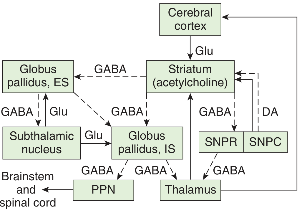
FIGURE: Principal connections of the basal ganglia. Solid lines = excitatory pathways; dashed lines = inhibitory pathways. DA = dopamine; Glu = glutamate; ES = external segment (GPe); IS = internal segment (GPi); SNPC = substantia nigra pars compacta; SNPR = substantia nigra pars reticulata; PPN = pedunculopontine nuclei. (Ganong's Review of Medical Physiology)
Direct Pathway (facilitates movement)
Cortex (Glu) → Striatum D1-MSNs → GPi/SNr (GABA ↓ inhibition) → Thalamus disinhibited → Cortex activated
- D1-MSNs use GABA + Substance P + Dynorphin
- Dopamine on D1 receptors excites this pathway (cAMP ↑)
- Net effect: movement facilitation
Indirect Pathway (suppresses movement)
Cortex (Glu) → Striatum D2-MSNs → GPe (GABA ↓ inhibition) → STN disinhibited → GPi/SNr activated (GABA ↑) → Thalamus inhibited → Cortex suppressed
- D2-MSNs use GABA + Enkephalin
- Dopamine on D2 receptors inhibits this pathway (cAMP ↓)
- Net effect: movement suppression
Dopamine from SNc therefore promotes movement via BOTH pathways: exciting D1 (direct) and inhibiting D2 (indirect).
(Kandel's Principles of Neural Science; Ganong's, p. 251)
6. Functional Territories
The striatum is organized along a ventromedial-dorsolateral continuum into three functional domains:
| Territory | Input | Function |
|---|---|---|
| Ventral striatum (nucleus accumbens) | Limbic (amygdala, hippocampus, prefrontal cortex) | Reward, motivation, addiction |
| Associative striatum (caudate head, anterior putamen) | Prefrontal cortex | Cognition, executive function, working memory |
| Sensorimotor striatum (posterior putamen) | Sensorimotor cortex | Motor planning and execution |
The cellular architecture (MSNs + interneurons) is identical across all territories, suggesting the striatum applies the same computational operations to diverse inputs.
(Kandel's Principles of Neural Science)
7. Three Balanced Biochemical Systems
Three systems normally operate in balance; their disruption leads to movement disorders:
| System | Disorder when disrupted |
|---|---|
| Nigrostriatal dopaminergic (SNc → striatum) | Hypofunction → Parkinson disease (hypokinesia, rigidity, tremor) |
| Intrastriatal cholinergic (ACh interneurons) | Relative excess vs. dopamine → parkinsonian symptoms |
| GABAergic (striatum → globus pallidus/SNr) | Loss of GABAergic MSNs → Huntington disease (hyperkinesia, chorea) |
(Ganong's Review of Medical Physiology, p. 251)
Sources: Gray's Anatomy for Students, p. 1337 | Kandel's Principles of Neural Science, 6th ed. | Ganong's Review of Medical Physiology, 26th ed., p. 251 | Bradley & Daroff's Neurology in Clinical Practice, p. 2095 | Kaplan & Sadock's Comprehensive Textbook of Psychiatry, p. 9363

IMG_5616.jpg
Explain me this
This is a textbook excerpt about the neurons and receptors of the corpus striatum. Let me break it down point by point with full context.
Point 4: Corpus Striatum - Interneurons & Neurons
🔵 Interneurons release ACh (Acetylcholine)
The corpus striatum contains large aspiny interneurons (called Tonically Active Neurons or TANs) that release acetylcholine (ACh).
- These are only ~5% of striatal neurons
- They fire continuously (tonically) and keep a baseline level of ACh in the striatum
- ACh has an excitatory effect on striatal output neurons
- They act as a counterbalance to dopamine
🔵 Spiny Neurons release GABA
The medium spiny neurons (MSNs) are the dominant cell type (~95% of all striatal neurons). They are:
- Inhibitory - release GABA
- Have dendrites covered in spines (hence "spiny")
- Receive glutamate input from the cortex and dopamine from the substantia nigra
- They are the ones that carry information OUT of the striatum
Now here is the key split - these spiny neurons are divided into two equal populations (50/50):
The 50/50 Split - D1 vs D2 Receptors
✅ 50% express D1 receptor → Direct Pathway
Cortex (Glu) → Striatum D1-MSNs → GPi + SNr pars reticulata
                                          ↓ (GABA - inhibits)
                                        Thalamus (disinhibited)
                                          ↓ (Glu - excites)
                                        Cortex → MOVEMENT
- D1 neurons project to GPi (globus pallidus internal) and SNr (substantia nigra pars reticulata)
- D1 is an excitatory receptor - it is Gs-coupled → increases cAMP
- When dopamine hits D1 → it activates/excites these neurons
- These activated neurons then inhibit GPi/SNr (via GABA)
- GPi/SNr normally suppress movement - when they are inhibited, movement is released (facilitated)
- Co-express Substance P and Dynorphin
D1 + Dopamine = Direct pathway ON = Movement facilitated
✅ 50% express D2 receptor → Indirect Pathway
Cortex (Glu) → Striatum D2-MSNs → GPe (external)
                                         ↓ (GABA - inhibits GPe)
                                    GPe normally inhibits STN
                                    STN disinhibited → excites GPi
                                         ↓ (Glu)
                                        GPi/SNr more active
                                         ↓ (GABA - suppresses)
                                        Thalamus suppressed
                                         ↓
                                        Cortex → MOVEMENT SUPPRESSED
- D2 neurons project to GPe (globus pallidus external)
- D2 is an inhibitory receptor - it is Gi-coupled → decreases cAMP
- When dopamine hits D2 → it inhibits/suppresses these neurons
- This means GPe is less inhibited → GPe can now inhibit the STN
- STN activity drops → GPi/SNr suppression → thalamus freed → movement facilitated
- Co-express Enkephalin
D2 + Dopamine = Indirect pathway OFF = Movement facilitated
The Big Picture: How Dopamine Promotes Movement
Dopamine acts on both pathways simultaneously and the net effect is always pro-movement:
| Pathway | Receptor | Dopamine Effect | Net Result |
|---|---|---|---|
| Direct | D1 (excitatory) | Activates D1-MSNs | Direct pathway ON → movement ✅ |
| Indirect | D2 (inhibitory) | Suppresses D2-MSNs | Indirect pathway OFF → movement ✅ |
Both actions work synergistically to promote motor activity.
ACh vs Dopamine Balance
The last point is critical:
| Neurotransmitter | Effect on Striatum | Effect on Movement |
|---|---|---|
| Dopamine (from SNc) | Excites D1, inhibits D2 | Facilitates movement |
| ACh (from interneurons) | Excites MSNs | Opposes movement / brakes |
These two must be balanced for normal movement. When this balance is broken:
| Imbalance | Disease | Symptoms |
|---|---|---|
| Dopamine ↓ (or ACh relatively ↑) | Parkinson disease | Hypokinesia, rigidity, tremor |
| Dopamine ↑ (or ACh relatively ↓) | Huntington disease / chorea | Hyperkinesia, involuntary movements |
This is why anticholinergic drugs (like benztropine) help in Parkinson disease - they restore the balance by reducing ACh to compensate for lost dopamine.
Simple Memory Aid
D1 = Direct = Do movement D2 = inDirect = Dopamine inhibits this to allow movement ACh = brakes on movement; Dopamine = accelerator
Explain me short
Corpus Striatum - Short Summary
Two types of neurons:
| Neuron | Releases | Role |
|---|---|---|
| Interneurons (aspiny) | ACh | Opposes movement |
| Spiny neurons (MSNs) | GABA | Carry output signal |
Spiny neurons split 50/50:
D1 receptor (50%) → Direct pathway
- Dopamine hits D1 → excites → facilitates movement ✅
D2 receptor (50%) → Indirect pathway
- Dopamine hits D2 → inhibits → also facilitates movement ✅
Dopamine promotes movement via BOTH pathways simultaneously.
Key balance:
- Dopamine = accelerator (pro-movement)
- ACh = brake (anti-movement)
- Both must be balanced for normal movement
Dopamine ↓ → Parkinson (too little movement)
Dopamine ↑ → Chorea/Huntington (too much movement)
No I understand
Got it! Let me know whenever you have a new question or topic you want to explore. 😊
This is a shared conversation. Sign in to Orris to start your own chat.