
Power grading in motor system 1-5
- 0 - No contraction at all
- 1 - Flicker or trace of contraction, but no movement
- 2 - Active movement with gravity eliminated (can move limb on a flat surface)
- 3 - Active movement against gravity, but not against resistance
- 4 - Active movement against gravity and some resistance (subdivided as 4-, 4, 4+ in some systems)
- 5 - Normal power against full resistance
MRC muscle power grading scale 0 to 5 motor system

This diagnostic image set consists of six T1-weighted axial MRI scans of the human thigh, demonstrating the Modified Mercuri scale for grading muscular fatty replacement. The series illustrates a progressive increase in signal hyperintensity within the skeletal muscle compartments, corresponding to adipose tissue infiltration. Score 0 shows normal muscle bulk and signal. Score 1 identifies an 'early moth-eaten' appearance with scattered hyperintense punctate areas. Scores 2 and 3 show increasing confluence of fatty signal, involving <30% and 30-60% of the muscle volume, respectively. Score 4 exhibits a 'faded' appearance with dominant fatty signal, and Score 5 represents end-stage disease where muscle is almost entirely replaced by adipose and connective tissue, with only thin fascia and neurovascular bundles remaining visible. This visual scale is essential for the clinical evaluation of neuromuscular disorders and myopathies, providing a standardized method to quantify disease progression and muscular involution.
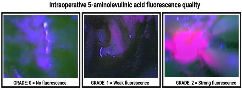
This diagnostic comparison chart illustrates the 3-stage classification system for intraoperative 5-aminolevulinic acid (5-ALA) fluorescence quality grading in neurosurgery, specifically for identifying high-grade glioma margins. The image comprises three side-by-side panels captured under 400 nm violet-blue excitation light. - Grade 0 (No fluorescence): Shows a predominantly green/dark background with minimal Protoporphyrin IX accumulation; no distinct pink or purple emission is visible. - Grade 1 (Weak fluorescence): Displays a subdued, localized purple/pink hue against a dark background, indicating low-level fluorescence that remains transparent enough to see underlying tissue texture. - Grade 2 (Strong fluorescence): Exhibits intense, vibrant lava-pink or solid purple fluorescence that is opaque and clearly demarcated from surrounding non-fluorescent tissue. This scale is a standard neurosurgical tool used during fluorescence-guided resection (FGR) to differentiate tumor tissue from healthy brain parenchyma, aiding in achieving gross total resection of glioblastomas.
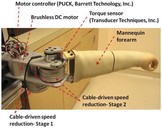
This clinical photograph displays a telerobotic master device designed for remote orthopedic and neurological assessment. The system serves as a haptic interface for a clinician to evaluate a patient's elbow joint remotely. Key components labeled include a mannequin forearm that mimics the patient's anatomy, which is connected to a mechanical drivetrain. The drive system consists of a brushless DC motor controlled by a motor controller (PUCK). Force transmission is managed through a low-friction, cable-driven speed reduction system featuring Stage 1 and Stage 2 pulley mechanisms. A high-precision torque sensor is integrated to measure the resistance and force feedback during passive range of motion (ROM) tests, muscle strength grading (MRC scale), and spasticity evaluation (Modified Ashworth Scale). The device is utilized in physical therapy and rehabilitation medicine to provide physical interaction between a clinician and a remote environment, enabling functional clinical assessments through bilateral control architecture.
MRC Scale - Motor Power Grading (0-5)
| Grade | Description |
|---|---|
| 0 | Complete paralysis - no contraction whatsoever |
| 1 | Minimal (flicker/trace) contraction - visible or palpable, but no movement produced |
| 2 | Active movement only with gravity eliminated (limb can move horizontally on a flat surface) |
| 3 | Full movement against gravity, but no resistance can be offered against manual opposition |
| 4 | Active movement against gravity AND resistance, but can be overcome by examiner |
| 5 | Normal strength - moves against full resistance |
Key Clinical Points
- 4- - Movement against gravity + minimal resistance (easily overcome)
- 4 - Movement against gravity + moderate resistance
- 4+ - Movement against gravity + strong resistance, barely overcome (near-normal)
- Ask the patient to contract the muscle maximally while the examiner opposes with graded resistance (isokinetic method), OR
- Ask for maximal contraction and try to "break" it (isometric/MVIC method)
- For Grade 2: position the limb so gravity is eliminated (e.g., test hip abduction in the supine position)
- Large trunk/girdle muscles may mask slight weakness - supplement with functional tasks (squatting, walking on toes/heels)
A patient with tectal plate meningioma with obstructive hydrocephalus was taken for mpvp shunt & post shunt edh happened which was evacuated now the patient has left eye esotropia what could be the differential diagnosis
Left Eye Esotropia Post VP Shunt + EDH Evacuation - Differential Diagnosis
Priority 1: Left CN VI (Abducens) Palsy - Most Likely
- CN VI palsy is a classic false localizing sign of raised ICP from any source (hydrocephalus, EDH, shunt dysfunction). It occurs because the abducens nerve stretches over the apex of the petrous bone as the brainstem descends with raised ICP.
- CN VI palsy can appear after VP shunt insertion itself - specifically documented in Adams and Victor's: "rarely, it may appear after...insertion of a ventricular shunt."
- The EDH event would have caused acute ICP elevation, further stressing the nerve.
- In hydrocephalus: "In mild or slowly developing cases, only a sixth-nerve palsy may be seen...hydrocephalus may affect the sixth nerve of one or both eyes" - Neuroanatomy through Clinical Cases.
Priority 2: Shunt Over-drainage / Slit Ventricle Syndrome
- Traction on cranial nerve roots as the brain sags with low ICP
- CN VI palsy is the most common cranial nerve manifestation
- Can paradoxically cause new neurological symptoms despite a working shunt
- Occurs in ~10% of shunted patients
Priority 3: Residual/Recurrent Obstructive Hydrocephalus (Shunt Malfunction)
- Raised ICP reaccumulates
- The dilated third ventricle/suprapineal recess pushes downward on the tectal plate and collicular plate
- This directly compresses tectal structures AND stretches CN VI
- Must be excluded urgently with CT (shunt series + ventricular size assessment)
Priority 4: Direct Tectal/Midbrain Injury - Dorsal Midbrain Syndrome (Parinaud's)
- Convergence retraction nystagmus (adducting saccades on attempted upgaze)
- Tonic convergence spasm - produces esotropia as part of the dorsal midbrain syndrome
- Supranuclear upgaze palsy
- Lid retraction (Collier sign)
- Light-near dissociation
Priority 5: EDH-Related CN VI Traction/Compression
- Transtentorial herniation with uncal pressure on CN III (causing CN III palsy - but CN III palsy would cause exotropia + ptosis, not pure esotropia)
- Brainstem displacement stretching CN VI over the petrous ridge
- Post-evacuation, the nerve may still be recovering
Priority 6: Divergence Paralysis / Acute Acquired Comitant Esotropia (AACE)
- Esotropia greater at distance than near
- Associated with severe head injury or raised ICP
- Bilateral symmetrical esotropia, no true abduction deficit
- Mechanism: disruption of divergence center (likely in the midbrain/pons)
- Relevant given the posterior fossa/midbrain involvement in this patient
Priority 7: Midbrain Pseudo-sixth Nerve Palsy
- A midbrain lesion can mimic CN VI palsy without actual CN VI involvement
- Produced by internuclear or paramedian pontine reticular formation (PPRF) dysfunction
- Differentiated from true CN VI palsy by MRI
Priority 8: Post-surgical CN VI Neuropraxia
- Shunt catheter placement (frontal or parietal approach)
- Bone work during EDH evacuation
- Retraction or traction on the base of skull/tentorium
Summary Table
| Diagnosis | Key Feature | Urgency |
|---|---|---|
| CN VI palsy (ICP-related) | Limited left abduction, esotropia worse at distance | Investigate ICP |
| Shunt over-drainage | Symptoms worse upright, low-pressure headache | CT + shunt check |
| Shunt malfunction / re-raised ICP | Progressive symptoms, papilledema | Urgent CT |
| Dorsal midbrain syndrome | Upgaze palsy + convergence spasm + lid retraction | MRI brain |
| AACE / Divergence paralysis | Comitant ET, worse at distance | MRI brain |
| Post-surgical CN VI neuropraxia | Subacute onset, may resolve | Observe + MRI |
| Midbrain pseudo-sixth palsy | No abduction deficit on testing | MRI midbrain |
Recommended Workup
- CT brain (urgent) - ventricle size, shunt catheter position, EDH recurrence, residual hematoma
- Formal ocular motility exam - is abduction truly limited? (incomitant = CN VI; comitant = central/divergence)
- MRI brain with midbrain cuts - tectal plate, periaqueductal grey, CN VI course, petrous apex
- Shunt series X-ray - catheter continuity
- Fundoscopy - papilledema (raised ICP) vs. no papilledema (over-drainage or direct nerve injury)