
Congenital heart disease
I'll start by reading the medical library skill, then search for comprehensive information on congenital heart disease.
Reading File
Now let me search the medical library for congenital heart disease content in parallel:
Finding Sources
Finding Sources
Excellent results. Let me now read the most authoritative sources - Robbins Pathology for pathology/classification, Braunwald's for clinical details, and Goldman-Cecil for a clinical overview, all in parallel:
Reading File
Reading File
Reading File
Now let me read the detailed pathology content from Robbins and the Braunwald's clinical section for specific defects:
Reading File
Finding Sources
Searching PubMed
"congenital heart disease" AND management
Excellent - I have rich content. Let me now fetch the key diagram image and read more of the Robbins content covering specific defects (tetralogy, transposition, etc.):
Loading Image
Reading File
I need to read the specific defect sections (ASD, VSD, PDA, Tetralogy, etc.) from Robbins. Let me read the right section:
Reading File
I now have comprehensive data. Let me compile the full, detailed answer:
Congenital Heart Disease (CHD)
Definition & Epidemiology
Congenital heart diseases are structural abnormalities of the heart or great vessels present at birth. They account for 20-30% of all birth defects and affect nearly 1% of all newborns (~40,000 infants per year in the United States). Incidence is higher in premature infants and stillborns, approximately one-fourth of whom have significant cardiac malformations. Thanks to advances in surgical technique, an estimated 1.5 million individuals now live with CHD in the United States alone. In 25% of cases, surgical intervention is required for survival in the first year of life.
- Robbins & Kumar Basic Pathology, p. 347
Pathogenesis
CHD most commonly arises from faulty embryogenesis during gestational weeks 3-8, when major cardiovascular structures develop. The cause is unknown in approximately 90% of cases.
Identified risk factors include:
-
Prematurity
-
Family history
-
Maternal conditions: diabetes, hypertension, obesity, phenylketonuria, thyroid disorders, connective tissue disorders
-
Maternal drug exposure: phenytoin, retinoic acid, alcohol, smoking
-
Assisted reproductive technology (e.g., in vitro fertilization)
-
Chromosomal/genetic disorders: Trisomy 21 (Down syndrome), Trisomy 18 (Edwards), Trisomy 13 (Patau), Turner syndrome (45,X)
-
In utero infections: rubella, cytomegalovirus
-
Robbins & Kumar Basic Pathology, p. 347
Classification by Hemodynamic Consequence
Structural anomalies in CHD fall into three major groups:
| Group | Direction of Shunt | Cyanosis | Key Examples |
|---|---|---|---|
| Left-to-right shunts | L → R (initially) | Absent initially; late "Eisenmenger" cyanosis | ASD, VSD, PDA |
| Right-to-left shunts | R → L | Early, prominent | Tetralogy of Fallot, Transposition of Great Arteries |
| Obstructive lesions | No shunt | Usually absent | Coarctation of aorta, Pulmonary stenosis, Aortic stenosis |
- Robbins & Kumar Basic Pathology, p. 348
Frequency of Common Malformations
| Malformation | % of CHD |
|---|---|
| Ventricular septal defect (VSD) | 42% |
| Atrial septal defect (ASD) | 10% |
| Pulmonary stenosis | 8% |
| Patent ductus arteriosus (PDA) | 7% |
| Tetralogy of Fallot | 5% |
| Coarctation of aorta | 5% |
| Atrioventricular septal defect | 4% |
| Aortic stenosis | 4% |
| Transposition of great arteries | 4% |
| Truncus arteriosus | 1% |
| Total anomalous pulmonary venous connection | 1% |
| Tricuspid atresia | 1% |
These 12 entities account for 85% of all CHD. - Robbins & Kumar Basic Pathology, p. 347
Left-to-Right Shunt Lesions
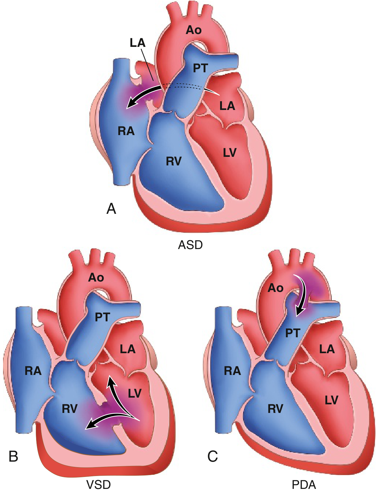
Fig. 9.2 - Common L→R shunt defects: (A) ASD, (B) VSD, (C) PDA. Ao=Aorta; PT=Pulmonary trunk; LA=Left atrium; LV=Left ventricle; RA=Right atrium; RV=Right ventricle. (Robbins & Kumar Basic Pathology)
Left-to-right shunts increase pulmonary blood flow. They are not initially cyanotic, but prolonged shunting can lead to pulmonary hypertension and eventual shunt reversal (Eisenmenger syndrome) with late-onset cyanosis. This is why early surgical correction is the rule.
Atrial Septal Defect (ASD)
- Fixed opening in the atrial septum, allowing unrestricted interatrial blood flow
- Types:
- Ostium secundum (90%): near the foramen ovale; most common
- Ostium primum (5%): lowest atrial septum; associated with mitral/tricuspid valve abnormalities
- Sinus venosus (5%): high in the atrial septum; often associated with anomalous pulmonary venous drainage
- PFO (Patent Foramen Ovale): present in ~20% of adults; flaps are sealed but not fused - allows paradoxical embolism during raised intrathoracic pressure
- Clinical features: Usually asymptomatic until adulthood (most common CHD first diagnosed in adults); right ventricular hypertrophy and dilation; risk of paradoxical embolism
- Management: Surgical or catheter-based (intravascular) closure; low mortality
Ventricular Septal Defect (VSD)
- Most common CHD diagnosed at birth (42%)
- Defects in ventricular septum, most commonly at the membranous (perimembranous) region (~90%)
- Many small VSDs close spontaneously; large defects require surgical repair
- Clinical: systolic murmur; if large, left ventricular overload, pulmonary hypertension, and eventual Eisenmenger syndrome
Patent Ductus Arteriosus (PDA)
- Persistence of the ductus arteriosus (connecting the pulmonary artery to the aorta) after birth
- Normally closes within hours to days postnatally in response to rising arterial O2 tension and falling prostaglandin levels
- Medical closure: Indomethacin (prostaglandin inhibitor) in premature infants
- Surgical/catheter closure for symptomatic or large PDAs
- Continuous ("machinery") murmur is characteristic
Right-to-Left Shunt Lesions (Cyanotic CHD)
These cause early-onset cyanosis and are associated with:
- Polycythemia (compensatory)
- Hypertrophic osteoarthropathy
- Paradoxical embolization (venous thrombi reach systemic circulation)
- Risk of cerebral abscess
Tetralogy of Fallot (TOF)
The most common cyanotic CHD. Four features:
- VSD (large)
- Pulmonary stenosis (right ventricular outflow obstruction)
- Overriding aorta (aorta straddles the VSD)
- Right ventricular hypertrophy (secondary to obstruction)
- Direction of shunt: right-to-left (through VSD) due to pulmonary stenosis
- "Tet spells": paroxysmal hypercyanotic episodes relieved by squatting (increases systemic vascular resistance)
- CXR: "boot-shaped" heart (coeur en sabot)
- Management: complete surgical repair; palliative Blalock-Taussig (BT) shunt in neonates
Transposition of the Great Arteries (TGA)
- Aorta arises from the right ventricle; pulmonary artery arises from the left ventricle
- Creates two parallel, non-mixing circuits - incompatible with life unless there is a mixing lesion (ASD, VSD, or PDA)
- D-TGA is the most common type presenting as a cyanotic emergency in neonates
- Management: arterial switch operation (Jatene procedure) in the neonatal period
Truncus Arteriosus
- Single great vessel arising from the heart, supplying both systemic and pulmonary circulations
- Associated with large VSD; always requires surgery
Total Anomalous Pulmonary Venous Connection (TAPVC)
- All four pulmonary veins drain anomalously into the right atrium (or systemic veins) rather than into the left atrium
- Must have an ASD for survival
- Management: surgical reconnection of pulmonary veins to left atrium
Tricuspid Atresia
- Complete absence of the tricuspid valve; no direct communication from RA to RV
- Survival depends on ASD (RA to LA) and VSD or PDA (for pulmonary flow)
- Management: staged palliative surgeries (Glenn → Fontan procedure)
Obstructive Lesions
Coarctation of the Aorta
- Narrowing of the aorta, most commonly just distal to the left subclavian artery (juxtaductal/postductal)
- Two patterns:
- Preductal (infantile): proximal to ductus - duct-dependent; presents in infancy with heart failure
- Postductal (adult): distal - collateral vessels develop; presents with hypertension in upper extremities, weak/absent femoral pulses
- Associated with bicuspid aortic valve (most common associated defect), Turner syndrome
- Classic sign: rib notching on CXR (from collateral intercostal arteries)
- Management: surgical resection or balloon angioplasty/stenting
Pulmonary Stenosis
- Obstruction of right ventricular outflow; most cases are valvular (domed, fused cusps)
- Mild to moderate: well tolerated, often asymptomatic; severe: exertional dyspnea, RV failure
- Management: balloon valvuloplasty (preferred for valvular PS)
Aortic Stenosis
- Congenital (usually bicuspid aortic valve), supravalvular, or subvalvular
- Bicuspid aortic valve: most common congenital cardiac abnormality overall (1-2% of population), though often not counted among "CHD" in classic tables
- Progressive stenosis leads to LV hypertrophy, angina, syncope, and heart failure
Eisenmenger Syndrome
When any left-to-right shunt causes sustained pulmonary hypertension, the rising pulmonary vascular resistance eventually exceeds systemic resistance, reversing the shunt from right-to-left. This produces late-onset central cyanosis. At this stage, structural defects are considered irreversible - the window for corrective surgery has passed. This underscores the rationale for early intervention in all large shunt lesions.
Key Associations
| Defect | Association |
|---|---|
| ASD | Down syndrome, Holt-Oram syndrome |
| VSD | Down syndrome, DiGeorge syndrome |
| Tetralogy of Fallot | DiGeorge syndrome (22q11 deletion), Down syndrome |
| PDA | Rubella, prematurity, prostaglandin E |
| Coarctation | Turner syndrome (45,X), bicuspid aortic valve |
| TGA | Maternal diabetes |
| AVSD (AV canal) | Down syndrome (trisomy 21) - most classic association |
| Supravalvular aortic stenosis | Williams syndrome (7q11 deletion) |
Clinical Approach
Neonatal presentation (duct-dependent lesions):
- Present in first days to weeks when ductus closes
- Includes: TGA, pulmonary atresia, hypoplastic left heart syndrome, severe coarctation
- Emergency management: prostaglandin E1 infusion to keep ductus open while awaiting surgery
Diagnosis:
- Echocardiography: gold standard for structural diagnosis
- ECG, chest X-ray, pulse oximetry (critical CHD screening)
- Cardiac catheterization: for hemodynamic assessment, interventional procedures
- MRI/CT angiography: complex anatomy delineation
Surgical principles:
- Most defects are repaired in infancy or childhood
- Palliative procedures (e.g., BT shunt, Glenn, Fontan) used for single-ventricle physiology
- Adults with CHD (ACHD) are a growing population requiring specialized care; complex issues include arrhythmias, heart failure, pregnancy planning, and endocarditis prophylaxis
Sources: Robbins & Kumar Basic Pathology, pp. 347-350; Braunwald's Heart Disease, 12th Ed.; Goldman-Cecil Medicine
Recent literature note: A 2024 systematic review (PMID 39609780) confirmed significantly elevated stroke risk in patients with CHD, reinforcing the importance of anticoagulation and paradoxical embolism surveillance in this population.
Pyloric stenosis
Reading File
Searching the library for pyloric stenosis in parallel across headings and paragraphs:
Finding Sources
Finding Sources
Searching PubMed
"pyloric stenosis" AND infant
Excellent results. Now reading the most comprehensive sources in parallel - Robbins (pathology), Schwartz's (surgery), Grainger (radiology/imaging), and Harriet Lane (pediatrics):
Reading File
Reading File
Reading File
Reading File
I have rich content. Let me now read the radiology imaging criteria and the surgical details, and fetch the ultrasound image:
Reading File
Reading File
Reading File
I now have comprehensive, authoritative content from multiple textbooks. Let me fetch the key ultrasound/anatomy image and compile the full answer:
Loading Image
Loading Image
I have everything needed for a thorough, well-illustrated answer.
Hypertrophic Pyloric Stenosis (HPS)
Definition
Pyloric stenosis (most commonly infantile hypertrophic pyloric stenosis, HPS) is a condition caused by progressive hypertrophy of the pyloric muscularis propria, leading to gastric outlet obstruction. It is not a true congenital anomaly - the pylorus is normal at birth and the hypertrophy develops postnatally. It is the most common cause of vomiting requiring surgery in infants.
Epidemiology
| Feature | Detail |
|---|---|
| Incidence | ~1 in 300-900 live births (~0.2% overall) |
| Sex ratio | Males : Females = 4-5 : 1 |
| Age of onset | 3-6 weeks of age; rare after 3 months |
| Birth order | More common in first-born children |
| Race | More common in white infants; slightly lower in black infants |
| Genetics | Concordance in monozygotic twins; ~6% recurrence in siblings of affected patients (vs 0.2% in general population) |
- Schwartz's Principles of Surgery, p. 1749; Robbins Pathologic Basis of Disease, p. 703
Pathology & Pathogenesis
The exact cause is unknown. Hyperplasia of the pyloric muscularis propria (circular > longitudinal muscle) causes progressive narrowing and elongation of the pyloric canal. Mucosal and submucosal edema and inflammation may worsen the obstruction. The result is a firm, pale, 1-2 cm ovoid mass - the classic "olive" palpable in the right upper quadrant.
Risk factors / associations:
-
Family history (strong genetic component; GWAS studies have identified loci on chromosome 11q23.3 and genes related to GI development)
-
Prematurity and young maternal age
-
Macrolide antibiotic exposure (erythromycin or azithromycin) - either orally or via breast milk - in the first 2 weeks of life is strongly associated
-
Turner syndrome and trisomy 18 (increased risk)
-
First-born male
-
Robbins, Cotran & Kumar Pathologic Basis of Disease, p. 703
Clinical Presentation
Symptom onset: Typically 3-6 weeks of age, with a progressive course over days to weeks.
Key features:
- Forceful, projectile, non-bilious vomiting - immediately after or during feeding. Non-bilious because obstruction is proximal to the ampulla of Vater.
- Persistent hunger - infant feeds eagerly despite repeated vomiting ("hungry vomiter")
- Progressive dehydration - decreased wet diapers, sunken fontanelle, dry mucous membranes
- Visible gastric peristaltic waves - left-to-right across the upper abdomen, just before emesis (now less commonly seen as infants present earlier)
- Palpable "olive" - firm, mobile, ovoid mass in the right upper quadrant (epigastrium). Previously found in up to 90% on examination; now found in <30% as infants present earlier before severe symptoms develop
- Jaundice - unconjugated hyperbilirubinemia occurs in ~2-3% (mechanism unclear; possibly related to decreased glucuronyl transferase activity from starvation)
- Schwartz's Principles of Surgery; Grainger & Allison's Diagnostic Radiology
Metabolic Consequences
Repeated vomiting of gastric contents (HCl) causes a characteristic electrolyte disturbance:
Hypochloremic, Hypokalemic Metabolic Alkalosis
Mechanism:
- Loss of H⁺ and Cl⁻ in gastric juice → metabolic alkalosis and hypochloremia
- Kidneys initially compensate by excreting HCO₃⁻ with Na⁺ and K⁺ → hypokalemia
- As hypochloremia worsens, the kidney preferentially exchanges H⁺ for Na⁺ in the distal tubule to conserve sodium → paradoxical aciduria (urine becomes acidic despite systemic alkalosis)
This is a medical problem, not a surgical emergency. Electrolyte correction must precede anesthesia and surgery.
Diagnosis
Ultrasound (Gold Standard)
US has replaced barium studies as the investigation of choice. Sensitivity and specificity are up to 98% and 100%, respectively.
Diagnostic criteria:
| Measurement | Abnormal (HPS) |
|---|---|
| Pyloric muscle wall thickness | ≥ 4 mm (some sources use ≥ 3 mm in younger infants) |
| Pyloric channel length | > 15-17 mm |
US signs:
- Shoulder sign - hypertrophic muscle bulges into the fluid-filled gastric antrum
- Nipple sign - double-layered hyperechoic mucosa protrudes into the stomach
- Cervix sign - hypertrophied pylorus resembles the uterine cervix on US
- Exaggerated, failed peristaltic waves visible in real-time
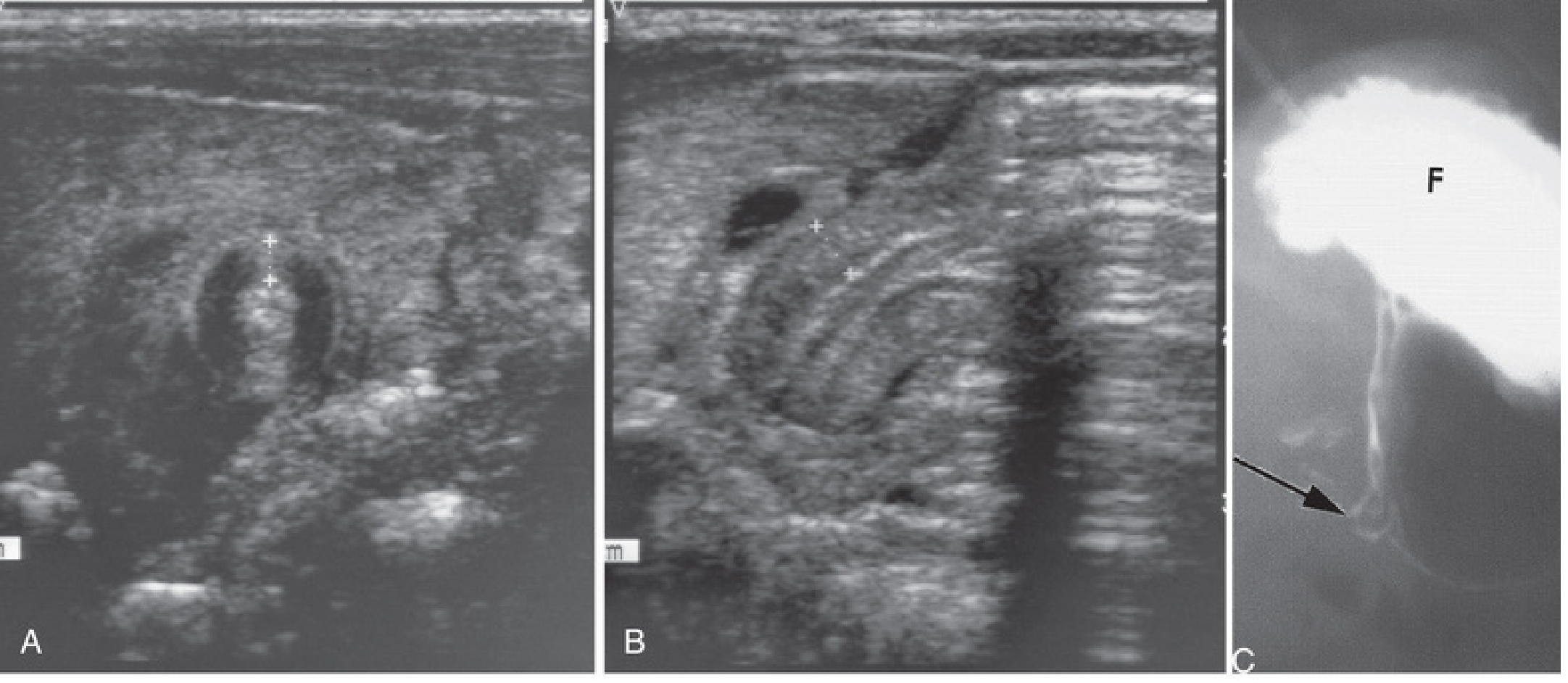
Fig. 11.4 - (A) Transverse US: pyloric muscle wall thickness >4 mm. (B) US: pyloric channel length >14 mm. (C) Contrast radiograph: narrowed pyloric canal (arrow) and distended stomach fundus (F). (The Developing Human - Clinically Oriented Embryology)
A 2023 meta-analysis (PMID 37722950) confirmed that point-of-care ultrasound combined with clinical findings (vomiting + palpable mass) has excellent diagnostic accuracy for HPS.
Upper GI Contrast Study (if US equivocal)
-
"String sign" or "railroad track sign" - elongated, narrow pyloric channel
-
Delayed gastric emptying
-
Used when US is technically inadequate or results are equivocal
-
Grainger & Allison's Diagnostic Radiology; Harriet Lane Handbook, 23rd ed.
Treatment
Step 1: Medical Stabilization (MANDATORY before surgery)
- IV fluid resuscitation: typically 5% dextrose + 0.45% NaCl + 2-4 mEq/kg/day KCl at ~150-175 mL/kg/day
- Goal: urine output > 2 mL/kg/hour, correction of alkalosis and electrolytes
- Surgery is safe to proceed when serum chloride > 100 mEq/L and HCO₃⁻ < 30 mEq/L
Step 2: Fredet-Ramstedt Pyloromyotomy (Definitive)
The operation involves splitting the hypertrophied pyloric muscle longitudinally down to the submucosal base, without entering the mucosa, allowing the mucosa to herniate through the incision and relieve the obstruction.
- Incision extends from just proximal to the pyloric vein of Mayo to the gastric antrum (1-2 cm)
- Can be performed via open (umbilical or right upper quadrant transverse incision) or laparoscopic approach
- Two RCTs have shown both approaches are equally safe; laparoscopic approach offers superior cosmetic results
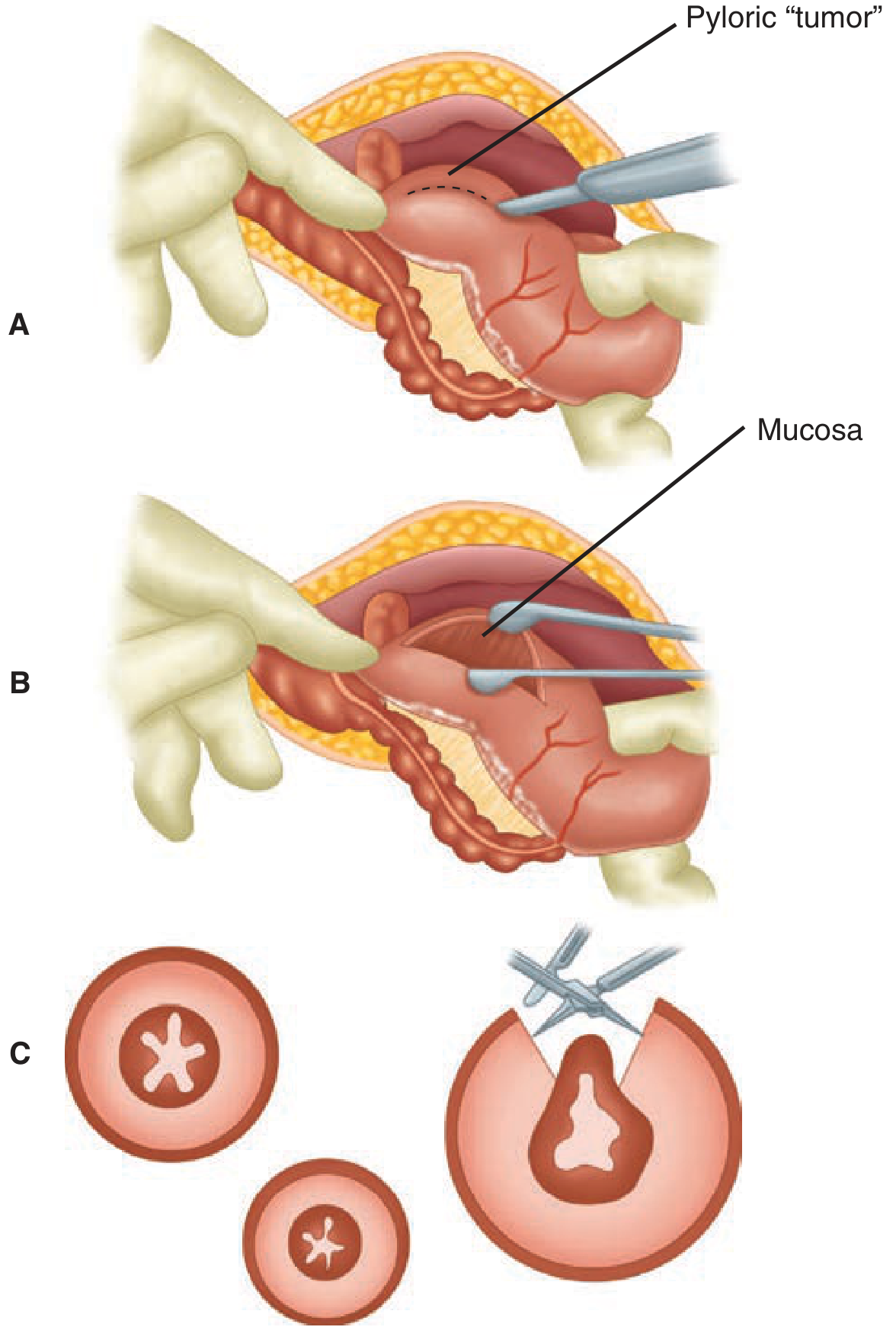
Figure 39-12 - Fredet-Ramstedt pyloromyotomy. (A) Pylorus delivered and seromuscular layer incised. (B) Muscle separated to permit mucosa to herniate. (C) Cross-section showing depth of incision and spreading. (Schwartz's Principles of Surgery, 11th ed.)
A 2024 meta-analysis (PMID 38935193) comparing umbilical versus right upper transverse incisions found no significant difference in complication rates between the two open approaches.
Postoperative Care
- IV fluids continued for several hours post-op
- Oral feeds reintroduced gradually (Pedialyte → formula/breast milk, advancing to 60 cc every 3 hours)
- Discharge within 24-48 hours; ad lib feeding protocols may shorten hospital stay
Complications of Pyloromyotomy
| Complication | Rate |
|---|---|
| Mucosal perforation | 1-3% |
| Incomplete myotomy / recurrence | Rare |
| Wound infection | Uncommon |
| Bleeding | Rare |
- If mucosal perforation occurs, it is repaired primarily with a stitch; NG tube left 24 hours
- A 2026 systematic review (PMID 41196118) addressed recurrent HPS after pyloromyotomy, noting early recognition and re-operation are key
Acquired Pyloric Stenosis (Adults)
Distinct from infantile HPS. Occurs secondary to:
-
Antral gastritis or peptic ulcers close to the pylorus (fibrosis/scarring)
-
Carcinoma of the distal stomach or pancreas (malignant infiltration/narrowing)
-
Unlike infantile HPS, treatment targets the underlying cause (endoscopic dilation, surgical bypass, or resection)
-
Robbins, Cotran & Kumar Pathologic Basis of Disease, p. 703
Key Summary Points
| Feature | Detail |
|---|---|
| Type of vomiting | Non-bilious, projectile (no bile = above ampulla of Vater) |
| Classic age | 3-6 weeks |
| Sex | Males >> Females (4-5:1) |
| Metabolic picture | Hypochloremic, hypokalemic metabolic alkalosis + paradoxical aciduria |
| Classic sign | Palpable "olive" in RUQ (now <30%) |
| Imaging | Ultrasound first line (muscle ≥4 mm; canal >16-17 mm) |
| Management | Correct electrolytes first → Fredet-Ramstedt pyloromyotomy |
| Prognosis | Excellent; most discharged within 48 hours |
Sources: Robbins, Cotran & Kumar Pathologic Basis of Disease, p. 703; Schwartz's Principles of Surgery, 11th ed., pp. 1749-1750; Grainger & Allison's Diagnostic Radiology; The Developing Human - Clinically Oriented Embryology; Harriet Lane Handbook, 23rd ed.
This is a shared conversation. Sign in to Orris to start your own chat.