
50 year old female known diabetic presenting with history of evening rise of temperature associated with severe low back pain which spontaneously recovers for > 1 month, previously she was evaluated and found to have raised crp. What may be the cause
vertebral osteomyelitis spondylodiscitis MRI spine infection
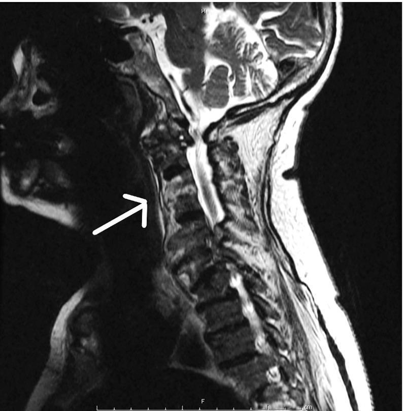
This diagnostic image is a sagittal T2-weighted MRI of the cervical spine illustrating a case of vertebral osteomyelitis and spondylodiscitis spanning from the C5 to C7 levels. The primary findings include abnormally high signal intensity (hyperintensity) within the C5, C6, and C7 vertebral bodies, characteristic of bone marrow edema and infection. The intervertebral discs at these levels demonstrate loss of normal architecture and altered signal intensity, confirming disc involvement (spondylodiscitis). A white arrow highlights the region where cortical endplate irregularities and blurring are most prominent. The prevertebral space anterior to these segments shows increased thickness and signal changes, suggesting associated inflammatory phlegmon or soft tissue edema. The spinal cord is visible posteriorly; while structurally intact, it shows mild signs of extrinsic compression secondary to the vertebral inflammation. This image serves as a clinical example of hematogenous spinal infection, in this instance related to Rat-bite fever (Streptobacillus moniliformis), emphasizing the radiological features of infectious spinal processes in an adult patient.
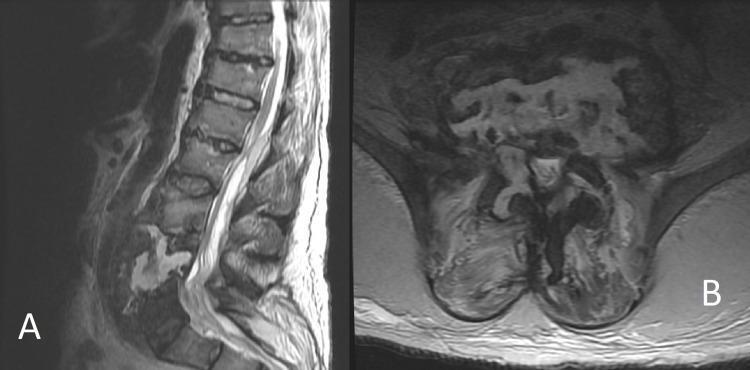
Diagnostic Magnetic Resonance Imaging (MRI) of the lumbar spine featuring T2-weighted sagittal (A) and axial (B) views. The images demonstrate severe pyogenic spondylodiscitis with associated osteomyelitis. In the sagittal view, there is significant osseous destruction of the lower lumbar vertebral bodies (specifically L3-L5), characterized by a near-complete loss of normal vertebral height and the presence of heterogeneous, high T2 signal intensity indicative of marrow edema and necrosis. A large, hyperintense prevertebral and epidural collection is visible, consistent with a complex abscess extending from L1 to L4. The axial view (B) highlights the loss of anatomical integrity of the vertebral body, replaced by ill-defined, fluid-signal intensity regions. There is extensive involvement of the paraspinal soft tissues and distortion of the thecal sac, illustrating advanced vertebral body erosion and the inflammatory spread characteristic of a systemic infection originating from sources such as decubitus ulcers. This clinical image is a primary educational resource for recognizing the radiological hallmarks of spinal infection and destructive spinal pathology.
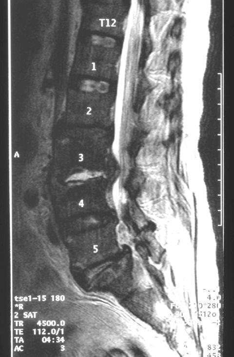
This sagittal T2-weighted magnetic resonance imaging (MRI) scan of the lumbar spine demonstrates clinical findings diagnostic of infectious spondylodiscitis at the L3/L4 level. The image shows significant disruption of the intervertebral disc space between L3 and L4, characterized by increased signal intensity and loss of normal disc morphology. Adjacent vertebral bodies (L3 and L4) exhibit signal alterations at the endplates, consistent with osteomyelitis. A prominent finding is the presence of an epidural abscess, visible as a hyperintense collection within the spinal canal at the L3/L4 level, causing potential compression of the thecal sac. Furthermore, there is evidence of extensive inflammatory changes in the surrounding soft tissues, including the paravertebral regions and psoas muscles. In contrast, the superior segments (T12 through L2) and the inferior L5 segment maintain relatively normal vertebral height, alignment, and signal characteristics. This educational image illustrates the classic imaging triad of discitis, osteomyelitis, and associated abscess formation in the setting of spinal infection.
Pott's disease tuberculosis spine X-ray MRI
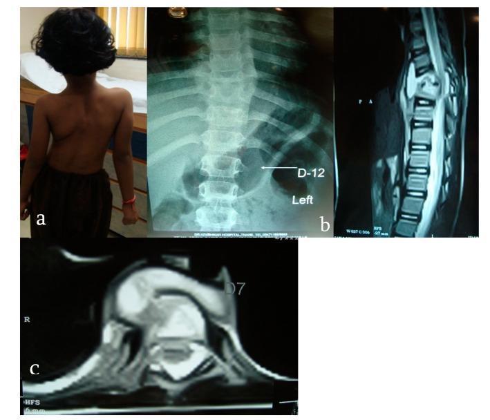
This composite figure illustrates a case of spinal tuberculosis (Pott's disease) in a pediatric patient through clinical photography, radiography, and MRI. Image (a) is a posterior clinical photograph of a 6-year-old girl showing significant lateral spinal curvature (scoliosis) and rib prominence. Image (b) is an anteroposterior X-ray of the dorsal spine demonstrating a collapse of the D8 vertebral body and a characteristic fusiform soft tissue shadow on the left side at the D12 level, suggestive of a paravertebral cold abscess. Image (c) consists of sagittal and axial MRI views of the dorsal spine. These scans reveal a wedge-shaped collapse of the D8 vertebral body with associated marrow edema and significant prevertebral and paravertebral fluid collections. There is evidence of subligamentous and epidural extension of the abscess, leading to spinal canal encroachment and compression of the neural elements. The findings are highly characteristic of granulomatous spinal infection, typically caused by Mycobacterium tuberculosis, highlighting the progression from vertebral destruction to deformity and potential neurological deficit.
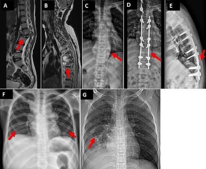
This composite figure displays diagnostic imaging of skeletal and pulmonary tuberculosis (Pott's disease). Images A-E showcase spinal involvement: (A) Sagittal T2-weighted MRI of the lumbar spine reveals L2 vertebral destruction with associated psoas and paravertebral collections. (B) Sagittal T2-weighted MRI of the thoracic spine shows destruction of T6-T7 with epidural and subligamentous extension. (C) Anteroposterior (AP) radiograph demonstrates a large paravertebral abscess at T10. (D) AP and (E) lateral radiographs show post-operative posterior stabilization using pedicle screws and rods for spinal decompression. Images F and G illustrate pulmonary manifestations: (F) An AP chest X-ray shows diffuse bilateral nodular consolidations, consistent with miliary or disseminated tuberculosis. (G) A thoracic CT scout or radiograph indicates localized consolidation with cystic changes and branching 'tree-in-bud' nodular opacities in the right lower lobe. The collection serves as an educational tool for identifying various stages and modalities used in diagnosing and treating disseminated Mycobacterium tuberculosis infections in pediatric and adult populations.
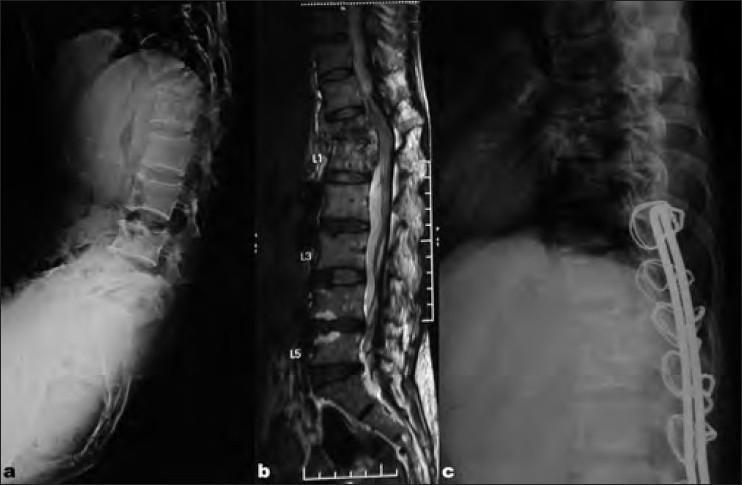
This composite figure displays diagnostic and postoperative imaging of the thoracolumbar spine in a patient with Pott’s disease (spinal tuberculosis). Image (a) is a lateral preoperative X-ray showing a sharp 38° kyphotic angulation at the D12 level, caused by a complete collapse of the D12 vertebral body. Image (b) is a midsagittal T2-weighted MRI, highlighting the marrow signal changes and anterior panvertebral involvement at D12, resulting in focal spinal angulation and narrowing of the spinal canal. Image (c) is a lateral postoperative X-ray demonstrating surgical correction of the kyphosis. The reconstruction features a tricortical bone graft placed anteriorly to restore vertebral height and a posterior Hartshill rectangle fixation system. This hardware consists of two vertical rods secured to the posterior elements by multiple sublaminar wires. The images illustrate the surgical management of spinal tuberculosis through single-stage anterior decompression, bone grafting, and posterior stabilization.
Clinical Analysis: 50F Diabetic with Evening Fever + Severe Low Back Pain + Raised CRP for >1 Month
Most Likely Diagnosis: Pyogenic Vertebral Osteomyelitis / Spondylodiscitis
- "The most common presenting symptom of spinal infection is localized back pain or neck pain... Fever is present in only about 50% of patients but when present should immediately raise suspicion of a spinal infection." - Campbell's Operative Orthopaedics, 15th Ed.
- "C-reactive protein is almost always elevated in adult spinal infection." - Campbell's Operative Orthopaedics
- "An increased ESR or CRP has been reported in 98% and 100% of cases, respectively." - Harrison's Principles, 22E
- Diabetes mellitus is a major risk factor for both acquiring spinal infection AND for failure of non-operative treatment.
Differential Diagnoses (Ranked by Likelihood)
1. Pott's Disease (Spinal Tuberculosis) - HIGH on the differential
- Insidious onset, dull aching back pain, evening/night fever, constitutional symptoms (anorexia, weight loss, night sweats) are classic for TB spine.
- Accounts for ~50% of skeletal TB.
- Thoracolumbar junction is most commonly involved.
- Raised CRP/ESR expected.
- The subacute >1-month course with apparent partial recovery fits TB well - granulomatous infections have a more indolent course than pyogenic infections.
- Bradley & Daroff's Neurology: "The patient often has symptoms of systemic infection such as weight loss, fever, night sweats, or malaise. Pott disease classically presents with destruction of vertebral bodies."
2. Brucellosis of the Spine
- In endemic areas (Middle East, Mediterranean, South Asia, Latin America), brucellosis is a common cause of infective spondylitis.
- Classic presentation: undulating (evening/night) fever, sweats, back pain, headache.
- The "undulating fever" pattern (fever peaking in evening, then subsiding) is a hallmark of Brucella.
- Rheumatology (Elsevier 2022): "The acute presentation is characterized by undulating fever, sweats, headaches, lymph node enlargement, hepatosplenomegaly, back pain, and weakness."
- Must be tested for if patient has exposure to livestock, raw dairy, or unpasteurized products.
3. Spinal Epidural Abscess
- Can accompany vertebral osteomyelitis or occur independently.
- Diabetes is listed as a specific risk factor for treatment failure.
- Back pain + fever + raised CRP; neurological deficits are a warning sign.
- CRP >115 mg/L specifically increases risk in diabetics.
4. Seronegative Spondyloarthropathy (Ankylosing Spondylitis / Reactive Arthritis)
- Can cause inflammatory low back pain (worse at rest, better with activity), morning stiffness, and raised CRP.
- However, the pattern here - evening fever + severe pain with apparent spontaneous recovery (vs. chronic stiffness) - makes this less likely than infection.
- Still, Harrison's lists "seronegative spondylitis (ankylosing spondylitis, psoriasis, reactive arthritis, enteropathic arthritis)" in the differential for febrile back pain.
5. Spinal Metastases / Multiple Myeloma
- Pathological fracture or bony metastases can cause severe back pain. Fever (paraneoplastic) and raised CRP can occur.
- Less likely in the absence of a known primary, but must be excluded.
Key Distinguishing Features
| Feature | Pyogenic VO | TB Spine | Brucellosis | SpA |
|---|---|---|---|---|
| Onset | Acute-subacute | Insidious (months) | Subacute | Chronic |
| Fever | Intermittent, ~50% | Evening/night fever | Undulating (evening) | Low-grade or absent |
| Back pain | Severe, sharp | Dull, aching | Moderate to severe | Worse in AM, better with activity |
| CRP/ESR | Almost always raised | Raised | Raised | Raised |
| Diabetes link | Strong risk factor | Less direct | Less direct | Not related |
| Spontaneous fluctuation | Can fluctuate | Yes | Yes - classic | Common |
Investigations to Order
- MRI spine (with gadolinium) - gold standard; shows disc/endplate involvement, epidural extension, paraspinal abscess
- Blood cultures x3 (before antibiotics if stable)
- ESR, CRP serial monitoring
- Mantoux / IGRA (Quantiferon-TB) for TB
- Brucella serology (serum agglutination test, ELISA)
- CBC, blood glucose, HbA1c
- CT-guided biopsy if blood cultures negative - send for aerobic/anaerobic/fungal/mycobacterial cultures + histopathology
- Urine culture (common source of hematogenous seeding in diabetics)