
Left bundle branch block
Reading File
Finding Sources
Searching PubMed
"left bundle branch block" diagnosis management
Finding Sources
Reading File
Reading File
Reading File
Reading File
Loading Image
Finding Sources
Reading File
Reading File
I now have comprehensive information from Harrison's, Goldman-Cecil, and other major textbooks. Let me compile the full answer.
Left Bundle Branch Block (LBBB)
Anatomy and Pathophysiology
The left bundle branch (LBB) is a broad sheet of conduction tissue that fans out below the aortic valve over the left side of the interventricular septum. It normally subdivides into:
- Left anterior fascicle - supplies the anterosuperior left ventricle
- Left posterior fascicle - supplies the inferoposterior left ventricle
When conduction through the LBB is blocked, depolarization of the left ventricle must occur via slower, cell-to-cell myocardial conduction from the right ventricle. This causes:
- Reversal of septal depolarization - normally left-to-right, now proceeds right-to-left
- Delayed LV activation - overall QRS vector shifts leftward and posteriorly
- Ventricular dyssynchrony - RV contracts before LV
(Harrison's Principles of Internal Medicine, 22E)
ECG Criteria for Complete LBBB
| Feature | Finding |
|---|---|
| QRS duration | ≥ 120 ms (complete); 110-120 ms (incomplete) |
| Lead V1 | Wide, predominantly negative (QS or rS) complex |
| Lead V5/V6 | Broad, entirely positive (monophasic R) complex; no septal q wave |
| T-wave | Discordant - opposite polarity to last QRS deflection (secondary repolarization change) |
| Axis | Usually left axis or normal |
The figure below (from Harrison's 22E) illustrates the classic waveform differences between normal, RBBB, and LBBB in V1 and V6:
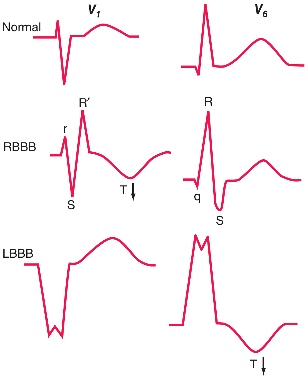
Note the characteristic wide, deep QS in V1 and broad monophasic R in V6 with a discordant T-wave inversion in V6 for LBBB.
Causes / Etiology
LBBB is rarely seen in structurally normal hearts (unlike RBBB, which is common as a normal variant). When present, it is a marker of one of four underlying conditions:
- Coronary artery disease (most common; frequently with impaired LV function)
- Hypertensive heart disease
- Aortic valve disease (including after transcatheter aortic valve replacement - TAVR)
- Cardiomyopathy (dilated, hypertrophic, infiltrative)
LBBB may be:
- Chronic (stable, established)
- Intermittent / rate-related (appears above a critical heart rate)
(Harrison's Principles of Internal Medicine, 22E, p.1915)
Clinical Features
LBBB itself is typically asymptomatic. However, the dyssynchrony it creates can:
- Precipitate or worsen heart failure (delayed LV contraction, sometimes accompanied by mitral regurgitation)
- Represent the first sign of underlying ischemic or structural heart disease
Key clinical scenarios:
New LBBB + Suspected ACS
A new (or presumed new) LBBB in the setting of clinical/laboratory findings suggesting acute MI is associated with high mortality and should be treated equivalently to STEMI - immediate reperfusion triage. The Sgarbossa criteria help identify superimposed MI patterns within LBBB morphology (concordant ST elevation ≥1 mm, concordant ST depression ≥1 mm in V1-V3, or excessively discordant ST elevation ≥5 mm).
(Goldman-Cecil Medicine, p.3412)
LBBB + Syncope/Presyncope
Strongly suggests intermittent high-degree heart block - requires urgent evaluation.
Workup / Evaluation
- Compare with prior ECGs - determine if new or chronic
- Echocardiography - assess LV function, structural disease
- Stress test (echo or nuclear) - exclude ischemic heart disease
- Cardiac MRI - if infiltrative cardiomyopathy is suspected
- Rhythm monitoring (Holter/event monitor) - if associated with PR prolongation, alternating bundle branch blocks, or presyncope/syncope
(Goldman-Cecil Medicine)
Treatment
Treatment is directed at the underlying cause. Rhythm-specific interventions:
Cardiac Resynchronization Therapy (CRT / Biventricular Pacing)
The most important LBBB-specific treatment. Indicated for patients with:
- Heart failure with LVEF ≤ 35%
- NYHA class III or ambulatory class IV (or class I/II with LVEF ≤ 30%)
- QRS ≥ 130-150 ms, especially with LBBB morphology
CRT reduces hospitalizations by ~30% and mortality by 24-36% in appropriately selected patients. LBBB morphology with QRS >150 ms predicts the best response. CRT-D (combined with ICD) is appropriate when both indications coexist.
Pacemaker
- New bifascicular block (LBBB alone or RBBB + fascicular block) + PR prolongation: prophylactic pacing often placed given high risk of progression to complete heart block
- Bifascicular block + syncope: pacemaker preferred over implantable cardiac monitor
- LBBB + bradycardia management: CRT pacing to avoid dyssynchrony when >40% pacing expected and LVEF ≤ 50%
His-bundle pacing and left bundle branch area pacing (LBBAP) are newer physiologic options whose evidence base continues to grow - a 2026 systematic review in Europace (PMID 42152765) found clinical results match or exceed biventricular pacing.
Prognosis
- LBBB without structural heart disease: mortality minimally increased if stress testing and cardiac MRI are normal
- LBBB with underlying heart disease: risk of all-cause mortality increased ~6-fold
- LBBB overall population: mortality increased approximately 2-3 fold compared with those without LBBB
(Goldman-Cecil Medicine)
Fascicular Blocks (Hemiblocks)
Partial blocks in the left system do NOT prolong QRS substantially, but cause frontal plane axis shifts:
| Block | QRS Axis | Notes |
|---|---|---|
| Left anterior fascicular block (LAFB) | More negative than -45° | Most common cause of marked left axis deviation in adults |
| Left posterior fascicular block (LPFB) | More rightward than +110-120° | Rare in isolation; must exclude other causes of right axis deviation |
Sources: Harrison's Principles of Internal Medicine 22E (2025); Goldman-Cecil Medicine International Edition; Tintinalli's Emergency Medicine; Miller's Anesthesia 10E
This is a shared conversation. Sign in to Orris to start your own chat.