Now I have enough rich content for a comprehensive answer. Let me compile it.
Hepatocellular Carcinoma (HCC) - Full Details
Definition & Overview
Hepatocellular carcinoma (HCC) is the most common primary malignant tumor of the liver, arising from malignant transformation of hepatocytes. It is the third leading cause of cancer death worldwide, accounting for approximately 900,000 new cases and 830,000 deaths annually. More than 80% of cases occur in developing countries. Rates are rising in developed nations due to chronic hepatitis C, non-alcoholic steatohepatitis (NASH), and alcohol-related cirrhosis.
- Goldman-Cecil Medicine, p. 2063
Epidemiology & Risk Factors
| Risk Factor | Notes |
|---|
| Hepatitis B virus (HBV) | Can cause HCC without cirrhosis via direct viral DNA integration into host genome |
| Hepatitis C virus (HCV) | Typically causes HCC after long-standing cirrhosis |
| Cirrhosis (any cause) | Alcohol, NASH, hemochromatosis, primary biliary cirrhosis |
| Aflatoxin | Dietary carcinogen (mold toxin); major factor in sub-Saharan Africa and Asia |
| NASH/NAFLD | Rising cause in Western countries |
| Alcohol-related liver disease | Via cirrhosis |
| Hemochromatosis (iron overload) | Direct hepatotoxic + carcinogenic effect |
Geographic distribution closely parallels HBV and HCV prevalence. In Western countries, incidence is increasing as a direct consequence of the hepatitis C epidemic and the obesity/NASH epidemic.
- Grainger & Allison's Diagnostic Radiology, p. 628
- Current Surgical Therapy 14e, p. 418
Pathobiology & Molecular Mechanisms
HCC is an epithelial neoplasm arising from a multi-step carcinogenesis process triggered by chronic liver injury:
- Chronic inflammation, necrosis, and regeneration lead to progressive accumulation of somatic mutations
- Oncogene activation or tumor suppressor gene inactivation occurs over years
Key molecular alterations:
- TERT promoter mutations - seen in ~45% of cases (activation of telomerase reverse transcriptase)
- TP53 inactivation - seen in ~30% of cases
- CTNNB1 (β-catenin) activation - seen in ~25% of cases
- Additional alterations in Wnt signaling, cell cycle, and chromatin-remodeling pathways
HBV-specific mechanism: Direct viral DNA integration into the host genome at specific loci, particularly activating the TERT promoter, bypassing the usual cirrhosis-dependent pathway.
- Goldman-Cecil Medicine, p. 2063 (Pathobiology section)
Morphological Patterns
- Solitary (most common in non-cirrhotic settings)
- Multifocal - in up to 40% of cases (particularly common in the Far East)
- Diffuse (rare)
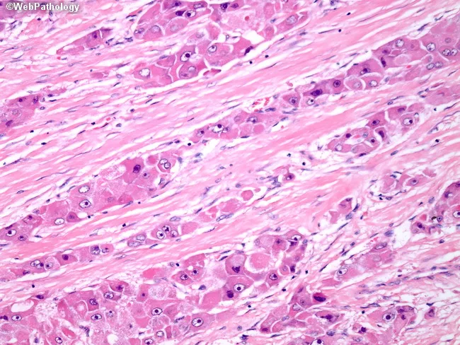
- Fibrolamellar HCC (FL-HCC) - a distinct variant with thick lamellar bands of dense eosinophilic collagen separating broad cords of neoplastic hepatocytes; arises in younger patients and non-cirrhotic livers; distinct biology and often better prognosis than conventional HCC
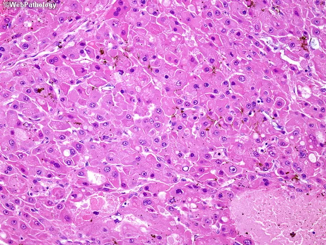
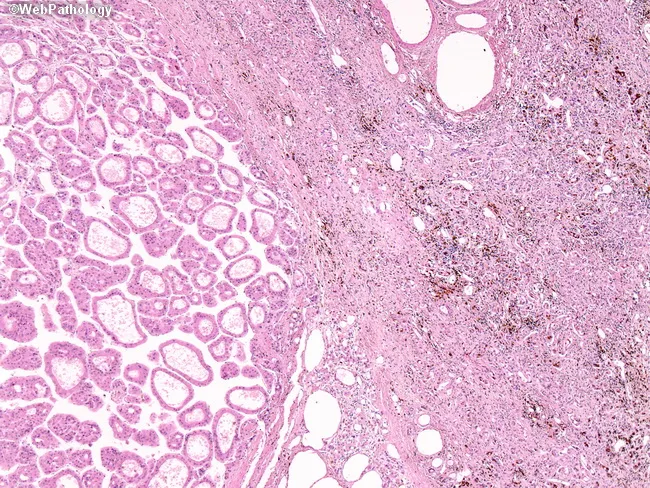
Histologically, HCC shows trabecular, acinar (pseudoglandular), or solid growth patterns. Classic features include hepatocyte-like cells with abundant eosinophilic cytoplasm, prominent nucleoli, bile production, and absence of portal tracts within the tumor. HepPar-1 and glypican-3 are immunohistochemical markers supporting hepatocellular origin.
Clinical Manifestations
Asymptomatic: Commonly detected via surveillance imaging in at-risk patients (cirrhosis, chronic viral hepatitis).
Symptomatic presentations:
- Right upper quadrant pain or a palpable mass
- Nausea, early satiety (mass effect)
- Weight loss, fevers, night sweats
- Jaundice (from biliary obstruction)
- Signs of portal hypertension
- Hemorrhage from tumor rupture (especially in large lesions)
Paraneoplastic syndromes (rare):
- Hypercalcemia of malignancy
- Erythrocytosis (from erythropoietin production by tumor)
- Hypoglycemia
Metastatic sites: Lung (most common), peritoneum, bone, spleen, adrenal gland, brain.
- Goldman-Cecil Medicine, Clinical Manifestations section
Diagnosis
Tumor Markers
| Marker | Notes |
|---|
| Alpha-fetoprotein (AFP) | Primary marker; markedly elevated levels are diagnostic even without imaging confirmation. However, HCC can occur with a normal AFP |
| CA 19-9 | Add when diagnosis is unclear (to rule out cholangiocarcinoma) |
| CEA | Add when diagnosis is unclear |
Imaging
Ultrasound (US):
- Used as primary screening modality
- Small HCCs may be hyper- or hypoechoic relative to surrounding parenchyma
- Large lesions show internal heterogeneity (hemorrhage, necrosis, fat)
- Portal vein tumor thrombus - pathognomonic of advanced HCC
- High-velocity Doppler signals reflect arterioportal shunting
- Limited sensitivity (60-80%) for lesions ≤1 cm in cirrhotic livers
CT (Multiphasic):
- Unenhanced CT: ill-defined low-attenuation lesions; calcification in ~7.5%; fat accumulation possible
- Classic pattern: Homogeneous arterial phase hyperenhancement (APHE) with portal venous/delayed phase washout - virtually diagnostic of HCC
- "Mosaic" enhancement pattern seen in some lesions
- Late arterial phase imaging is essential - many lesions visible only in this phase
MRI (gold standard for sensitivity):
- Most sensitive technique for HCC detection
- T1-weighted: typically decreased signal (some high T1 signal from glycogen accumulation)
- T2-weighted: moderately increased signal with internal heterogeneity
- Contrast enhancement pattern mirrors CT: arterial phase enhancement with washout
- 5-10% of HCCs are atypical (relatively avascular)
- Hepatobiliary-phase agents (Gd-EOB-DTPA) improve characterization
LI-RADS Classification
The Liver Imaging Reporting and Data System (LI-RADS) standardizes terminology for liver lesions in patients with chronic liver disease at risk for HCC:
| LI-RADS Category | Meaning |
|---|
| LR-1 | Definitely benign |
| LR-2 | Probably benign |
| LR-3 | Intermediate probability |
| LR-4 | Probably HCC |
| LR-5 | Definitely HCC (diagnostic) |
| LR-M | Probably malignant, not HCC-specific |
A LI-RADS 5 lesion is diagnostic of HCC - biopsy is not required in the appropriate clinical context.
Biopsy
Not indicated in the typical setting of chronic liver disease with classic imaging findings. May be needed for:
- Atypical imaging characteristics
- Molecular analysis to guide systemic therapy selection
Staging Systems
Three main staging systems are used in clinical practice:
1. AJCC/TNM Staging
Standard oncologic staging based on tumor size, vascular invasion, lymph node involvement, and metastasis.
2. Barcelona Clinic Liver Cancer (BCLC) Staging
Most widely used. Incorporates tumor burden, liver function, and performance status. Directly links stage to treatment strategy:
| BCLC Stage | Description | Recommended Treatment |
|---|
| 0 (Very early) | Single tumor ≤2 cm, no vascular invasion, Child-Pugh A | Resection, ablation, transplant |
| A (Early) | 1-3 nodules ≤3 cm, Child-Pugh A-B | Resection, ablation, transplant |
| B (Intermediate) | Multinodular, preserved liver function | TACE |
| C (Advanced) | Vascular invasion or extrahepatic spread | Sorafenib/lenvatinib ± immunotherapy |
| D (Terminal) | Poor liver function (Child-Pugh C) | Best supportive care |
3. Okuda Staging
Older system incorporating tumor size, ascites, albumin, and bilirubin.
Child-Turcotte-Pugh Score (for liver function assessment)
| Measure | 1 Point | 2 Points | 3 Points |
|---|
| Total bilirubin (mg/dL) | <2 | 2-3 | >3 |
| Serum albumin (g/L) | >35 | 28-35 | <28 |
| PT/INR | <1.7 | 1.71-2.30 | >2.30 |
| Ascites | None | Mild | Moderate to severe |
| Hepatic encephalopathy | None | Grades I-II | Grades III-IV |
- Class A: 5-6 points (well-compensated)
- Class B: 7-9 points (significant compromise)
- Class C: 10-15 points (decompensated)
Treatment
HCC management requires simultaneous management of the underlying chronic liver disease. Treatment is multidisciplinary.
1. Liver Transplantation (Preferred in cirrhotic patients)
- Addresses both the malignancy AND the underlying liver disease
- Eliminates the "field defect" (entire cirrhotic liver removed, preventing de novo HCC)
- Milan Criteria for transplant eligibility: single tumor ≤5 cm, or up to 3 tumors each ≤3 cm, no vascular invasion, no extrahepatic disease
- Limited by organ availability and immunosuppression risks
- Best option for patients with advanced cirrhosis and limited HCC
2. Surgical Resection
- Excellent option for limited liver disease and/or tumors beyond transplant criteria
- Requires Child-Pugh A (and absence of portal hypertension) for major hepatectomy
- Perioperative mortality <5% in Child-Pugh A; significantly higher in B or C
- Preferred: anatomic resection (sectionectomy, segmentectomy) - HCC spreads along portal venous tributaries; associated with improved recurrence-free and overall survival
- No proven adjuvant systemic therapy post-resection
3. Thermal Ablation
- Excellent for local tumor control in non-surgical candidates
- Radiofrequency ablation (RFA) or microwave ablation for tumors <2-3 cm
- Randomized trials show long-term outcomes similar to surgical resection for small HCC, with less morbidity
- Also used as bridge to transplantation
4. Intra-Arterial (Locoregional) Therapies
Used when curative options are not available; also used as bridge to transplantation:
- TACE (Transarterial Chemoembolization) - standard of care for intermediate-stage (BCLC-B)
- HAE (Hepatic Arterial Embolization, "bland")
- TARE / SIRT (Transarterial Radioembolization with Yttrium-90) - for tumors 3-5 cm or portal vein thrombosis
- Reserved when transplant, resection, or ablation are not feasible
5. Systemic Therapy (Advanced/Metastatic HCC - BCLC-C)
First-line options:
| Agent | Class | Notes |
|---|
| Atezolizumab + Bevacizumab | PD-L1 inhibitor + anti-VEGF | Current preferred first-line combination |
| Sorafenib | Multikinase inhibitor (anti-VEGFR, RAF) | First approved systemic agent |
| Lenvatinib | Multikinase inhibitor | Non-inferior to sorafenib in first-line |
| Durvalumab + Tremelimumab | Anti-PD-L1 + anti-CTLA-4 | Approved first-line option |
Second-line options:
| Agent | Class |
|---|
| Regorafenib | Multikinase inhibitor |
| Cabozantinib | Multikinase inhibitor (anti-MET, VEGFR) |
| Ramucirumab | Anti-VEGFR-2 (AFP ≥400 ng/mL) |
| Nivolumab ± ipilimumab | Anti-PD-1 ± anti-CTLA-4 |
| Pembrolizumab | Anti-PD-1 |
- Current Surgical Therapy 14e, p. 418; Goldman-Cecil Medicine (Abstract section)
Surveillance
In high-risk patients (cirrhosis, chronic HBV, chronic HCV with advanced fibrosis):
- Abdominal ultrasound every 6 months ± AFP
- Recommended by AASLD and EASL guidelines
- MRI preferred for surveillance in patients with poor ultrasound windows (obesity, NASH)
Prognosis
- Overall 5-year survival <30% for all comers
- Early-stage (BCLC 0-A) treated with curative intent: 5-year survival 50-75%
- Advanced-stage (BCLC-C) with systemic therapy: median overall survival ~13-19 months (improved with immunotherapy combinations)
- Portal vein tumor thrombus, extrahepatic metastasis, and poor liver function are major adverse prognostic factors
Key Images
HCC Histology - Solid Pattern (H&E):
Solid sheets of monomorphic hepatocyte-like cells with abundant eosinophilic cytoplasm, prominent nucleoli, and intracellular bile. Portal tracts are absent - characteristic of HCC.
Fibrolamellar HCC (H&E):
Thick lamellar bands of dense eosinophilic collagen separating large polygonal tumor cells with granular cytoplasm - the hallmark of fibrolamellar HCC, arising in younger patients without cirrhosis.
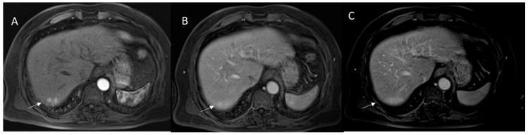
MRI - Combined HCC/Cholangiocarcinoma showing arterial hyperenhancement:
Axial MRI: Panel A (arterial phase) shows APHE; Panel B (portal venous phase) lacks classic washout; Panel C (delayed) shows progressive enhancement - illustrating how atypical enhancement raises suspicion for combined HCC-cholangiocarcinoma.
Recent Evidence (2024-2026)
A 2024
systematic review of clinical practice guidelines for HCC management found that guidelines consistently recommend surveillance ultrasound ± AFP, LI-RADS for imaging characterization, BCLC staging for treatment allocation, and the atezolizumab-bevacizumab combination as preferred first-line systemic therapy for advanced HCC. A 2025
systematic review on molecular targeting highlights the shift toward combined immunotherapy plus anti-angiogenic regimens and the role of biomarker-driven treatment selection in HCC.
Summary Table
| Feature | Key Point |
|---|
| Most common primary liver cancer | Yes - 900,000 cases/year globally |
| Primary risk factors | HBV, HCV, cirrhosis (any cause), aflatoxin, NAFLD/NASH |
| Classic imaging | Arterial hyperenhancement + portal venous washout |
| Diagnostic imaging classification | LI-RADS (LR-5 = diagnostic) |
| Curative options | Transplant, resection, ablation |
| Bridge/palliative locoregional | TACE, TARE (Y-90) |
| First-line systemic | Atezolizumab + bevacizumab (preferred); sorafenib; lenvatinib |
| Key molecular alterations | TERT (45%), TP53 (30%), CTNNB1 (25%) |
| Surveillance | US ± AFP every 6 months in at-risk patients |
| Prognosis (overall 5-year) | <30% |