
Cause of cancers
- Carcinogens (tobacco, radiation, chemicals)
- Oncogene activation and tumor suppressor gene loss (e.g., TP53, RB1)
- Viral infections (HPV, HBV, EBV, H. pylori)
- Inherited mutations (BRCA1/2, Lynch syndrome genes)
- Chronic inflammation and immune dysregulation
causes of cancer carcinogenesis chemical radiation viral genetic diagram
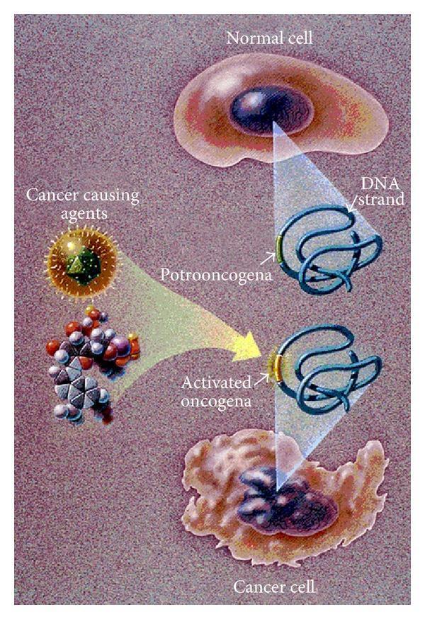
This medical illustration depicts the pathophysiology of carcinogenesis, focusing on the transformation of a normal cell into a cancer cell through oncogene activation. At the top, a normal cell is shown with a regular, rounded plasma membrane and a smooth, oval nucleus. Below it, a cancer cell exhibits characteristic pleomorphism, featuring an irregular, ruffled membrane and a misshapen, hyperchromatic nucleus with an uneven border. The central diagram illustrates the molecular mechanism where 'cancer-causing agents' (represented by viral and chemical structures) interact with a proto-oncogene within a DNA strand. This genetic insult results in the formation of an 'activated oncogene,' represented by a highlighted modification on the DNA coil. The illustration serves as an educational tool to compare normal cellular morphology with malignant alterations and to link these changes to genetic mutations. Key concepts included are cellular ruffling, nuclear atypia, and the transition from proto-oncogene to oncogene as a primary driver of uncontrolled cell growth.
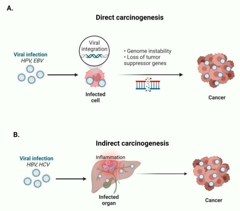
A pathophysiology diagram illustrating two distinct mechanisms of viral-induced oncogenesis. Panel A, titled 'Direct carcinogenesis,' depicts the process for viruses like HPV and EBV. It shows viral particles infecting a cell, followed by 'Viral integration' into the host genome. This leads to molecular consequences including 'Genome instability' and 'Loss of tumor suppressor genes' (visualized by a damaged chromosome icon), ultimately resulting in a cluster of neoplastic cells labeled 'Cancer.' Panel B, titled 'Indirect carcinogenesis,' shows the pathway for HBV and HCV. Here, the viral infection targets an 'Infected organ' (illustrated as a liver), which triggers a state of chronic 'Inflammation.' This inflammatory microenvironment is depicted as the driver that subsequently leads to the 'Cancer' phenotype. The diagram summarizes how different viral etiologies utilize either intracellular genetic disruption or organ-level inflammatory processes to achieve malignant transformation, serving as an educational resource for medical oncology and microbiology.
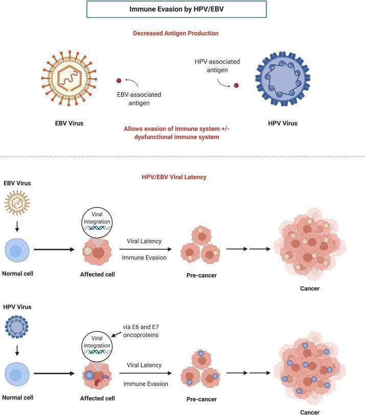
This pathophysiology diagram illustrates the mechanisms of immune evasion and viral carcinogenesis for Epstein-Barr Virus (EBV) and Human Papillomavirus (HPV). The top section compares the structural morphology of the two viruses: EBV is shown as an enveloped spherical virion with radiating surface spikes, while HPV is depicted as a non-enveloped icosahedral-like structure with a uniform, gear-shaped capsid. Both viruses are shown producing minimal antigens to evade host immune detection. The lower section details parallel oncogenic pathways. For both viruses, the process begins with a 'Normal cell' transitioning to an 'Affected cell' through 'Viral integration' of genetic material into the host genome. In HPV, this integration is specifically noted as occurring via E6 and E7 oncoproteins. This leads to 'Viral Latency' and 'Immune Evasion,' resulting in clonal expansion into a 'Pre-cancer' cluster of cells and ultimately progressing to a disorganized, densely packed 'Cancer' cell mass. The diagram highlights the clinical significance of viral latency and reduced antigen expression as key factors in the development of virus-associated malignancies like oropharyngeal and nasopharyngeal carcinomas.
Causes of Cancer (Carcinogenesis)
- Robbins & Kumar Basic Pathology, p. 250
- Schwartz's Principles of Surgery, p. 346
1. Genetic Basis of Cancer
- Oncogene activation - gain-of-function mutations in proto-oncogenes (e.g., RAS, MYC, HER2) that drive uncontrolled cell proliferation
- Tumor suppressor gene loss - loss-of-function mutations in "brakes" on cell division (e.g., TP53, RB1, BRCA1/2, APC)
2. Chemical Carcinogens
| Agent | Cancer Type |
|---|---|
| Tobacco smoke (benzo[a]pyrene) | Lung cancer |
| β-Naphthylamine (aniline dyes) | Bladder cancer |
| Aflatoxin B1 (Aspergillus mold on grains/nuts) | Hepatocellular carcinoma |
| Nitrosamines (food preservatives) | GI cancers |
| Asbestos | Mesothelioma, lung cancer |
| Vinyl chloride, arsenic, chromium | Various occupational cancers |
| Alkylating agents (chemotherapy drugs) | Secondary leukemia |
3. Radiation Carcinogenesis
- Atomic bomb survivors (Hiroshima/Nagasaki): markedly increased leukemia (latent period ~7 years), plus elevated thyroid, breast, colon, and lung cancer rates
- Radioactive miners: 10-fold increased lung cancer incidence
- Chernobyl: ongoing elevated cancer rates in surrounding regions
- Therapeutic head/neck irradiation: papillary thyroid cancers years later
- Ionizing radiation: causes chromosome breaks, translocations, inversions, and point mutations. Double-stranded DNA breaks are the most mutagenic form.
- UV radiation: forms pyrimidine dimers in DNA (normally repaired by nucleotide excision repair). With excessive UV exposure (sunbathing, tanning beds), repair systems are overwhelmed - leading to melanoma, squamous cell, and basal cell carcinomas. Patients with xeroderma pigmentosum (defective nucleotide excision repair) have dramatically elevated skin cancer risk. - Robbins & Kumar Basic Pathology, p. 251
4. Viral and Microbial Oncogenesis
| Agent | Cancer | Mechanism |
|---|---|---|
| HPV (types 16, 18) | Cervical, oropharyngeal | E6 inactivates TP53; E7 inactivates RB1 |
| EBV (Epstein-Barr virus) | Burkitt lymphoma, nasopharyngeal carcinoma, Hodgkin lymphoma | Viral integration, genome instability |
| HBV / HCV | Hepatocellular carcinoma | Chronic inflammation, cirrhosis → cancer |
| HTLV-1 | Adult T-cell leukemia/lymphoma (ATLL) | Infects CD4+ T cells; Tax protein activates NF-κB and PCNA |
| Helicobacter pylori (bacterium) | Gastric adenocarcinoma, MALT lymphoma | Chronic inflammation, CagA oncoprotein |
- Robbins & Kumar Basic Pathology, p. 252
5. Inherited (Germline) Mutations
| Gene | Syndrome | Cancers |
|---|---|---|
| BRCA1/BRCA2 | Hereditary breast/ovarian cancer | Breast, ovary, pancreas |
| TP53 | Li-Fraumeni syndrome | Breast, sarcoma, brain, adrenal, leukemia |
| APC | Familial adenomatous polyposis | Colorectal cancer |
| MLH1/MSH2 | Lynch syndrome (HNPCC) | Colorectal, endometrial |
| RB1 | Hereditary retinoblastoma | Retinoblastoma, osteosarcoma |
| VHL | von Hippel-Lindau syndrome | Renal cell, hemangioma |
| PTEN | Cowden syndrome | Breast, thyroid, endometrium |
| STK11 | Peutz-Jeghers syndrome | GI, ovarian, pancreatic |
6. Chronic Inflammation
- Chronic hepatitis (HBV/HCV) → hepatocellular carcinoma
- H. pylori gastritis → gastric cancer
- Chronic ulcerative colitis → colorectal cancer
- Barrett's esophagus (chronic acid reflux) → esophageal adenocarcinoma
Summary: The Hallmarks of Cancer
- Self-sufficiency in growth signals
- Insensitivity to anti-growth signals
- Evasion of apoptosis
- Limitless replicative potential
- Sustained angiogenesis
- Tissue invasion and metastasis
- Reprogramming of energy metabolism
- Evasion of immune destruction