
Explain in simpler terms the attachment of ligaments of hip joint
I'll look up the medical library skill first, then retrieve detailed information about hip joint ligaments.
Reading File
Now let me search the library for hip joint ligaments content in parallel:
Finding Sources
Finding Sources
Finding Sources
Finding Sources
Excellent! Found great sources. Now let me read the key sections in parallel:
Reading File
Reading File
Reading File
Now let me read the Gray's Anatomy ligaments section and fetch the relevant diagrams:
Reading File
Loading Image
Now let me fetch the diagram specifically showing the three main ligaments:
Finding Sources
Loading Image
I now have all the information needed. Let me also look for the image showing the three ligaments from the Gray's Anatomy figure 6.33:
Reading File
Loading Image
I now have everything needed for a thorough, simplified explanation. Here is the complete answer:
Ligaments of the Hip Joint - Simplified
The hip joint is a ball-and-socket joint where the head of the femur (thigh bone) sits inside the acetabulum (the "cup" of the pelvis). Three major ligaments wrap around the outside of the joint capsule to hold it together, plus one ligament inside the joint itself.
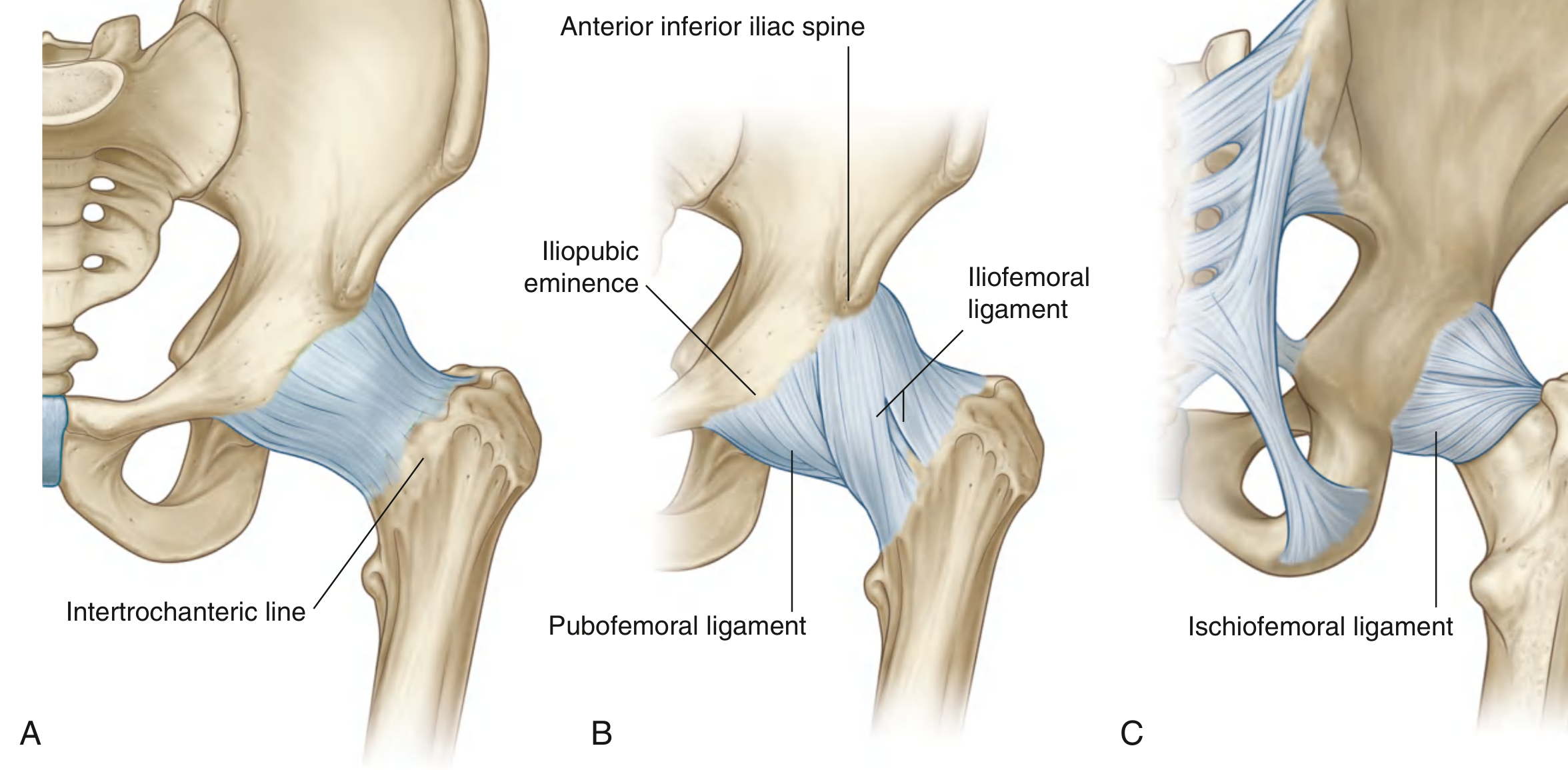
Fig. 6.33 Fibrous Membrane and Ligaments of the Hip Joint. (A) Fibrous capsule, anterior view. (B) Iliofemoral and pubofemoral ligaments, anterior view. (C) Ischiofemoral ligament, posterior view. - Gray's Anatomy for Students
1. Iliofemoral Ligament ("Y-ligament" or "Ligament of Bigelow")
This is the strongest ligament in the entire body. It sits on the front of the hip joint.
- Shape: Triangular/Y-shaped
- Proximal attachment (top/origin): The anterior inferior iliac spine (AIIS) of the ilium and the rim of the acetabulum - think of this as the "tip" of the Y
- Distal attachment (bottom/insertion): The intertrochanteric line on the front of the femur - the two limbs of the Y spread out and attach to the upper and lower parts of this line
- What it does: Prevents the hip from extending (bending backward) too far. When you stand up straight, this ligament becomes tight and locks the hip - this is why you can stand without much muscle effort.
Simple way to remember: "Ilium to femur, front of hip, Y-shape, stops over-extension."
2. Pubofemoral Ligament
This ligament sits on the front-lower (anteroinferior) side of the hip joint, below the iliofemoral ligament.
- Shape: Triangular
- Proximal attachment (medial/origin): The iliopubic eminence, adjacent pubic bone, and obturator membrane - the bony bump where the ilium meets the pubis
- Distal attachment (lateral/insertion): It blends into the joint capsule and merges with the deep surface of the iliofemoral ligament - it does not have a distinct separate insertion on the femur
- What it does: Controls excessive abduction (moving the leg out to the side) and also limits extension. Acts like a "floor" to the iliofemoral ligament.
Simple way to remember: "Pubis to femur area (via capsule), front-bottom of hip, controls abduction."
3. Ischiofemoral Ligament
This ligament reinforces the back (posterior) of the hip joint.
- Shape: Spiral band
- Proximal attachment (medial/origin): The ischium, just behind and below the acetabulum
- Distal attachment (lateral/insertion): The greater trochanter of the femur, deep to the iliofemoral ligament
- What it does: Prevents over-extension from the back. Also limits internal rotation (turning the leg inward).
Simple way to remember: "Ischium to greater trochanter, back of hip, prevents over-extension + limits internal rotation."
4. Ligament of the Head of Femur (Ligamentum Teres)
This ligament lives inside the joint capsule, not outside.
- Proximal attachment (origin): The fovea capitis (a small pit on the head of the femur)
- Distal attachment (insertion): The acetabular fossa, the transverse acetabular ligament, and the margins of the acetabular notch (all part of the socket)
- What it does: Carries a small blood vessel (branch of the obturator artery) to the femoral head. Its mechanical role in stabilizing the joint is minor in adults.
Simple way to remember: "Fovea on femoral head to inside of acetabulum - mainly carries blood supply."
Quick Summary Table
| Ligament | Location | From (Proximal) | To (Distal) | Main Function |
|---|---|---|---|---|
| Iliofemoral | Front | AIIS + acetabular rim | Intertrochanteric line (upper + lower) | Prevents over-extension; strongest ligament |
| Pubofemoral | Front-bottom | Iliopubic eminence + pubis + obturator membrane | Blends with capsule / iliofemoral lig. | Limits abduction and extension |
| Ischiofemoral | Back | Ischium (behind acetabulum) | Greater trochanter | Prevents over-extension; limits internal rotation |
| Ligamentum Teres | Inside joint | Fovea on femoral head | Acetabular fossa + transverse lig. | Carries blood to femoral head |
Key Concept - The Spiral Trick
All three external ligaments run in a spiral fashion around the hip. This means when you extend the hip (stand up straight), all three ligaments tighten simultaneously, locking the joint passively. This is why humans can stand for long periods without the muscles working hard - the ligaments do much of the work.
Sources: Gray's Anatomy for Students, p. 661-663 | Imaging Anatomy Vol. 3, p. 739
This is a shared conversation. Sign in to Orris to start your own chat.