
Difference between B and T lymphocytes both physiological and clinical
I'll start by reading the medical library skill, then search for comprehensive information on B and T lymphocytes.
Reading File
Now let me search the medical library for B and T lymphocytes in parallel:
Finding Sources
Finding Sources
Excellent results. Now let me read the key sections in parallel - Guyton (physiology), Roitt's Immunology, and Cellular & Molecular Immunology:
Reading File
Reading File
Reading File
I have a great image URL from Roitt's. Let me now fetch the key comparison image and read more of Guyton's physiology content on T vs B lymphocytes, plus search for clinical differences:
Loading Image
Reading File
Finding Sources
Excellent! Let me now get the Guyton content on T-lymphocyte activation/function, plus read the immunodeficiency chapter from Cellular & Molecular Immunology for clinical disorders:
Reading File
Reading File
The table from Cellular & Molecular Immunology is exactly what I need for clinical differences. Let me read more of it:
Reading File
Reading File
I now have very rich content. Let me also get the surface markers content from Histology for a complete picture, and check for the key clinical table that was partially cut off:
Reading File
Finding Sources
I now have comprehensive information from multiple authoritative textbooks. Let me compile the full, detailed answer.
B Lymphocytes vs T Lymphocytes: Physiological and Clinical Differences
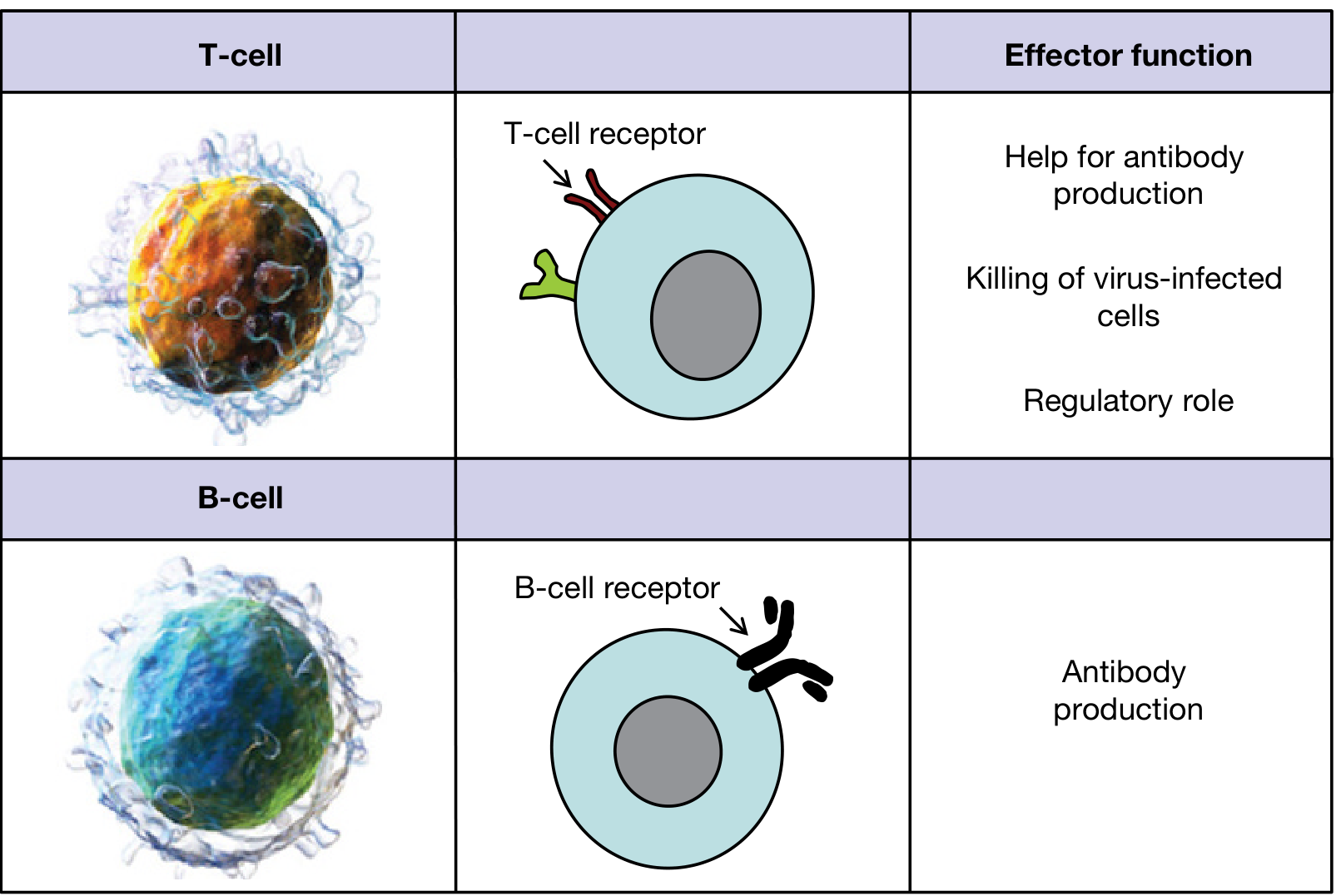
1. Origin and Preprocessing
| Feature | B Lymphocytes | T Lymphocytes |
|---|---|---|
| Common origin | Bone marrow stem cells | Bone marrow stem cells |
| Preprocessing site | Liver (mid-fetal life), then bone marrow (late fetal + post-birth) | Thymus gland |
| Mature in | Bone marrow | Thymus |
| Named after | Bursa (of Fabricius in birds) / Bone marrow | Thymus |
In the thymus, T lymphocytes undergo rapid proliferation and develop diversity for specific antigens. Crucially, up to 90% of developing T cells are destroyed by negative selection - those that react against self-antigens are eliminated before release. B lymphocytes in the bone marrow undergo a parallel process of negative selection against self-antigens. - Guyton and Hall Textbook of Medical Physiology
2. Percentage in Peripheral Blood
-
T cells: 60-80% of circulating lymphocytes
-
B cells: 20-30% of circulating lymphocytes
-
NK cells: 5-15% (neither T nor B)
-
Histology: A Text and Atlas With Correlated Cell and Molecular Biology
3. Surface Markers (CD Markers)
| Marker | B Cells | T Cells |
|---|---|---|
| Pan-marker | CD19, CD20, CD21, CD22 | CD3 (pan-T marker) |
| Specific subsets | CD40, surface Ig (sIg) | CD4 (helper), CD8 (cytotoxic) |
| Receptor | BCR = membrane immunoglobulin | TCR (T-cell receptor) - alpha/beta or gamma/delta |
| Co-receptors | CD40 (interacts with CD154 on T cells) | CD28 (co-stimulation), CD154 (CD40L) |
| MHC restriction | Recognize free/soluble antigen | Recognize antigen only when bound to MHC on APCs |
Although T and B cells cannot be distinguished on the basis of morphology alone, their distinctive CD proteins identify them on immunolabeling. - Histology: A Text and Atlas
4. Antigen Recognition - A Key Physiological Difference
| Feature | B Cells | T Cells |
|---|---|---|
| What they recognize | Free/intact antigen (3D conformational epitopes) | Peptide fragments presented on MHC molecules |
| Presented by | Not required (direct antigen binding) | Antigen-presenting cells (macrophages, dendritic cells, B cells) |
| MHC restriction | None | CD4+ T cells: MHC Class II; CD8+ T cells: MHC Class I |
| Receptor structure | BCR = surface immunoglobulin | TCR = alpha/beta heterodimer (95%) or gamma/delta |
"Although B lymphocytes recognize intact antigens, T lymphocytes respond to antigens only when they are bound to specific molecules called MHC proteins on the surface of antigen-presenting cells." - Guyton and Hall, p.468
5. Subtypes and Their Physiological Functions
T Lymphocyte Subtypes
| Subtype | Marker | Function |
|---|---|---|
| T-helper (Th) | CD4+ | >75% of T cells. Master regulator - secretes lymphokines (IL-2, IL-4, IL-5, IFN-γ, etc.) that activate B cells, macrophages, and CTLs. Subsets: Th1, Th2, Th17, Tfh |
| Cytotoxic T cells (CTL) | CD8+ | Kill virus-infected cells, tumor cells, and allograft cells. Recognize antigen on MHC I. Release perforins and granzymes causing target cell lysis |
| Regulatory T cells (Treg) | CD4+CD25+FOXP3+ | Suppress excessive immune responses. Prevent autoimmunity. Important in tumor immunology and tolerance |
| γδ T cells | TCR γδ | Innate-like function; found especially in gut and skin; respond to non-peptide antigens |
B Lymphocyte Subtypes
| Subtype | Function |
|---|---|
| Naive B cells | Circulate in follicles; not yet antigen-activated |
| Plasma cells | Terminally differentiated B cells; secrete large amounts of antibody (>2000 Ig molecules/second) |
| Memory B cells | Long-lived; responsible for rapid secondary immune response |
| Regulatory B cells (Breg) | Secrete IL-10; suppress inflammatory responses |
6. Effector Mechanisms
| Feature | B Cells | T Cells |
|---|---|---|
| Type of immunity | Humoral (antibody-mediated) | Cell-mediated |
| Effector product | Antibodies (IgM, IgG, IgA, IgE, IgD) | Activated T cells released into lymph + circulation |
| Target | Extracellular pathogens, toxins, soluble antigens | Intracellular pathogens, viruses, tumor cells, transplanted tissues |
| Memory | Long-lived memory B cells | Long-lived memory T cells |
| Secondary response | Faster, higher-affinity antibodies (class switching) | Faster, larger expansion of antigen-specific T cells |
"The principal difference is that instead of releasing antibodies, whole activated T cells are formed and released into the lymph... circulate again and again throughout the body, sometimes lasting months or even years." - Guyton and Hall, p.467
7. Anatomic Location in Lymph Nodes
- B cells reside in follicles in the cortex. Primary follicles = naive B cells. Secondary follicles with germinal centers = activated B cells undergoing somatic hypermutation and affinity maturation
- T cells reside in the paracortical cords (deep cortex / T-cell zone), packed around high endothelial venules (HEVs). ~70% are CD4+, ~30% CD8+
Their segregation is maintained by chemokines - CXCL13 (by follicular dendritic cells) attracts B cells; CCL19/CCL21 (by fibroblastic reticular cells) attract T cells. - Cellular and Molecular Immunology, p.112-115
8. Interaction Between B and T Cells
B cells require T-cell help (for T-dependent antigens) to undergo:
- Class switch recombination (IgM → IgG/IgA/IgE)
- Affinity maturation in germinal centers
- Memory cell formation
This help is provided by follicular helper T cells (Tfh) through CD40L-CD40 interaction and secretion of IL-21, IL-4. T-independent antigens (e.g., polysaccharides) can activate B cells without T-cell help but produce only IgM and no memory.
9. Clinical Differences (Immunodeficiency Patterns)
This table from Cellular and Molecular Immunology summarizes the clinical pattern perfectly:
| Feature | B Cell Deficiency | T Cell Deficiency |
|---|---|---|
| Infections | Pyogenic bacteria (otitis, pneumonia, meningitis, osteomyelitis), enteric bacteria/viruses, some parasites | Pneumocystis jirovecii, viruses, fungi, intracellular bacteria, protozoa |
| Organisms missed | Encapsulated organisms (Strep. pneumoniae, H. influenzae, Neisseria) | Intracellular pathogens, herpesviruses, CMV, EBV, Candida |
| Example diseases | X-linked agammaglobulinemia (Bruton's), Common Variable Immunodeficiency (CVID), IgA deficiency, Hyper-IgM syndrome | DiGeorge syndrome, SCID (T-cell component) |
| Diagnosis | Low serum Ig, absent B cells on flow cytometry, poor antibody responses to vaccination | Reduced blood T cells, poor lymphoproliferative response to PHA, absent DTH reactions, absent TRECs on neonatal screen |
| Age of onset | After 6 months (maternal IgG wanes) | Infancy (shortly after birth) |
| Cancer risk | Lymphoma (EBV-associated) | EBV-related lymphoma, HPV-associated cancers |
- Cellular and Molecular Immunology, Table 21.1, pp.1362-1363
10. Key Clinical Diseases Linked to Each
B Cell-Associated Clinical Conditions
- X-linked agammaglobulinemia (Bruton's): Absent B cells due to BTK mutation; recurrent bacterial infections after 6 months
- Common Variable Immunodeficiency (CVID): B cells present but fail to differentiate into plasma cells; low IgG/IgA/IgM
- Hyper-IgM syndrome: Defect in CD40L (T cells) or CD40 (B cells) - impairs class switching; elevated IgM, absent IgG/IgA/IgE
- Multiple myeloma: Malignant plasma cell proliferation
- B-cell ALL/CLL: Malignancies of B-cell lineage (CD19+, CD20+, CD10 in precursor B-ALL)
T Cell-Associated Clinical Conditions
- DiGeorge syndrome: Thymic aplasia → absent T cells → viral/fungal infections; associated with 22q11 deletion
- SCID: Combined loss of T and B function (often due to ADA or γ-chain deficiency); screened by TRECs at birth
- AIDS (HIV): Selective destruction of CD4+ T helper cells → CD4 count <200/µL defines AIDS; susceptibility to Pneumocystis, CMV, toxoplasma
- T-cell ALL: Malignancy of T-cell precursors (CD3+, CD7+, TdT+)
- Autoimmune diseases: Failure of Treg function or T-cell tolerance (e.g., Type 1 DM, MS, SLE)
11. Quick Summary Table
| Parameter | B Lymphocytes | T Lymphocytes |
|---|---|---|
| Processing site | Bone marrow (liver in fetus) | Thymus |
| % in blood | 20-30% | 60-80% |
| Surface markers | CD19, CD20, sIg | CD3, CD4 or CD8 |
| Receptor | BCR (immunoglobulin) | TCR (alpha/beta or gamma/delta) |
| Antigen recognition | Free/intact antigen | Peptide + MHC complex |
| Effector product | Antibodies (humoral immunity) | Activated T cells (cell-mediated immunity) |
| Main function | Fight extracellular pathogens, toxins | Fight intracellular pathogens, viruses, tumors |
| Location in lymph node | Follicles (cortex) | Paracortex |
| Memory | Yes (memory B cells) | Yes (memory T cells) |
| Deficiency pattern | Recurrent bacterial infections | Opportunistic viral/fungal infections |
| Key clinical disease | Agammaglobulinemia, CVID | DiGeorge, AIDS, SCID |
Sources:
- Guyton and Hall Textbook of Medical Physiology, pp. 462-470
- Cellular and Molecular Immunology (Abbas et al.), pp. 112-115, 1360-1363
- Roitt's Essential Immunology, pp. 37-38
- Histology: A Text and Atlas with Correlated Cell and Molecular Biology, pp. 716-717
This is a shared conversation. Sign in to Orris to start your own chat.