
Tuberculosis
tuberculosis chest X-ray and pathology
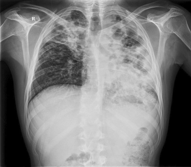
This posteroanterior (PA) chest X-ray demonstrates extensive bilateral pulmonary pathology, highly characteristic of advanced post-primary tuberculosis with superinfection. The left lung field shows a significantly distorted architecture with large, irregular, and oval-shaped lucencies in the upper and middle zones, consistent with pulmonary cavitation. The left lower zone exhibits dense alveolar opacification with visible air bronchograms and spotty shadowing, indicating consolidation. The right lung displays inhomogeneous opacities in the upper and middle lobes, accompanied by volume loss evidenced by the retraction of the right hilum and horizontal fissure. The mediastinal structures appear shifted, and there is visible distortion of the normal hilar anatomy. This diagnostic image illustrates severe parenchymal destruction, cavitary disease, and bilateral infiltrates, which are key clinical markers for evaluating disease severity and treatment response in infectious respiratory medicine.
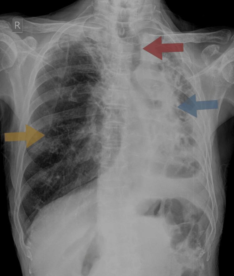
This chest X-ray (posteroanterior view) demonstrates significant pulmonary pathology consistent with left lung collapse and architectural distortion, likely as a sequela of pulmonary tuberculosis. Key findings include an opaque left hemithorax with a marked loss of volume (blue arrow), causing a significant ipsilateral shift of the mediastinum and trachea (red arrow). The ribs on the left side appear crowded, further reflecting the volume loss. In contrast, the right lung shows signs of compensatory hyperinflation (yellow arrow), evidenced by increased radiolucency and expanded volume to compensate for the contralateral dysfunction. The cardiac silhouette is obscured by the mediastinal shift and left-sided opacification. This radiological presentation is characteristic of chronic fibro-cavitary changes or total lung collapse, frequently observed in advanced or treated tuberculosis cases, and illustrates the physical pull exerted by parenchymal destruction and scarring on midline structures.
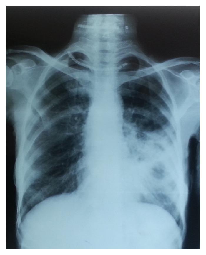
This diagnostic image is a posteroanterior (PA) chest X-ray illustrating a case of moderately advanced pulmonary tuberculosis. The primary pathology is localized in the right lung field, characterized by disseminated heterogeneous opacities of slight to moderate density. These confluent lesions extend across approximately one-third of the right lung volume. Notably, there are visible radiolucent areas within the opacified regions, indicating the presence of pulmonary cavitation, with the largest cavity measuring less than 4 cm in diameter. The left lung field remains relatively clear with normal bronchovascular markings. The mediastinal silhouette and cardiac contour are within normal limits, and the visible bony structures, including the ribs and clavicles, appear intact. This radiograph serves as a classic educational example for the classification of tuberculosis severity based on the extent of parenchymal involvement and the presence of cavitary disease, demonstrating the typical upper and middle zone predilection of secondary tuberculosis.
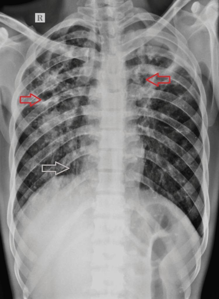
Posteroanterior (PA) chest X-ray demonstrating extensive bilateral pulmonary pathology, highly characteristic of chronic or post-primary pulmonary tuberculosis. The imaging shows widespread, asymmetrical air-space opacities scattered throughout both lung fields, with a greater density and involvement on the right side. Multiple well-defined cavitary lesions are visible, predominantly located in the bilateral upper zones and the right mid-zone (highlighted by red arrows), appearing as radiolucent areas surrounded by thick, irregular walls. In the right lower and upper zones, there is evidence of tractional bronchiectasis (highlighted by white arrows), indicated by tubular lucencies and architectural distortion. The trachea shows a mild shift toward the right, suggesting volume loss or fibrotic changes in the right lung parenchyma. These findings illustrate the chronic sequelae of mycobacterial infection, including lung destruction, cavitation, and secondary airway remodeling.
"tuberculosis"[MeSH Terms] AND "diagnosis OR treatment"
"tuberculosis"[MeSH Terms] AND "treatment"[MeSH Terms]
Tuberculosis (TB)
1. Etiology and Transmission
2. Pathogenesis
3. Clinical Presentations
Pulmonary TB (most common)
- Productive cough (often prolonged >3 weeks)
- Hemoptysis
- Fever and night sweats
- Anorexia and weight loss (hence the historical term "consumption")
- Fatigue and malaise
- Reactivation TB: Upper lobe cavitary lesions, fibrosis, and scarring
- Primary TB: Consolidation at the apex of lower lobe or base of upper lobe, with hilar/mediastinal lymphadenopathy
- Miliary TB: Diffuse 1-2 mm nodular ("millet seed") opacities throughout both lung fields
Extrapulmonary TB (10-25% of cases worldwide)
| Form | Site / Syndrome |
|---|---|
| Miliary / Disseminated | Widespread organ seeding (lung, liver, spleen, bone marrow) |
| Serosal TB | Tuberculous pleurisy, meningitis, pericarditis, peritonitis, arthritis |
| Solid organ TB | Renal TB, adrenal TB (formerly a common cause of Addison's disease), hepatic TB |
| Skeletal TB | Pott's disease (vertebral TB with potential cord compression) |
| TB meningitis | Most dangerous serosal form - difficult to diagnose, can cause permanent brain damage |
HIV and TB Co-infection
- TB may be more severe, with atypical clinical findings
- Chest X-ray is occasionally normal
- Rapid molecular-based DST is strongly recommended to guide treatment
Endobronchial TB
4. Diagnosis
Testing for Latent TB Infection (LTBI)
| Cut-off for Positivity | Population |
|---|---|
| ≥5 mm | HIV-positive; recent TB contact; fibrotic changes on CXR consistent with old TB; organ transplant recipients; immunosuppressed |
| ≥10 mm | High-risk populations (healthcare workers, residents of high-prevalence areas, IV drug users, lab personnel, persons with clinical conditions increasing TB risk) |
| ≥15 mm | Otherwise low-risk persons |
- QuantiFERON-TB Gold and T-SPOT.TB
- Advantages: single blood draw, greater specificity for M. tuberculosis, no anamnestic (booster) response on repeat testing
- Preferred over TST in BCG-vaccinated populations
Testing for Active TB
- Sputum smear (AFB microscopy): Rapid but less sensitive; 3 serial morning specimens recommended
- Sputum culture: Gold standard, takes 2-6 weeks (liquid media) or 4-8 weeks (solid media)
- Molecular tests (e.g., GeneXpert MTB/RIF): Rapid detection of M. tuberculosis AND rifampin resistance simultaneously - strong recommendation for HIV-positive patients
- Drug Susceptibility Testing (DST): Mandatory for all previously treated TB patients; focus at least on isoniazid and rifampin sensitivity
5. Treatment
Latent TB Infection (LTBI)
| Regimen | Duration | Notes |
|---|---|---|
| Isoniazid (INH) | 6-9 months | ~0.6% risk of clinical hepatitis |
| Rifampin (RIF) | 4 months | Effective alternative |
| Rifampin + Pyrazinamide | 2 months | No longer recommended - excess liver toxicity |
Active Pulmonary TB
- Intensive phase (months 1-2): Isoniazid (H) + Rifampin (R) + Pyrazinamide (Z) + Ethambutol (E) = HRZE
- Continuation phase (months 3-6): Isoniazid + Rifampin = HR
- Cavitary lesions
- Persistent positive cultures after 2 months of therapy
Drug-Resistant TB
- MDR-TB: Resistant to at least isoniazid AND rifampin. Requires second-line agents (fluoroquinolones, aminoglycosides, bedaquiline, linezolid) for 18-24 months.
- XDR-TB (Extensively Drug-Resistant): MDR-TB plus resistance to fluoroquinolones and at least one injectable agent.
Directly Observed Therapy (DOT)
6. Prevention
- BCG Vaccine: Live attenuated Mycobacterium bovis strain; given at birth in high-prevalence countries. Provides good protection against disseminated TB (miliary, meningitis) in children; less effective against adult pulmonary TB.
- Infection control: Airborne precautions (negative pressure rooms, N95 respirators) for known or suspected active TB
- LTBI screening: Targeted testing of high-risk populations (healthcare workers, HIV-infected, close contacts of TB cases, immigrants from high-prevalence countries)
- Contact investigation: All close contacts of an index case should be screened
7. High-Risk Groups for TB Reactivation
- HIV/AIDS
- Diabetes mellitus
- Chronic renal failure / dialysis
- Silicosis
- Malnutrition / low body weight
- Malignancy, especially head and neck
- Immunosuppressive therapy (TNF-alpha inhibitors, corticosteroids, post-transplant)
- Substance abuse (alcohol, IV drugs)
- Recent TB infection (within 2 years)
Sources
- Textbook of Family Medicine, 9th Edition (Rakel), Ch. 16
- Murray & Nadel's Textbook of Respiratory Medicine, Ch. 71