
Shiw me tge images of atlas and axis of cervical vertebrae
Reading File
This is a visual anatomy question. Let me search the medical library for atlas and axis images.
Finding Sources
Finding Sources
Found great sources. Let me read the relevant sections from Gray's Anatomy and the THIEME Atlas to get the images.
Reading File
Reading File
Reading File
I have multiple great images. Let me fetch them all in parallel.
Loading Image
Loading Image
Loading Image
Reading File
Reading File
Now let me fetch the additional THIEME atlas images of atlas and axis:
Loading Image
Loading Image
Loading Image
Loading Image
Loading Image
Here are all the images of the Atlas (C1) and Axis (C2) cervical vertebrae from authoritative anatomy textbooks:
Atlas (C1) and Axis (C2) - Cervical Vertebrae
Fig. 1 - Complete Overview: Atlas, Axis, and Atlanto-occipital Joint
(Gray's Anatomy for Students, Fig. 8.8)
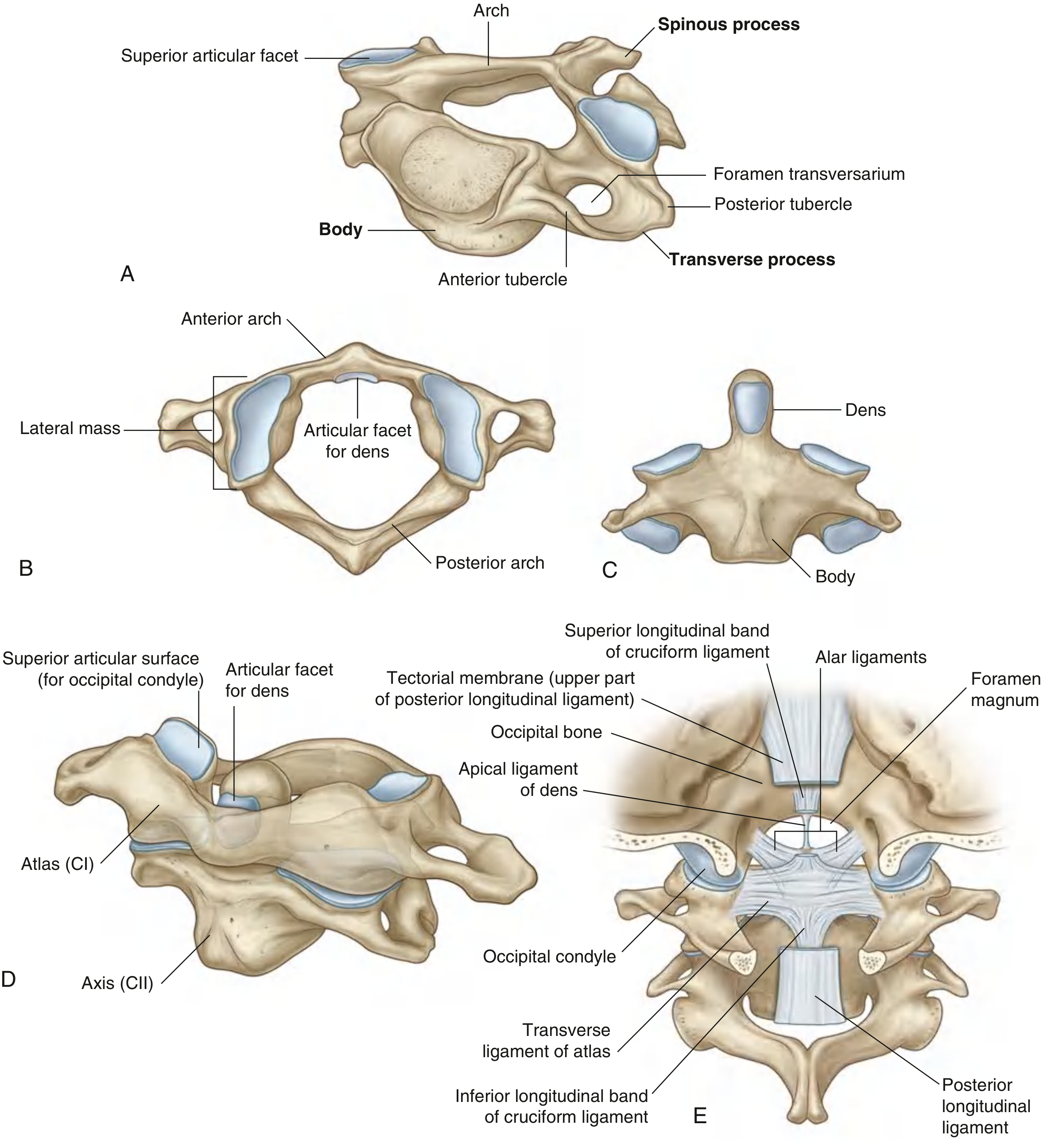
(A) Typical cervical vertebra | (B) Atlas (CI) - superior view | (C) Axis (CII) - superior view | (D) Atlas and axis together - anterolateral view | (E) Atlanto-occipital joint - posterior view showing ligaments
Fig. 2 - Atlas (C1) - Superior View (Bone specimen)
(THIEME Atlas of Anatomy)
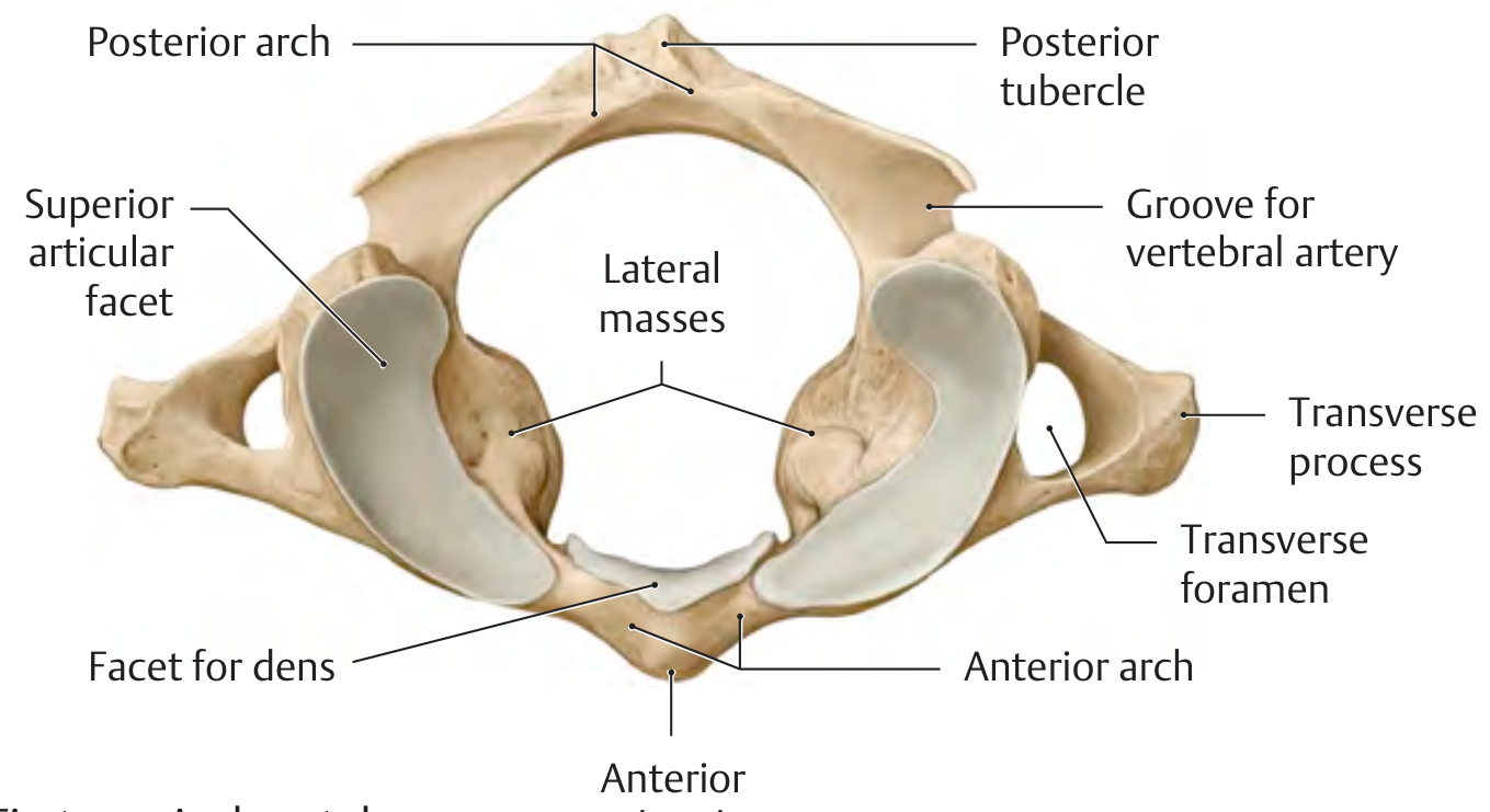
Key features visible: lateral masses, anterior arch, posterior arch, posterior tubercle, superior articular facets, facet for dens, groove for vertebral artery, transverse foramen
Fig. 3 - Atlas (C1) - Inferior/Anterior View (Bone specimen)
(THIEME Atlas of Anatomy)
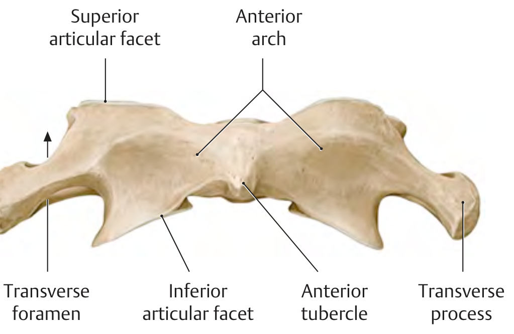
Fig. 4 - Axis (C2) - Superior View (Bone specimen)
(THIEME Atlas of Anatomy)
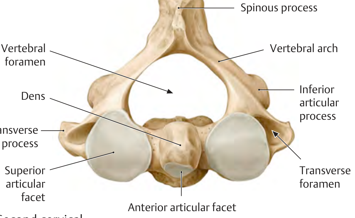
Key feature: the dens (odontoid process) projecting superiorly - this is the defining feature of the axis, formed by the fused body of C1
Fig. 5 - Axis (C2) - Anterior/Superior View (Bone specimen)
(THIEME Atlas of Anatomy)
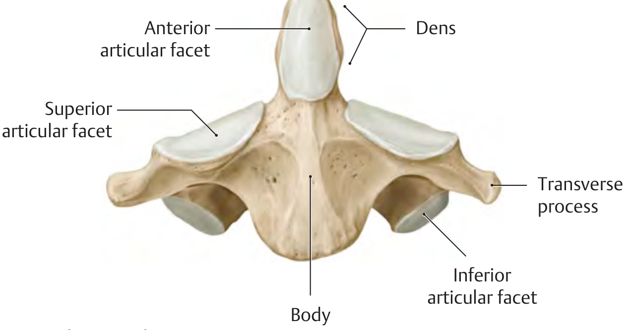
Fig. 6 - Atlas and Axis with Ligaments
(Gray's Anatomy for Students, Fig. 2.20B)
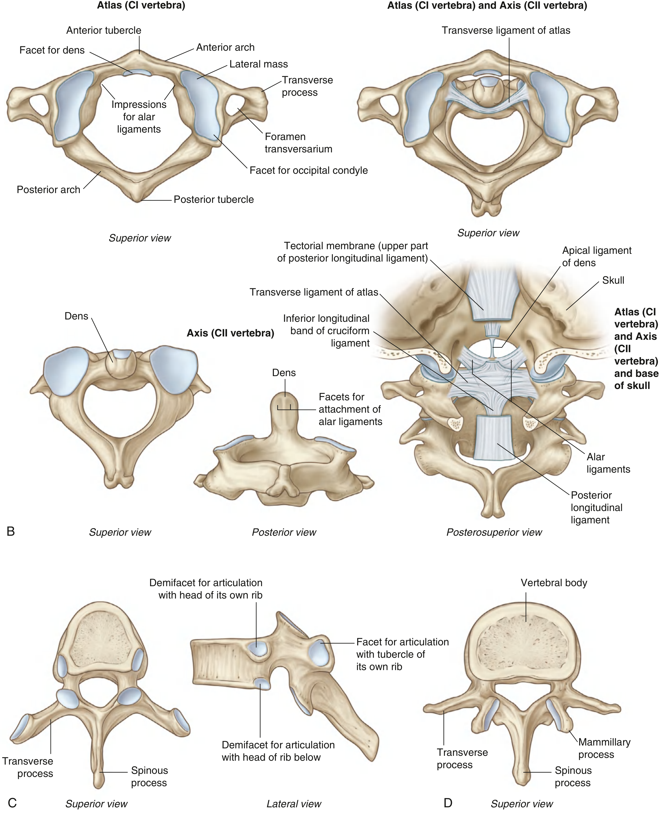
Shows: atlas superior view, axis superior and posterior views, and the posterosuperior view with the cruciform ligament complex and alar ligaments
Fig. 7 - Radiograph: Open-mouth (Odontoid peg) view
(Gray's Anatomy for Students, Fig. 2.21)
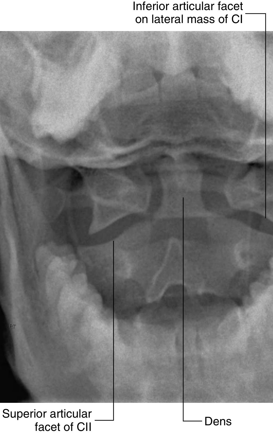
This is the classic open-mouth AP X-ray used clinically to visualize the dens (odontoid peg) and the lateral masses of C1 relative to C2.
Key Anatomical Points
Atlas (C1):
- Has NO vertebral body (fused onto C2 during development to become the dens)
- Ring-shaped with anterior and posterior arches
- Two lateral masses with superior concave articular facets (for occipital condyles) and inferior flat facets (for C2)
- Facet for dens on the posterior surface of the anterior arch
- Transverse ligament of atlas holds the dens in place
- Allows nodding (flexion/extension) of the head at the atlanto-occipital joint
Axis (C2):
- Defined by the prominent dens (odontoid process) projecting superiorly
- The dens acts as a pivot allowing the atlas + head to rotate side to side
- Has a true vertebral body, vertebral arch, spinous process (bifid)
- Alar ligaments attach from the dens to the occipital condyles, limiting excessive rotation
Sources: Gray's Anatomy for Students (9780323934237); THIEME Atlas of General Anatomy and Musculoskeletal System (9781626237186)
This is a shared conversation. Sign in to Orris to start your own chat.