
Poliovirus pathogenesis,prophylaxis, vaccine micro
Reading File
Now let me search the library for poliovirus content in parallel:
Finding Sources
Finding Sources
Good results found. Let me read the key sections in parallel:
Reading File
Reading File
Reading File
Excellent content retrieved. Let me continue reading the Harrison's section and get the vaccine/prophylaxis sections:
Reading File
Reading File
Let me get the vaccine/prophylaxis and clinical features sections:
Reading File
Finding Sources
Reading File
Reading File
Reading File
Reading File
I now have comprehensive content. Let me fetch the pathogenesis diagram image that was mentioned:
Loading Image
Reading File
I now have comprehensive material from multiple authoritative textbooks. Let me compile the full answer.
Poliovirus: Pathogenesis, Prophylaxis, and Vaccines
1. Classification and Structure
Poliovirus belongs to the family Picornaviridae, genus Enterovirus. There are 3 serotypes (types 1, 2, and 3). It is a non-enveloped, single-stranded positive-sense RNA virus with an icosahedral capsid composed of four viral proteins (VP1, VP2, VP3, VP4). The genome encodes a single polyprotein cleaved by viral proteases into structural and enzymatic components. The virus is:
-
Stable in acid (survives stomach), resistant to alcohol and detergents
-
Susceptible to chlorine-containing disinfectants
-
Can persist for days at room temperature
-
Sherris & Ryan's Medical Microbiology, 8th Ed., p. 463
-
Harrison's Principles of Internal Medicine, 22E, p. 1675
2. Pathogenesis
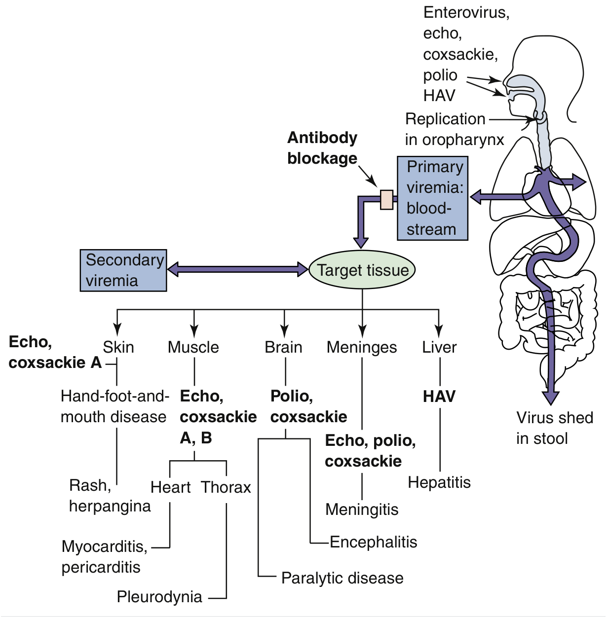
Fig. 46.4 - Pathogenesis of enterovirus infection showing poliovirus target tissues (Medical Microbiology 9e)
Step-by-step sequence:
Step 1 - Entry and primary replication
Virus enters via the fecal-oral route. After ingestion, it infects epithelial cells in the oropharyngeal mucosa, then passes to the gut. Virus binds to the poliovirus receptor (PVR) / CD155, an immunoglobulin-superfamily receptor expressed only in primates (explaining primate-limited tropism). Replication occurs in the mucosa and lymphoid tissue of the tonsils, Peyer's patches, and intestinal enterocytes. Virus is shed in oral secretions for up to 3 weeks and in stool for up to 12 weeks (>20 years in hypogammaglobulinemic patients).
Step 2 - Primary viremia
Virus enters regional lymph nodes, then the bloodstream. Viremia is detectable 3-5 days after infection. The virus spreads to reticuloendothelial cells of the lymph nodes, spleen, and liver. If the host immune response is adequate, infection is contained here - this accounts for the majority (90%+) of subclinical infections.
Step 3 - Secondary viremia and CNS invasion
In a subset of patients, a second round of replication drives secondary viremia. The CNS is reached via two routes:
- Hematogenous - crosses the blood-CNS barrier, favored by reflex dilation of capillaries supplying motor centers of the anterior horn
- Neural (axonal transport) - virus enters skeletal muscle at the neuromuscular junction (where PVR is expressed at the endplate), then travels retrogradely along peripheral nerve axons to the spinal cord - analogous to rabies virus spread. Sciatic nerve transaction in monkeys prevents CNS infection after IM injection, supporting this route.
Step 4 - Motor neuron destruction
Poliovirus is cytolytic for anterior horn cells (lower motor neurons) of the spinal cord and brainstem motor nuclei. The virus shuts down host protein synthesis by cleaving eIF4-G (the cap-binding protein of ribosomes), preventing cellular mRNA translation. Up to 100,000 virions per cell are produced before lysis. The location and number of neurons destroyed determines the extent of paralysis.
- Harrison's Principles of Internal Medicine, 22E, pp. 4455-4457
- Sherris & Ryan's Medical Microbiology, 8th Ed., pp. 2958-2963
- Medical Microbiology 9e, pp. 2922-2924
3. Clinical Manifestations
| Presentation | Frequency | Features |
|---|---|---|
| Subclinical | ~90% | No symptoms; still sheds virus |
| Abortive poliomyelitis | ~5% | Fever, sore throat, myalgia, headache, GI symptoms; resolves in 2-3 days; no CNS involvement |
| Aseptic meningitis (nonparalytic) | ~1% | Meningeal signs (stiff neck, back pain) + abortive symptoms; CSF: lymphocytic pleocytosis, normal glucose, normal/mildly elevated protein; recovers fully within days |
| Paralytic poliomyelitis | <2% | Asymmetric flaccid paralysis with NO sensory loss; maximum extent within days; recovery up to 6 months; permanent thereafter |
Incubation period: 4-35 days (usually 7-14 days for paralytic disease; 3-6 days for minor illness).
Bulbar polio: brainstem involvement causes paralysis of cranial nerves and respiratory muscles - most life-threatening form.
Post-polio syndrome: decades after acute infection, previously affected patients develop new progressive muscle weakness, fatigue, and pain - thought to be due to loss of the enlarged motor units that had reinnervated muscle after original attack.
- Sherris & Ryan's Medical Microbiology, 8th Ed., pp. 2974-2984
- Harrison's Principles of Internal Medicine, 22E, pp. 4475-4477
4. Immunity
-
Poliovirus induces specific IgM (persists <6 months) and specific IgG (lifelong).
-
Capsid protein VP1 is the predominant neutralizing antibody target.
-
IgG eliminates viremia and prevents CNS spread.
-
Secretory IgA is key for eliminating virus from the GI tract and reducing fecal shedding.
-
Neutralizing antibody confers lifelong protection against the same serotype but does not prevent initial infection or shedding.
-
Patients with agammaglobulinemia develop severe disseminated disease - emphasizing the primacy of humoral immunity.
-
Breast milk IgA can protect neonates from infection.
-
Harrison's Principles of Internal Medicine, 22E, p. 4460
5. Vaccines (Prophylaxis)
Two vaccines exist, each containing all three serotypes (1, 2, 3):
A. Inactivated Polio Vaccine (IPV) - Salk Vaccine
- Developed by Jonas Salk, introduced 1955
- Killed (formalin-inactivated) virus; given by subcutaneous injection
- Primarily stimulates IgG antibodies (systemic humoral immunity)
- Eliminates virus during viremia, preventing CNS spread
- 4-dose schedule: ages 2 months, 4 months, 6-18 months, and 4-6 years - produces antibody in >98% of recipients
- No risk of vaccine-associated paralytic polio (VAPP)
- Currently used exclusively in the United States and most developed countries
- Does NOT produce strong mucosal IgA; less effective at eliminating intestinal shedding
B. Oral Polio Vaccine (OPV) - Sabin Vaccine
- Developed by Albert Sabin, licensed in the United States in 1963
- Live, attenuated virus prepared by serial passage in cell cultures
- Given orally at a lower dose than IPV (replicates in intestinal epithelium)
- Stimulates IgA at mucosal surfaces (GI tract) AND systemic IgG + cell-mediated immunity (CD4, CD8)
- Produces antibodies to all 3 serotypes in >95% of recipients
- Key advantage: Like wild-type virus, OPV replicates and spreads - providing "contact immunity" to unvaccinated persons in the community
- Key disadvantage: Rare vaccine-associated paralytic poliomyelitis (VAPP) - ~1 case per 2.4 million doses (higher risk with first dose and in immunocompromised recipients)
- Vaccine-derived poliovirus (VDPV): Attenuated strains can revert to neurovirulence during replication, especially type 2 - now a major global concern (526 VDPV cases in 2023 vs. 12 wild-type cases)
- WHO now recommends at least 1 IPV dose alongside OPV schedules
Comparison Table
| Feature | IPV (Salk) | OPV (Sabin) |
|---|---|---|
| Type | Killed | Live attenuated |
| Route | Subcutaneous injection | Oral |
| Immunity induced | IgG (systemic) | IgA (mucosal) + IgG + CMI |
| Mucosal protection | Weak | Strong |
| Herd immunity via spread | No | Yes |
| VAPP risk | None | ~1/2.4 million doses |
| VDPV risk | None | Yes (especially type 2) |
| Use in immunocompromised | Safe | Contraindicated |
| Current US use | Yes | No (discontinued 2000) |
- Sherris & Ryan's Medical Microbiology, 8th Ed., pp. 2994-3021
- Harrison's Principles of Internal Medicine, 22E, pp. 4583-4631
6. Eradication Progress
-
1952: 57,879 cases in the US (peak)
-
1955: IPV introduced; cases plummeted
-
1979: Last endogenous US case
-
1991: Last paralytic case from wild-type virus in Western Hemisphere
-
2015: Wild-type type 2 declared eradicated globally
-
2019: Wild-type type 3 declared eradicated globally
-
2023: Only 12 wild-type cases globally (all type 1, in Afghanistan and Pakistan); 526 vaccine-derived cases in 21+ countries
-
Remaining challenges: Political instability, vaccine hesitancy, VDPV outbreaks, long-term poliovirus shedding by immunocompromised individuals (>25 years documented)
-
Harrison's Principles of Internal Medicine, 22E, p. 4587
7. Treatment
There is no specific antiviral treatment for established poliomyelitis. Management is supportive:
- Respiratory support (ventilators for bulbar/respiratory paralysis)
- IV immunoglobulin for chronic enterovirus infections in hypogammaglobulinemic patients
- Glucocorticoids are contraindicated
- Experimental agents: pocapavir (showed rapid resistance development), vapendavir - still in trials
- Physiotherapy for paralytic sequelae
Quick Summary: Key Exam Points
- Receptor: CD155 (PVR) - immunoglobulin superfamily - expressed only in primates
- Virus shuts down host translation by cleaving eIF4-G
- CNS tropism: hematogenous AND axonal (retrograde) transport
- Paralysis: asymmetric flaccid, NO sensory loss, anterior horn cells (LMN lesion)
- IPV = IgG = systemic; OPV = IgA = mucosal
- VAPP is an OPV complication; IPV has NO risk
- VDPV (especially type 2) now causes more cases than wild-type poliovirus globally
- Agammaglobulinemia = severe polio risk (humoral immunity critical)
- Poliovirus is the model used to understand all enterovirus pathogenesis
Orthomixo virus antigen shift and drift and lab diagnosis micro
Orthomyxoviruses (influenza) are RNA viruses with segmented genomes - the segmentation is the key to antigenic shift. Antigenic drift involves point mutations in HA/NA, while antigenic shift involves reassortment of gene segments between different influenza strains. Lab diagnosis includes rapid antigen tests, PCR, viral culture, and serology.
Searching sources...
Finding Sources
Finding Sources
Reading File
Reading File
Reading File
Reading File
Reading File
Finding Sources
Reading File
Reading File
Loading Image
Reading File
Reading File
Loading Image
Reading File
I now have comprehensive, well-sourced content from multiple authoritative textbooks. Here is the complete answer:
Orthomyxoviruses (Influenza): Antigenic Shift, Antigenic Drift, and Laboratory Diagnosis
1. Classification and Structure
Family: Orthomyxoviridae
Types: Influenza A, B, C (and D - animals only)
Key structural features:
- Enveloped, pleomorphic, 80-120 nm spherical particle
- Single-stranded, negative-sense RNA in 8 separate gene segments (Influenza A & B); 7 segments in Influenza C (lacks neuraminidase gene)
- The segmented genome is the molecular basis for antigenic shift
- Nine structural proteins including:
- Hemagglutinin (HA) - surface spike glycoprotein, ~25% of viral protein; 18 subtypes (H1-H18)
- Neuraminidase (NA) - surface spike glycoprotein, ~5%; 11 subtypes (N1-N11)
- Nucleoprotein (NP) and Matrix (M) protein - internal; determine A/B/C typing
- PB1, PB2, PA - RNA polymerase complex
- M2 - ion channel (target of amantadine)
HA and NA are the two surface antigens that determine immune recognition, subtyping, vaccine composition, and the basis of antigenic variation.
- Jawetz Melnick & Adelbergs Medical Microbiology 28E, pp. 587-590
2. Antigenic Drift vs. Antigenic Shift
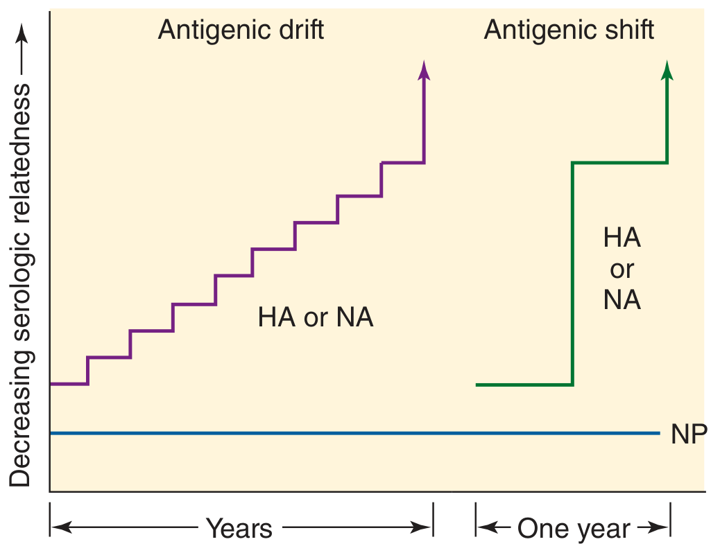
Fig. 39-3: Antigenic drift (gradual, stepwise change in HA/NA over years) vs. antigenic shift (abrupt, large change within one year). Internal proteins like NP remain constant. (Jawetz Melnick & Adelbergs, 28E)
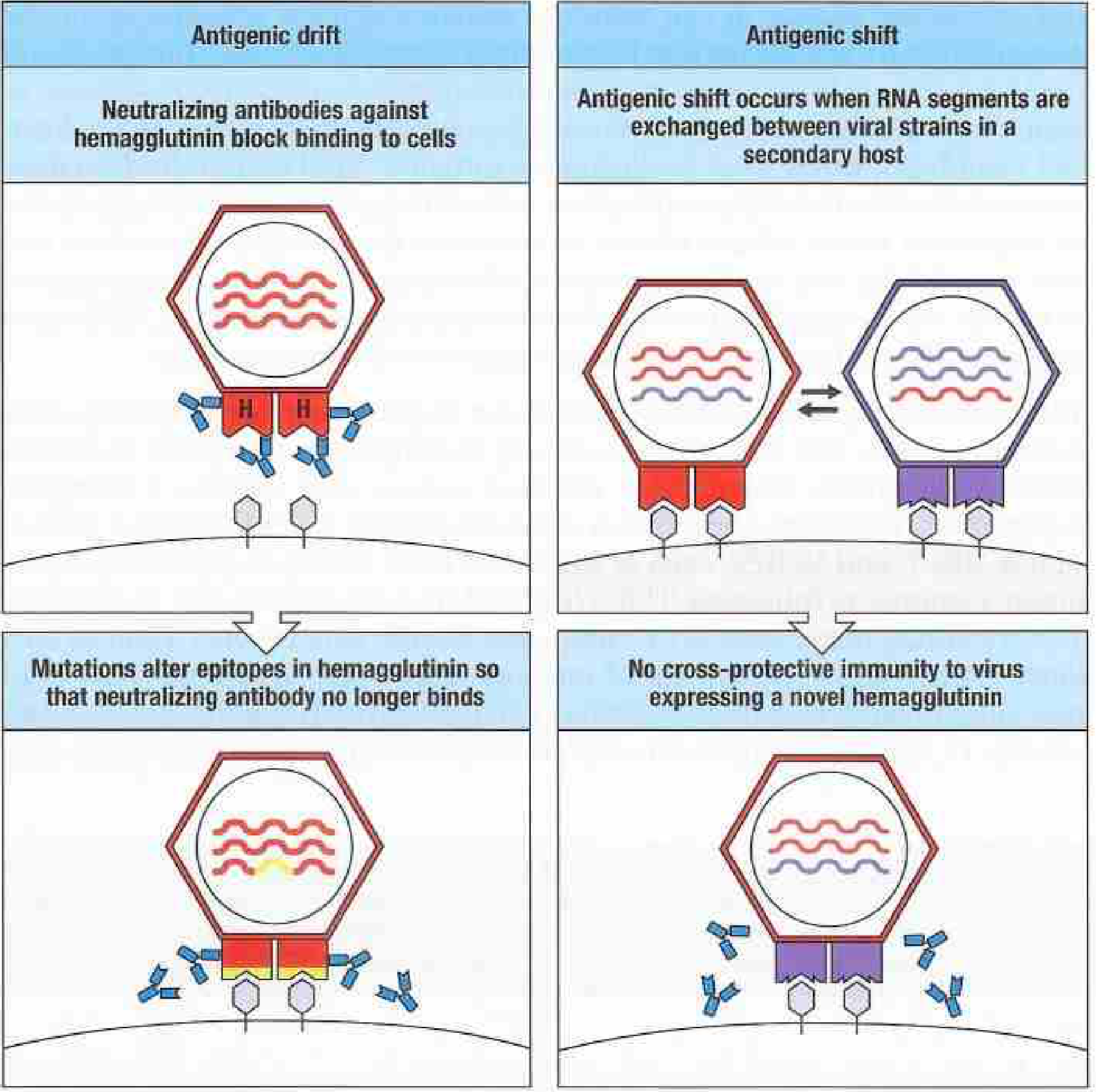
Fig. 12.45: Mechanism of antigenic drift and shift in influenza A. (Janeway's Immunobiology 10e)
A. Antigenic Drift
| Feature | Detail |
|---|---|
| Mechanism | Accumulation of point mutations in HA and/or NA genes |
| Result | Gradual amino acid changes at antigenic epitopes; virus "escapes" pre-existing antibody |
| Speed | Slow, stepwise over years |
| Viruses affected | All three types: A, B, and C |
| Epidemiologic consequence | Local/regional epidemics every 2-3 years (Influenza A); every 3-6 years (Influenza B) |
| Immune effect | Pre-existing antibody gives partial (reduced) protection - mild/modified disease, not complete failure |
| Minimum mutations | A variant must sustain 2 or more mutations before a new epidemiologically significant strain emerges |
Key concept: The immune system does not cause antigenic drift - it acts as a selection force that allows new antigenic variants (which evade existing antibody) to expand in the population.
- Jawetz Melnick & Adelbergs Medical Microbiology 28E, p. 590
B. Antigenic Shift
| Feature | Detail |
|---|---|
| Mechanism | Genetic reassortment of RNA gene segments during co-infection of a single cell by two different influenza A strains |
| Result | Abrupt, drastic replacement of HA and/or NA with an entirely novel subtype |
| Speed | Sudden - can occur within one year |
| Viruses affected | Influenza A ONLY (not B or C) |
| Epidemiologic consequence | Pandemic - globally immunologically naive population |
| Animal reservoir | Aquatic birds are the natural reservoir of all 18 HA subtypes; pigs serve as "mixing vessels" for human + avian strains |
Why only Influenza A?
Influenza B and C are restricted to humans (no significant animal reservoir), so there are no related animal strains to reassort with. Influenza A circulates in birds, pigs, horses, seals, whales, and humans - providing the diversity needed for reassortment.
The "mixing vessel" (pig) model:
Pig respiratory tract cells express receptors for both human influenza (SA α2,6 linkage) and avian influenza (SA α2,3 linkage). When a pig is simultaneously infected with a human and an avian strain, the 8 RNA segments from both viruses can randomly assort into progeny virions - generating a hybrid "reassortant" virus with a novel HA or NA.
- Medical Microbiology 9e, p. 4733
- Jawetz Melnick & Adelbergs Medical Microbiology 28E, pp. 590, 595
Comparison Table: Drift vs. Shift
| Feature | Antigenic Drift | Antigenic Shift |
|---|---|---|
| Mechanism | Point mutations | Genetic reassortment |
| Speed | Gradual (years) | Abrupt (sudden) |
| Magnitude of change | Minor | Major (new subtype) |
| Virus types | A, B, C | A only |
| Animal reservoir involved | No | Yes (birds, pigs) |
| Epidemiologic outcome | Epidemic | Pandemic |
| Pre-existing immunity | Partial protection | Little to no protection |
| Frequency | Continuous | Every 10-40 years |
| Gene segments changed | HA or NA point mutations | Entire HA and/or NA segment swapped |
Historical Pandemics (Antigenic Shift)
| Year | Subtype | Common Name |
|---|---|---|
| 1918 | H1N1 | Spanish flu (~50 million deaths) |
| 1957 | H2N2 | Asian flu |
| 1968 | H3N2 | Hong Kong flu |
| 1977 | H1N1 | Russian flu |
| 1997/2003 | H5N1 | Avian flu (China) |
| 2009 | H1N1 | Swine flu (quadruple reassortant: human + avian + 2 swine lineages) |
The 2009 H1N1 pandemic virus (A(H1N1)pdm09) was a quadruple reassortant containing gene segments from North American swine, Eurasian swine, avian, and human influenza viruses - a vivid example of multi-host reassortment.
- Jawetz Melnick & Adelbergs Medical Microbiology 28E, pp. 595-596
3. Nomenclature of Influenza A Strains
Format: Type / Host of origin / Geographic site / Strain number / Year (HA subtype N subtype)
Example: A/duck/Alberta/35/76 (H1N1) = Influenza A, isolated from a duck in Alberta, 1976, with H1 hemagglutinin and N1 neuraminidase.
For human strains, the host designation is omitted: A/California/04/2009 (H1N1)
Influenza B strains: no HA/NA subtype designation (no antigenic shift) - e.g., B/Singapore/3/64.
4. Key Antigens and Their Functions
| Protein | Function | Role in Immunity |
|---|---|---|
| HA (Hemagglutinin) | Binds sialic acid receptors on respiratory epithelium; mediates fusion; 18 subtypes | Primary target of neutralizing antibody; determines resistance to infection |
| NA (Neuraminidase) | Cleaves sialic acid; releases new virions; 11 subtypes | Antibody reduces severity and limits virus spread; target of oseltamivir/zanamivir |
| NP (Nucleoprotein) | Surrounds RNA; forms RNP complex | Determines A/B/C typing; no antigenic variation; target of cytotoxic T cells |
| M protein | Forms shell under lipid envelope | Determines A/B/C typing; no antigenic shift/drift |
| M2 | Ion channel; required for uncoating | Target of amantadine/rimantadine (Influenza A only) |
5. Laboratory Diagnosis
Specimens: Nasopharyngeal swabs, nasal aspirate, or nasal lavage - collected within 3 days of symptom onset for best yield.
A. Molecular Methods (Gold Standard)
RT-PCR (Reverse Transcriptase-PCR)
- Method of choice - rapid (<1 day), highly sensitive, highly specific
- Detects and distinguishes influenza A, B, and specific subtypes (e.g., H5N1, H3N2, H1N1pdm09)
- Multiplex RT-PCR panels simultaneously detect influenza A, B, RSV, adenovirus, and other respiratory viruses in a single run
- Can identify novel/pandemic strains and antiviral resistance mutations
- Specimens: nasopharyngeal swabs, nasal aspirate, bronchoalveolar lavage
B. Rapid Antigen Detection Tests (RIDTs)
- Results in <15-30 minutes at point of care
- Detect influenza A and B antigens (usually nucleoprotein) by immunochromatography
- Sensitivity: 50-70% (variable); Specificity: >90%
- A negative result does NOT rule out influenza - PCR must confirm if clinical suspicion high
- Useful for triage and infection control decisions; does not identify specific subtype
C. Viral Culture
- Cell lines: Primary monkey kidney cells or Madin-Darby canine kidney (MDCK) cells (preferred)
- Classic method: embryonated eggs (still used for vaccine production)
- CPE (cytopathic effects): often indistinct/subtle; not reliable alone
- Hemadsorption: Guinea pig erythrocytes added to infected cell monolayer will adhere to HA on infected cell surfaces - confirms virus presence even before CPE
- Hemagglutination (HA test): Added RBCs agglutinate in supernatant fluid due to viral HA - indicates virus present
- Culture takes 3-10 days; now largely replaced by molecular methods but still valuable for:
- Isolating strains for vaccine composition
- Antiviral susceptibility testing
- Definitive subtyping
D. Hemagglutination Inhibition (HI) Test
- Most important serologic method for strain identification and antibody measurement
- Principle: Specific antibody (in patient serum or reference antiserum) blocks HA from agglutinating RBCs
- Uses: (1) Identify virus type and subtype using reference antisera; (2) Measure patient antibody response
- Requires pretreatment of serum to destroy nonspecific mucoprotein inhibitors (receptor-destroying enzyme or kaolin)
- Fourfold or greater rise in titer between acute and convalescent sera = diagnostic
- HI titer ≥1:40 correlates with protective immunity
E. Immunofluorescence (DFA/IFA)
- Direct/indirect fluorescent antibody staining of respiratory epithelial cells in nasal aspirate
- Results in 2-4 hours; rapid but less sensitive than PCR
- Distinguishes influenza A from B using monoclonal antibodies
F. ELISA (Enzyme-Linked Immunosorbent Assay)
- Detects viral antigen in secretions or culture; more sensitive than HI for serology
- Serology: paired acute + convalescent sera; fourfold rise = infection
G. Complement Fixation (CF)
- Detects antibodies to internal proteins (NP, M) - type-specific but not subtype-specific
- Largely replaced by ELISA
Summary Table: Lab Diagnosis of Influenza
| Test | Detects | Time | Notes |
|---|---|---|---|
| RT-PCR | Viral RNA; type, subtype | <1 day | Gold standard; best sensitivity/specificity |
| Multiplex PCR | Multiple respiratory viruses | <1 day | Single specimen, broad panel |
| Rapid antigen test | HA/NP antigen | 15-30 min | Low sensitivity (~50-70%); quick triage only |
| Cell culture (MDCK) | Live virus | 3-10 days | Needed for vaccine strains; antiviral resistance |
| Hemadsorption | HA on infected cells | 2-5 days | Confirms culture positivity |
| Hemagglutination inhibition | Antibody/strain identity | Hours-days | Strain typing; immune status |
| Immunofluorescence | Viral antigen in cells | 2-4 hours | Moderate sensitivity |
| ELISA | Antigen or antibody | Hours | Sensitive serology |
| Complement fixation | Type-specific antibody | Days | Older method; type only |
- Medical Microbiology 9e (Murray), pp. 4859-4868
- Jawetz Melnick & Adelbergs Medical Microbiology 28E, pp. 5358-5395
6. Treatment and Prophylaxis (Brief Overview)
| Drug | Mechanism | Active Against |
|---|---|---|
| Oseltamivir (Tamiflu), Zanamivir, Peramivir | NA inhibitors - block virus release | Influenza A + B |
| Baloxavir marboxil | Inhibits PB2 cap-snatching polymerase | Influenza A + B |
| Amantadine, Rimantadine | M2 ion channel blockers | Influenza A only (widespread resistance; no longer recommended in US) |
Vaccines are reformulated annually based on WHO surveillance of circulating drift variants.
Quick Exam Memory Points
- Segmented genome (8 segments) = molecular basis of reassortment = antigenic shift
- Drift = mutations (slow, all types A/B/C) → epidemics
- Shift = reassortment (sudden, Type A ONLY) → pandemics
- Pig = mixing vessel (has both α2,3 and α2,6 sialic acid receptors)
- Birds = reservoir of all 18 HA subtypes
- HA subtype determines resistance to infection; NA subtype determines severity
- RT-PCR = gold standard for lab diagnosis
- HI test = identifies strain, measures protective antibody; fourfold rise = diagnosis
- Hemadsorption detects HA on culture before CPE develops
- Rapid antigen tests = fast but low sensitivity; negative does NOT rule out flu
- Original antigenic sin = anamnestic response to first-ever influenza subtype encountered, even when re-infected with a new drift variant
Orthomixo virus antigen shift and drift and lab diagnosis micro
This appears to be a duplicate of the previous question, which was just answered in full detail. Here is a concise summary of everything covered, for quick reference:
Orthomyxovirus: Antigenic Shift, Drift & Lab Diagnosis - Quick Reference
Structure Recap (Basis of Antigenic Variation)
- Family Orthomyxoviridae; enveloped, negative-sense ssRNA
- 8 segmented gene segments (Influenza A & B) - segmentation is the molecular basis of antigenic shift
- Key surface antigens: HA (Hemagglutinin) - 18 subtypes and NA (Neuraminidase) - 11 subtypes
- Internal antigens NP and M protein - determine A/B/C typing; do not undergo antigenic variation
Antigenic Drift
| Feature | Detail |
|---|---|
| Mechanism | Point mutations in HA or NA genes → amino acid changes at epitopes |
| Speed | Gradual, stepwise over years |
| Virus types | A, B, and C |
| Epidemiologic result | Local/regional epidemics (A: every 2-3 yrs; B: every 3-6 yrs) |
| Immune effect | Pre-existing antibody gives partial protection - mild/modified disease |
| Key point | Immune system acts as selection pressure - variants that escape antibody expand |
Antigenic Shift
| Feature | Detail |
|---|---|
| Mechanism | Genetic reassortment of RNA segments during co-infection with 2 different strains |
| Speed | Abrupt - can occur within one year |
| Virus types | Influenza A ONLY |
| Epidemiologic result | Pandemic - population has little/no pre-existing immunity |
| Animal reservoir | Aquatic birds (all 18 HA subtypes); pigs = "mixing vessel" |
Why only Type A? Types B and C are restricted to humans - no animal strains to reassort with. Influenza A circulates in birds, pigs, horses, seals - providing genomic diversity for reassortment.
Fig. 39-3: Antigenic drift vs. shift - HA/NA change gradually with drift, abruptly with shift; NP (internal protein) never changes. (Jawetz Melnick & Adelbergs, 28E)
Fig. 12.45: Mechanism of antigenic drift and shift. (Janeway's Immunobiology 10e)
Drift vs. Shift - Comparison Table
| Feature | Antigenic Drift | Antigenic Shift |
|---|---|---|
| Mechanism | Point mutations | Genetic reassortment |
| Speed | Gradual (years) | Sudden (months-year) |
| Change magnitude | Minor | Major (new subtype) |
| Virus types | A, B, C | A only |
| Animal reservoir | Not involved | Yes (birds, pigs) |
| Epidemic/Pandemic | Epidemics | Pandemics |
| Pre-existing immunity | Partial | None/minimal |
| Frequency | Continuous | Every 10-40 years |
Historical Pandemics (All Due to Antigenic Shift)
| Year | Subtype | Name |
|---|---|---|
| 1918 | H1N1 | Spanish flu (~50M deaths) |
| 1957 | H2N2 | Asian flu |
| 1968 | H3N2 | Hong Kong flu |
| 1977 | H1N1 | Russian flu |
| 2009 | H1N1 | Swine flu (quadruple reassortant: human + avian + 2 swine lineages) |
Laboratory Diagnosis
Specimen: Nasopharyngeal swab / nasal aspirate / nasal lavage - collected within 3 days of symptom onset
1. RT-PCR (Gold Standard)
- Detects viral RNA; <1 day turnaround
- Highly sensitive and specific
- Identifies type (A/B), subtype (H1N1, H3N2, H5N1), and antiviral resistance mutations
- Multiplex RT-PCR detects influenza + RSV + other respiratory viruses simultaneously
- Method of choice in current practice
2. Rapid Antigen Detection Tests (RIDTs)
- Results in 15-30 minutes
- Detects HA or NP antigen by immunochromatography
- Sensitivity: ~50-70%; Specificity: >90%
- Negative result does NOT rule out influenza - confirm with PCR if clinically suspected
- Useful for rapid triage and infection control
3. Viral Culture
- Cell lines: MDCK (Madin-Darby canine kidney) or primary monkey kidney cells; historically embryonated eggs
- Takes 3-10 days; CPE often indistinct
- Confirmed by hemadsorption (guinea pig RBCs adhere to HA on infected cell surface)
- or hemagglutination of supernatant fluid
- Still essential for: vaccine strain selection, antiviral susceptibility testing, pandemic surveillance
4. Hemagglutination Inhibition (HI) Test
- Most important serologic + strain-identification test
- Specific antibody blocks HA from agglutinating RBCs
- Uses: (1) Strain typing with reference antisera; (2) Measuring patient immune response
- Requires pre-treatment of serum to remove nonspecific inhibitors (receptor-destroying enzyme/kaolin)
- Diagnostic criterion: fourfold or greater rise in titer between acute and convalescent sera (collected 2-4 weeks apart)
- HI titer ≥1:40 = protective immunity
5. Immunofluorescence (DFA/IFA)
- Direct/indirect antibody staining of nasal epithelial cells
- Results in 2-4 hours; moderate sensitivity
- Distinguishes A from B; does not subtype
6. ELISA
- Detects antigen in secretions or culture supernatant
- More sensitive than HI for serology; widely used
7. Complement Fixation (CF)
- Detects antibodies to internal proteins (NP, M) - type-specific only (not subtype)
- Older method; largely replaced by ELISA and PCR
Lab Diagnosis Summary Table
| Test | Time | Sensitivity | Identifies Subtype | Use |
|---|---|---|---|---|
| RT-PCR | <1 day | Highest | Yes | Gold standard |
| Rapid antigen test | 15-30 min | Low (~50-70%) | No | Rapid triage |
| Viral culture (MDCK) | 3-10 days | High | With HI | Strain isolation, antiviral testing |
| Hemadsorption | 2-5 days | High | No (detects HA) | Culture confirmation |
| HI test | Hours | High | Yes | Strain ID, immunity testing |
| Immunofluorescence | 2-4 hours | Moderate | A vs B only | Rapid antigen in cells |
| ELISA | Hours | High | No | Sensitive serology |
| Complement fixation | Days | Moderate | Type only | Epidemiology |
Key Exam Memory Points
- 8 segments → reassortment → antigenic shift
- Drift = point mutations → gradual → A, B, C → epidemics
- Shift = reassortment → sudden → Type A ONLY → pandemics
- Pig = mixing vessel (expresses both α2,3 avian and α2,6 human sialic acid receptors)
- RT-PCR = gold standard lab diagnosis
- HI test = identifies strain + measures antibody; fourfold rise = acute infection
- Hemadsorption = guinea pig RBCs stick to HA on infected cells - detects culture before CPE
- Rapid antigen tests - negative does NOT rule out flu
- HA antibody → prevents infection; NA antibody → reduces severity
- Original antigenic sin - anamnestic response biased toward first influenza subtype ever encountered
- Jawetz Melnick & Adelbergs Medical Microbiology 28E
- Medical Microbiology 9e (Murray)
- Janeway's Immunobiology 10e
This is a shared conversation. Sign in to Orris to start your own chat.