
Fibronectin
I'll look up fibronectin from the medical library for a thorough, textbook-based answer.
Reading File
Now let me search for fibronectin content in parallel across headings and paragraphs:
Finding Sources
Finding Sources
Searching the Web
fibronectin structure function extracellular matrix clinical significance
Excellent results. Now let me read the key sections from Harper's Biochemistry (structure/function) and Junqueira's Histology, plus the rheumatology/clinical sections in parallel:
Reading File
Reading File
Reading File
Reading File
Now let me fetch the fibronectin structure image and read the clinical sections on fetal fibronectin and fibronectin glomerulopathy:
Loading Image
Loading Image
Reading File
Reading File
I now have rich, comprehensive content from multiple authoritative textbooks. Here is the full answer:
Fibronectin
Fibronectin (Latin: fibra = fiber, nexus = interconnection) is a high-molecular-weight multiadhesive glycoprotein of the extracellular matrix (ECM) that plays central roles in cell adhesion, migration, wound healing, and tissue organization.
Structure
Fibronectin is a dimer consisting of two nearly identical polypeptide subunits (~230-270 kDa each), joined by two disulfide bridges near their carboxyl terminals. The gene encoding fibronectin is very large (~50 exons), and its mRNA undergoes extensive alternative splicing - up to 20 different mRNA isoforms have been detected in various tissues.
Each monomer contains three types of repeating structural motifs organized into at least seven functional domains:
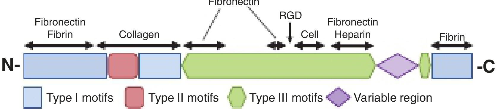
Structure of the fibronectin monomer (Harper's Illustrated Biochemistry, 32nd Ed., Fig. 50-4)
| Domain | Binding Partner |
|---|---|
| N-terminal Type I motifs | Fibrin, fibronectin itself |
| Type II motif region | Collagen / gelatin (denatured collagen) |
| Central Type III motifs (large) | Contains RGD sequence - binds integrins (primarily α3β1, also αvβ3) |
| Variable region | Alternatively spliced; tissue-specific |
| Heparin-binding domain (×2 per subunit, ~20 kDa each) | Heparan sulfate proteoglycans (e.g., syndecan) |
| C-terminal Type I motifs | Fibrin |
The RGD (Arg-Gly-Asp) sequence is the classic integrin-binding motif. Synthetic peptides containing RGD can competitively inhibit fibronectin-cell binding. - Harper's Illustrated Biochemistry, 32nd Ed., p. 616
Two Major Isoforms
| Form | Location | Function |
|---|---|---|
| Plasma fibronectin (soluble) | Circulates in blood and body fluids | Enhances blood clotting, wound healing, phagocytosis |
| Cellular fibronectin (insoluble fibrillar) | Assembled on cell surfaces, deposited in ECM | Tissue architecture, matrix assembly, cell guidance |
Fibrillar forms assemble on cell surfaces and are deposited into the ECM. The fibronectin fibrils on fibroblasts are coupled with intracellular actin stress fibers - the contractile actin-myosin cytoskeleton pulls on the fibronectin matrix and generates mechanical tension. - Sabiston Textbook of Surgery, p. 399
Synthesis
Fibronectin is synthesized largely by fibroblasts, though many cell types contribute. It is a 235-270 kDa dimer in connective tissue and forms insoluble fibrillar networks throughout connective tissue. - Junqueira's Basic Histology, 17e
Fibronectin-Integrin Signaling (Focal Adhesions)
The fibronectin-integrin receptor interaction does not merely anchor cells - it transmits signals from outside to inside the cell:
- Fibronectin binds transmembrane integrin receptors (heterodimers with α and β chains)
- Integrins connect intracellularly via attachment proteins: talin, vinculin, α-actinin, paxillin
- These complexes cluster into focal adhesions - large multiprotein complexes that:
- Anchor cells physically to the ECM
- Relay extracellular signals that influence cell behavior (proliferation, survival, differentiation)
- Focal adhesion kinases (FAK) provide a mechanism by which pulling forces trigger intracellular signaling cascades
This outside-in signaling is a key mechanism by which the ECM controls cell fate. - Harper's Illustrated Biochemistry, 32nd Ed., p. 616
Immunohistochemical Localization
The image below (immunohistochemistry, ×400) shows fibronectin forming a fine golden network throughout the ECM of connective tissue:
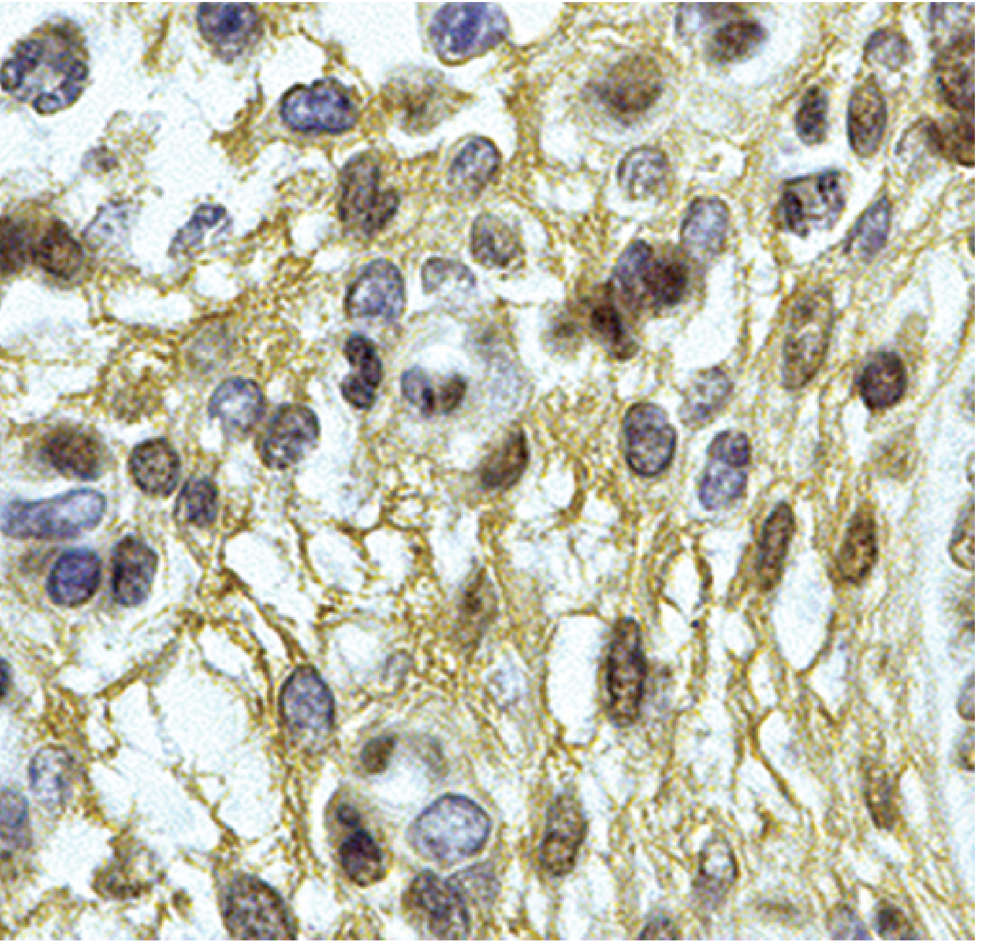
Fibronectin localization by immunohistochemistry (Junqueira's Basic Histology, 17e, Fig. 5-17)
Functions Summary
| Function | Mechanism |
|---|---|
| Cell adhesion | RGD domain binds integrins; fibrils provide substrate |
| Cell migration | Provides a molecular "rail" for cells to steer through ECM during development, repair, and metastasis |
| Wound healing | Soluble plasma fibronectin promotes clot formation and scaffolding |
| Phagocytosis | Acts as an opsonin (plasma form) |
| Matrix assembly | Guides orientation and assembly of ECM components |
| Signal transduction | Focal adhesions relay mechanical and biochemical signals via FAK and integrins |
| Embryogenesis | Essential for cell migration and tissue morphogenesis |
Role in Disease
1. Osteoarthritis (OA)
Fragments of fibronectin (not intact molecules) stimulate chondrocytes to produce matrix metalloproteinases (MMPs) and drive cartilage breakdown. Fibronectin is upregulated in articular cartilage at the earliest stages of OA in multiple species, possibly as part of an attempted repair response - but the resulting increased fibronectin availability means more substrate for fragmentation and further tissue degradation. - Rheumatology, 2-Volume Set (Elsevier 2022)
2. Cancer
Transformed/malignant cells have sharply reduced fibronectin around them, partly explaining their faulty interaction with the ECM, loss of contact inhibition, and enhanced migratory capacity. Abnormal fibronectin expression also facilitates tumor cell migration and metastasis.
3. Fibronectin Glomerulopathy
A rare, autosomal dominant familial glomerular disease caused by mutations in the FN1 gene (chromosome 2q32). Clinical features:
- Presents in early adolescence
- Proteinuria, microhematuria, hypertension
- Distal (type 4) renal tubular acidosis
- Slowly progressive to ESKD (between 2nd and 6th decades)
Pathology: Enlarged, lobular, normocellular glomeruli with PAS-positive, Congo red-negative material in mesangial and subendothelial areas. Immunofluorescence is strongly positive with anti-fibronectin antibody. EM shows granular to fibrillary subendothelial deposits. No effective treatment; can recur in renal allografts. - Comprehensive Clinical Nephrology, 7th Ed.
4. Fibrosis
Excessive fibronectin deposition is a hallmark of fibrotic diseases (liver cirrhosis, pulmonary fibrosis).
5. Microbial Pathogenesis
Streptococcus pyogenes has fibronectin-binding protein (Protein F) and fimbriae that bind fibronectin, facilitating bacterial adhesion to host tissues. Many pili also bind fibronectin. - Sherris & Ryan's Medical Microbiology, 8th Ed.
Fetal Fibronectin (Clinical Use in Obstetrics)
Fetal fibronectin acts as an adhesive glycoprotein at the maternal-fetal interface (choriodecidual junction). It is normally absent from cervicovaginal secretions between 22-35 weeks of gestation. Its presence indicates disruption of the maternal-fetal decidual attachment.
Clinical significance:
-
A positive fetal fibronectin test (cervicovaginal, before digital exam) predicts increased risk of preterm birth, especially within 2 weeks of the positive result
-
Sensitivity for spontaneous preterm birth <35 weeks at 22-24 weeks is ~25%, but rises to 65% for births <28 weeks
-
Antibiotic trials in women with positive fFN have been disappointing (no benefit, slightly higher preterm rates with antibiotics)
-
Screening of asymptomatic women is not recommended
-
Elastase-mediated degradation of glycosylated fetal fibronectin reduces its binding capacity, facilitating membrane separation from decidua - a mechanism in parturition
-
Creasy & Resnik's Maternal-Fetal Medicine, Principles and Practice
Key takeaway: Fibronectin is a multifunctional ECM dimer that integrates structural and signaling roles through its modular domain architecture and integrin binding, with critical roles in cell adhesion, migration, wound repair, and embryogenesis - and its dysregulation underlies several important diseases from osteoarthritis to cancer to rare glomerular disease.
This is a shared conversation. Sign in to Orris to start your own chat.