
Janus kinase receptor
Janus kinase JAK STAT signaling pathway diagram
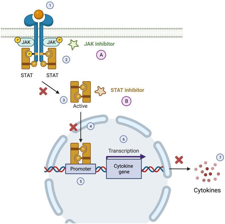
A pathophysiology diagram illustrating the JAK-STAT signaling pathway and points of therapeutic inhibition. The process begins at the cell membrane where a cytokine ligand (1) binds to a transmembrane receptor. This triggers Janus Kinases (JAK) to phosphorylate the receptor tails, facilitating the docking and phosphorylation of signal transducer and activator of transcription (STAT) proteins (2). Phosphorylated STATs then dissociate (3) and form an active dimer which translocates into the nucleus (4). Inside the nucleus, the STAT dimer binds to a specific promoter region on the DNA (5), initiating the transcription of cytokine genes (6) and resulting in the extracellular release of cytokines (7). The diagram highlights two pharmacological targets: JAK inhibitors (A), which act near the membrane to prevent phosphorylation; and STAT inhibitors (B), which are shown blocking the translocation of STAT dimers and the subsequent production of pro-inflammatory cytokines, indicated by red 'X' marks. This schematic is utilized to explain the mechanism of action for precision medicine in treating primary immune disorders and inflammatory conditions.
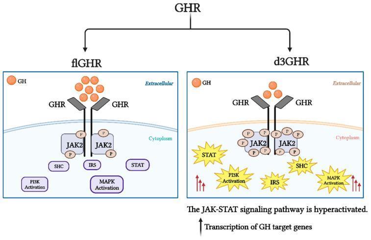
This pathophysiology diagram compares the intracellular signaling pathways of the full-length growth hormone receptor (flGHR) and the exon 3-deleted growth hormone receptor (d3GHR). Both pathways illustrate Growth Hormone (GH) binding to the extracellular domain of the GHR, leading to the recruitment and phosphorylation of Janus kinase 2 (JAK2) in the cytoplasm. Downstream signal transduction involves the activation of SHC, IRS, STAT, PI3K, and MAPK pathways. In the d3GHR variant, the diagram depicts hyperactivation of these signaling cascades, visually indicated by 'burst' icons around the cytoplasmic proteins (STAT, PI3K, IRS, MAPK), multiple phosphorylation markers on JAK2, and red upward arrows. This hyperactivation in the d3GHR pathway is associated with increased sensitivity of the JAK-STAT pathway, resulting in enhanced transcription of GH target genes compared to the standard flGHR isoform. The illustration serves as an educational tool for endocrinology and molecular biology to demonstrate how genetic variations in receptor structure influence hormonal signal intensity and gene expression.
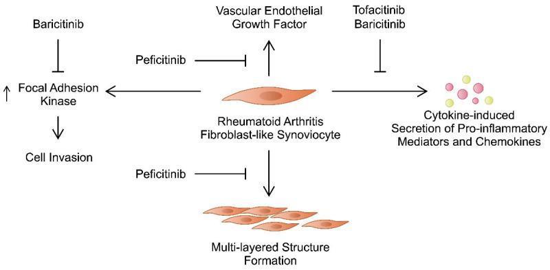
This pathophysiology diagram illustrates the signaling pathways and cellular mechanisms involving Rheumatoid Arthritis Fibroblast-like Synoviocytes (RA-FLS) and their inhibition by Janus kinase (JAK) inhibitors. The central element is an RA-FLS, which promotes several pathogenic processes: secretion of Vascular Endothelial Growth Factor (VEGF), cytokine-induced production of pro-inflammatory mediators and chemokines, and the formation of a multi-layered synovial lining structure. Additionally, the diagram shows a pathway where Focal Adhesion Kinase (FAK) activation leads to increased cell invasion. The educational focus is on the targeted therapeutic effects of specific drugs: Baricitinib is shown inhibiting FAK; Peficitinib is depicted inhibiting both VEGF production and the formation of the multi-layered structure; and both Tofacitinib and Baricitinib are shown inhibiting the secretion of pro-inflammatory mediators. Standard notation is used, with arrows representing stimulation/progression and perpendicular bars representing inhibition. This schematic is designed for medical education regarding the pharmacological management of rheumatoid arthritis at the molecular and cellular level.
Janus Kinase (JAK) Receptor System
What Is a JAK-Associated Receptor?
The JAK Family
| JAK | Primary Ligands / Functions |
|---|---|
| JAK1 | Broad - IL-6, IFN-γ, IL-2 family; inflammation, growth, T cell differentiation |
| JAK2 | EPO, GH, GM-CSF, G-CSF, IFN-γ; hematopoiesis, cell division, metabolism |
| JAK3 | γc-chain cytokines (IL-2, IL-4, IL-7, IL-9, IL-15, IL-21); T cell proliferation, immune response |
| TYK2 | IFN-α/β, IL-10, IL-12, IL-23; antiviral immunity, inflammation |
- Schwartz's Principles of Surgery, 11th ed.
- Firestein & Kelley's Textbook of Rheumatology, 2-Volume Set
Mechanism of Signaling (JAK-STAT Pathway)
- Cytokine binds to the extracellular domain of its specific receptor
- Receptor dimerizes (or oligomerizes), bringing constitutively bound JAKs into proximity
- Trans-phosphorylation - JAKs phosphorylate each other and then phosphorylate tyrosine residues on the receptor's cytoplasmic tail
- STAT recruitment - phosphorylated receptor tails act as docking sites for STAT proteins (via their SH2 domains)
- STAT phosphorylation by JAKs causes STATs to detach from the receptor
- STAT dimerization - activated STATs form homo- or heterodimers
- Nuclear translocation - STAT dimers enter the nucleus and bind promoter regions, driving transcription of cytokine-responsive genes
Cytokine-JAK-STAT Pairings
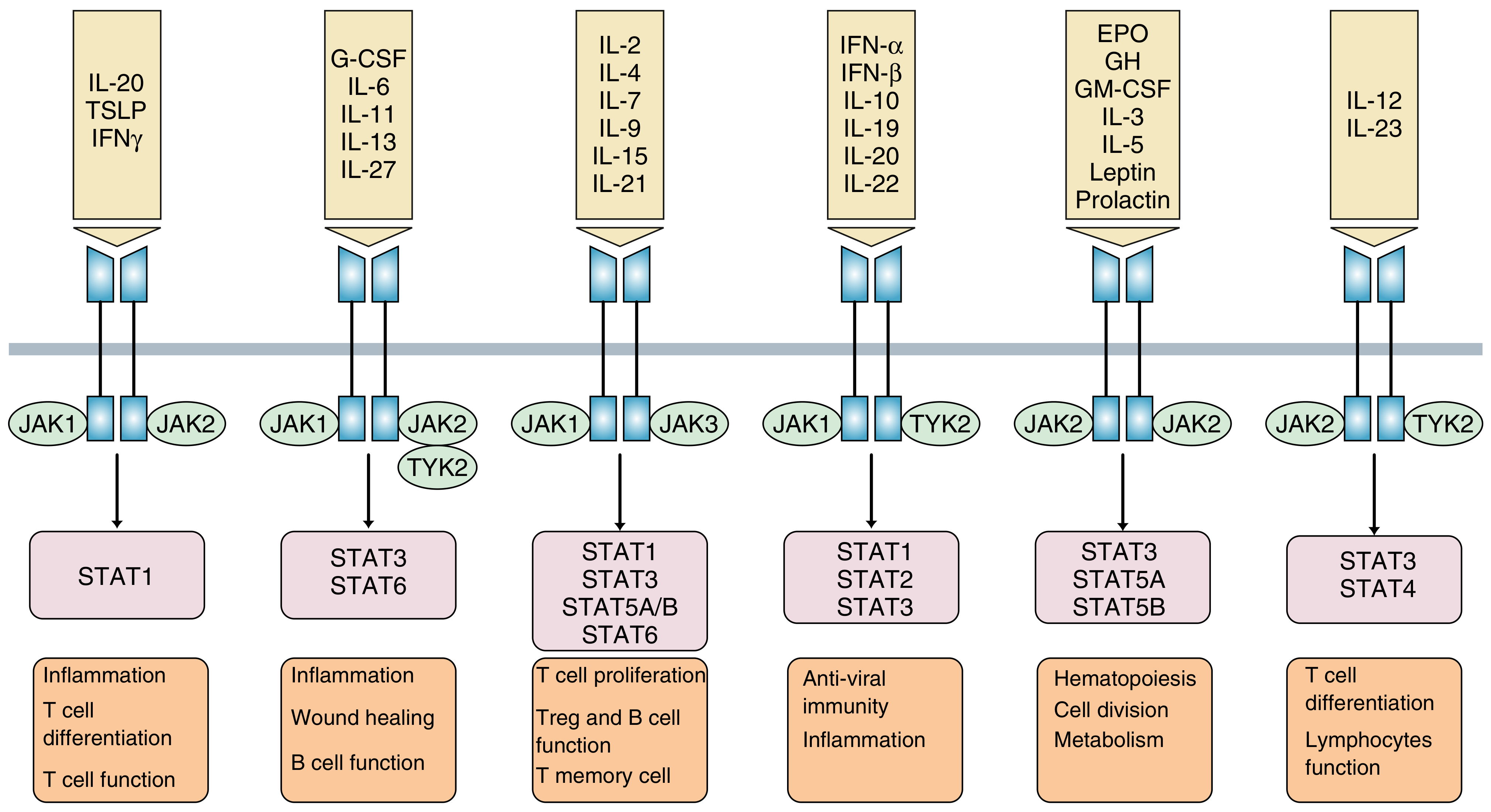
- IL-20, TSLP, IFN-γ → JAK1/JAK2 → STAT1 → inflammation, T cell differentiation
- IL-6, IL-11, IL-13, IL-27 → JAK1/JAK2 (+TYK2) → STAT3/STAT6 → inflammation, wound healing, B cell function
- IL-2, IL-4, IL-7, IL-15, IL-21 → JAK1/JAK3 → STAT1/3/5/6 → T cell proliferation, Treg/B cell function
- IFN-α/β, IL-10, IL-22 → JAK1/TYK2 → STAT1/2/3 → antiviral immunity
- EPO, GH, GM-CSF, Leptin, Prolactin → JAK2/JAK2 → STAT3/5A/5B → hematopoiesis, metabolism
- IL-12, IL-23 → JAK2/TYK2 → STAT3/STAT4 → T cell differentiation, lymphocyte function
The STAT Proteins
- An SH2 domain (for receptor docking)
- A DNA-binding domain
- A transactivation domain
-
STAT1 LOF → susceptibility to Salmonella and Mycobacterium
-
STAT1 GOF → autoimmunity + fungal susceptibility
-
STAT3 LOF (AD) → Hyper-IgE (Job) syndrome with failure to produce IL-17
-
STAT3 GOF → systemic autoimmunity
-
STAT4 polymorphisms → associated with RA, SLE, Sjögren syndrome
-
Rheumatology, 2-Volume Set (Elsevier 2022), pp. 127-128
Negative Regulation
- Phosphatases - dephosphorylate activated JAKs and STATs
- Nuclear export of STATs
- SOCS proteins (Suppressors of Cytokine Signaling): SOCS1-7 and CIS form a negative feedback loop - they are themselves induced by JAK/STAT signaling, creating an inhibitory circuit. Pattern recognition receptors (TLRs, C-type lectins) also activate SOCS.
Disease Associations (Genetic Evidence)
| Mutation | Consequence |
|---|---|
| JAK3 LOF | Autosomal recessive SCID (failure to signal γc cytokines) |
| TYK2 LOF | Susceptibility to mycobacterial and viral infections |
| JAK1 GOF | Systemic autoimmunity |
| JAK2 V617F GOF | Polycythemia vera and other myeloproliferative neoplasms (constitutive activation) |
| TYK2 polymorphisms | Associated with autoimmune diseases |
JAK Inhibitors (Jakinibs) - Clinical Applications
| Drug | JAK Selectivity | Approved Indications |
|---|---|---|
| Tofacitinib | JAK1 > JAK3 > JAK2, TYK2 | RA, psoriatic arthritis, juvenile arthritis, UC |
| Baricitinib | JAK1/JAK2 (moderate TYK2, minimal JAK3) | RA, COVID-19 (emergency use) |
| Upadacitinib | JAK1-selective | RA, UC, CD |
| Filgotinib | JAK1-selective | RA (Europe) |
| Ruxolitinib | JAK1/JAK2 | Myelofibrosis, polycythemia vera |
| Fedratinib, Pacritinib | JAK2-selective | Myelofibrosis |
- Harrison's Principles of Internal Medicine, 22nd ed.
- Goodman & Gilman's Pharmacological Basis of Therapeutics
- Rheumatology (Elsevier 2022), pp. 128-129