
Axilla anatomy
Reading File
Now let me search the library for axilla anatomy content in parallel:
Finding Sources
Finding Sources
Gray's Anatomy for Students has the most comprehensive coverage. Let me read the main AXILLA section and Contents section in parallel:
Reading File
Reading File
Excellent. Now let me read the walls section and continue the contents section, and fetch the key diagrams in parallel:
Reading File
Reading File
Loading Image
Now let me get the brachial plexus, axillary lymph nodes, and the key artery/branches diagram:
Reading File
Loading Image
Loading Image
Now let me get the brachial plexus section and axillary lymph nodes section:
Reading File
Finding Sources
Finding Sources
Reading File
Loading Image
Now let me get the axillary lymph nodes section from Gray's Anatomy and the posterior wall content:
Reading File
Reading File
Now let me get the axillary lymph nodes and the quadrangular/triangular spaces sections:
Reading File
Reading File
Now let me get the lymph nodes section from Gray's Anatomy:
Finding Sources
Reading File
Reading File
I now have comprehensive content. Let me fetch the lymph nodes diagram:
Loading Image
I now have all the information needed for a thorough, well-illustrated response. Here is the complete anatomy of the axilla:
Axilla Anatomy
The axilla is the gateway to the upper limb, providing the transition zone between the neck and the arm. It is an irregularly shaped pyramidal space formed by the clavicle, scapula, upper thoracic wall, humerus, and related muscles. All major structures passing to and from the upper limb travel through it.
- Gray's Anatomy for Students, p. 841
Boundaries
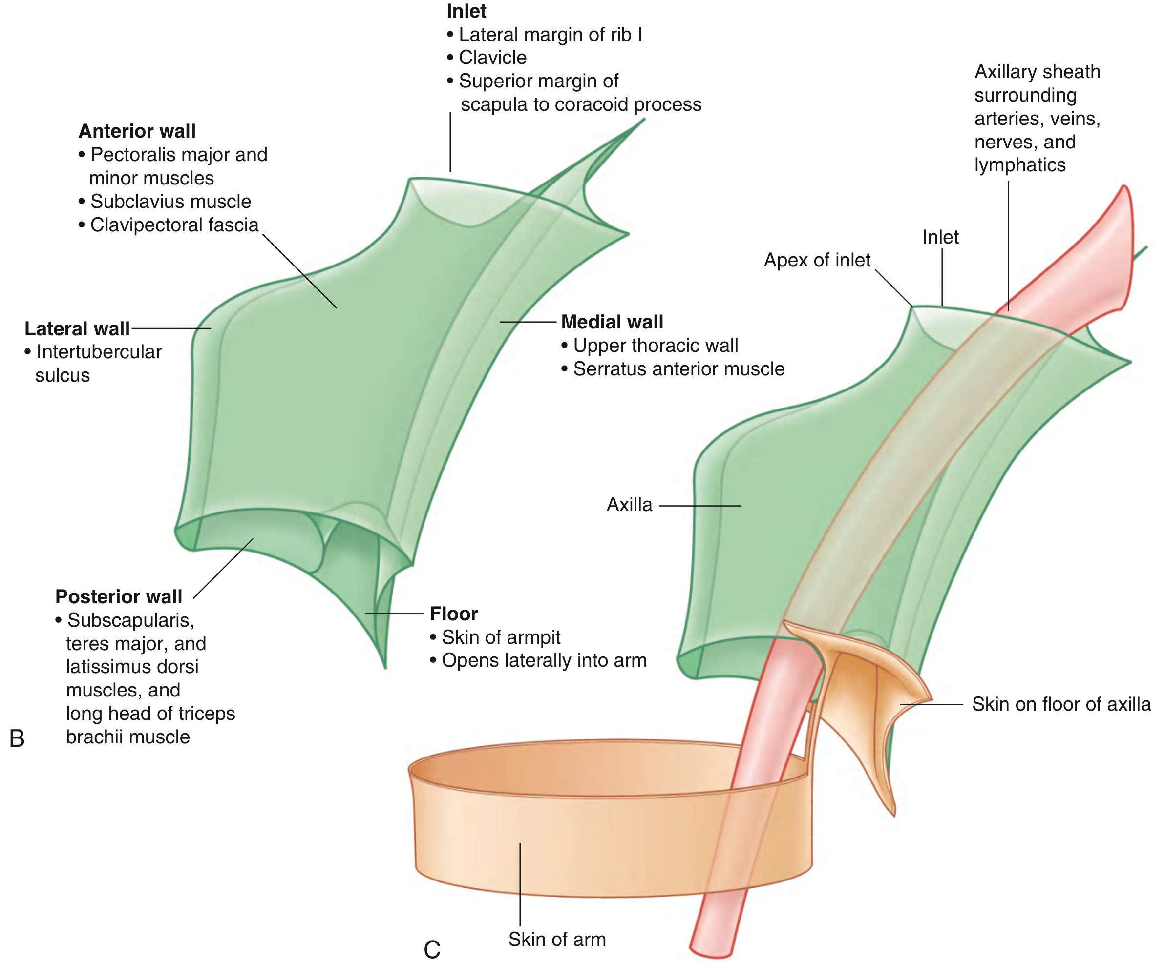
The axilla has:
- 1 inlet (apex) - superiorly continuous with the neck
- 4 walls - anterior, posterior, medial, lateral
- 1 floor (base) - inferiorly, opens into the arm
Axillary Inlet (Apex)
Triangular in shape, bounded entirely by bone:
- Medial margin: lateral border of rib I
- Anterior margin: posterior surface of clavicle
- Posterior margin: superior border of scapula to the coracoid process
- Apex (lateral point): medial aspect of the coracoid process
The subclavian artery/vein become the axillary artery/vein as they cross the lateral margin of rib I. At the inlet, the axillary vein is anterior, then the axillary artery, then the trunks of the brachial plexus posteriorly.
Walls
Anterior Wall
Formed by:
- Pectoralis major (largest, most superficial; its inferior margin underlies the anterior axillary fold) - innervated by lateral and medial pectoral nerves (C5-T1)
- Pectoralis minor (deep; pulls the shoulder tip down, protracts scapula) - innervated by medial and lateral pectoral nerves (C5-T1)
- Subclavius (small; stabilises the sternoclavicular joint) - innervated by nerve to subclavius (C5, C6)
- Clavipectoral fascia - spans between subclavius and pectoralis minor; pierced by the thoraco-acromial artery, cephalic vein, and lateral pectoral nerve
Medial Wall
Formed by:
- Upper thoracic wall (ribs 1-4 and intercostal muscles)
- Serratus anterior - originates from lateral surfaces of upper 8-9 ribs; innervated by the long thoracic nerve (C5, C6, C7); protracts and rotates the scapula
Posterior Wall
The most complex wall. Bone framework = costal surface of scapula. Muscles:
- Subscapularis - fills the subscapular fossa; medial rotator of arm; innervated by upper and lower subscapular nerves (C5, C6)
- Teres major - medial rotation and extension; innervated by lower subscapular nerve (C5, C6, C7)
- Latissimus dorsi - adduction, medial rotation, extension; innervated by thoracodorsal nerve (C6, C7, C8)
- Long head of triceps brachii - passes vertically through the wall from the infraglenoid tubercle
Gaps between these posterior wall muscles create three important apertures:
| Space | Boundaries | Contents |
|---|---|---|
| Quadrangular space | Inferior margin of subscapularis, surgical neck of humerus, superior margin of teres major, lateral margin of long head of triceps | Axillary nerve, posterior circumflex humeral artery/vein |
| Triangular space | Medial margin of long head of triceps, superior margin of teres major, inferior margin of subscapularis | Circumflex scapular artery/vein |
| Triangular interval | Lateral margin of long head of triceps, shaft of humerus, inferior margin of teres major | Radial nerve, profunda brachii artery |
Lateral Wall
The narrowest wall - formed by the intertubercular (bicipital) sulcus of the humerus.
Floor (Base)
Formed by fascia and a dome of skin spanning the inferior margins of all walls, supported by the clavipectoral fascia. The anterior axillary fold is more superior than the posterior axillary fold.
Contents
Axillary Artery
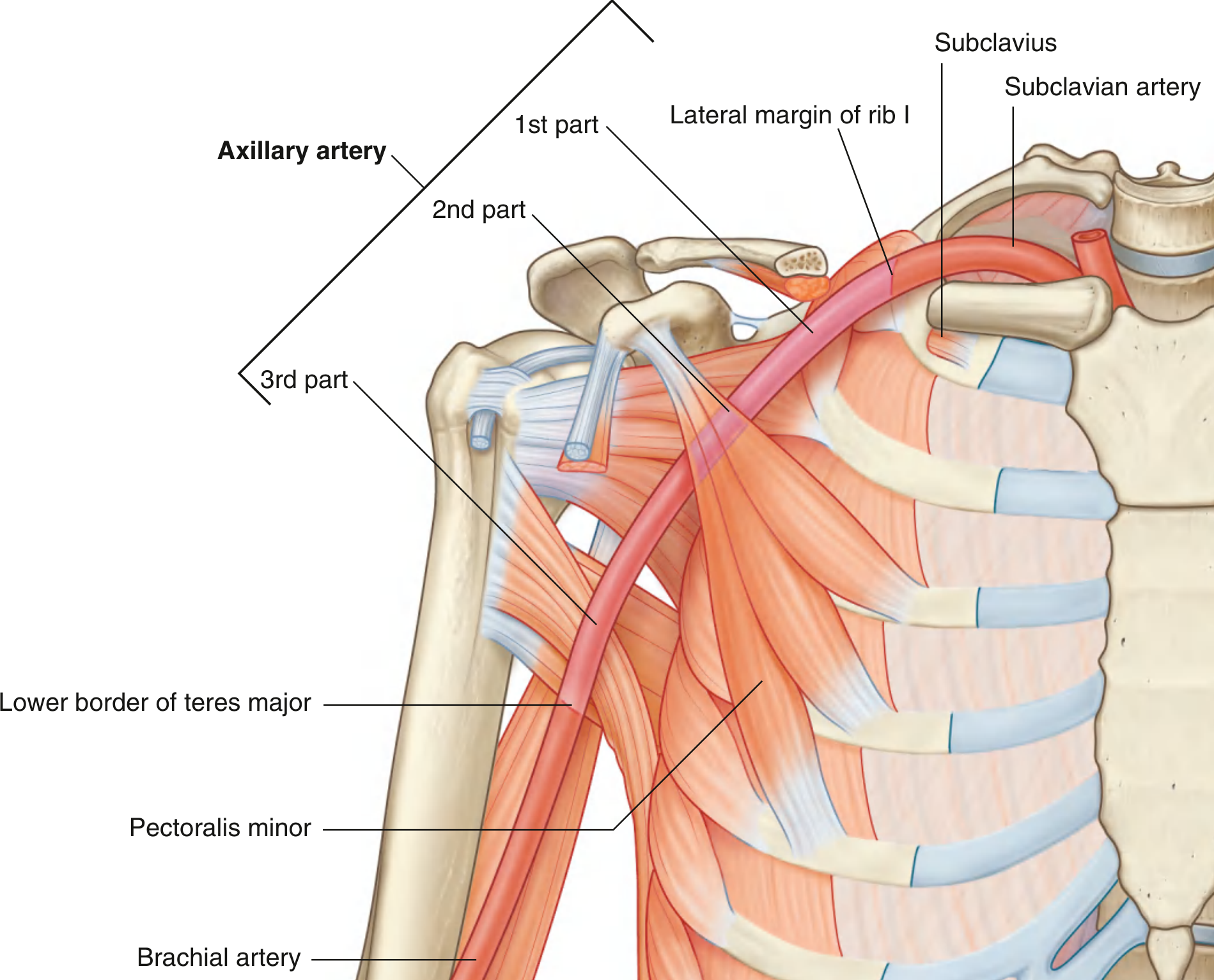
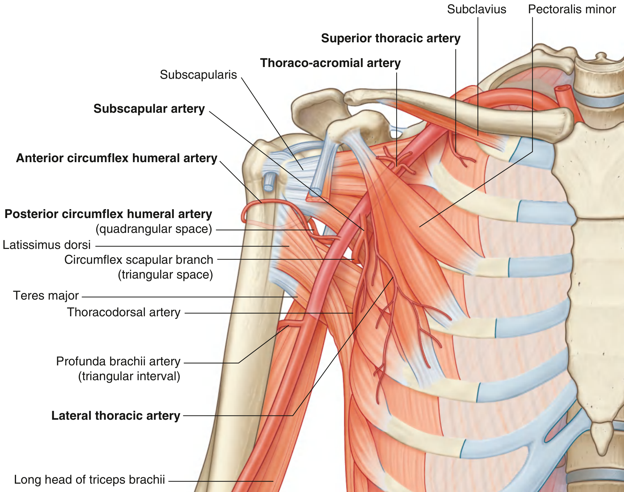
The subclavian artery becomes the axillary artery at the lateral margin of rib I, and becomes the brachial artery at the inferior margin of teres major. The pectoralis minor divides it into three parts:
| Part | Relation to Pec Minor | Branches (mnemonic: 1-2-3) |
|---|---|---|
| 1st | Proximal to it | Superior thoracic artery (1 branch) |
| 2nd | Posterior to it | Thoraco-acromial artery + Lateral thoracic artery (2 branches) |
| 3rd | Distal to it | Subscapular artery + Anterior circumflex humeral + Posterior circumflex humeral (3 branches) |
The subscapular artery (largest branch) divides into the circumflex scapular artery (passes through triangular space) and thoracodorsal artery (supplies latissimus dorsi).
Axillary Vein
Formed at the inferior margin of teres major by the union of the venae comitantes of the brachial artery and the basilic vein. It lies anterior to the axillary artery throughout. Becomes the subclavian vein as it crosses rib I.
Brachial Plexus

Formed by anterior rami of C5-C8 and T1. The sequence of components is: Roots → Trunks → Divisions → Cords → Branches (mnemonic: "Real Tourists Drink Cold Beer").
Trunks (over rib I):
- Superior = C5 + C6
- Middle = C7
- Inferior = C8 + T1
Divisions: Each trunk splits into anterior and posterior.
Cords (named by position relative to the axillary artery's second part):
- Lateral cord - from anterior divisions of superior and middle trunks
- Medial cord - from anterior division of inferior trunk
- Posterior cord - from all three posterior divisions
Terminal branches in the axilla:
| Cord | Branches |
|---|---|
| Lateral | Lateral pectoral nerve, musculocutaneous nerve, lateral root of median nerve |
| Medial | Medial pectoral nerve, medial cutaneous nerve of arm, medial cutaneous nerve of forearm, medial root of median nerve, ulnar nerve |
| Posterior | Superior subscapular nerve, thoracodorsal nerve, inferior subscapular nerve, axillary nerve (exits via quadrangular space), radial nerve (exits via triangular interval) |
Axillary Lymph Nodes
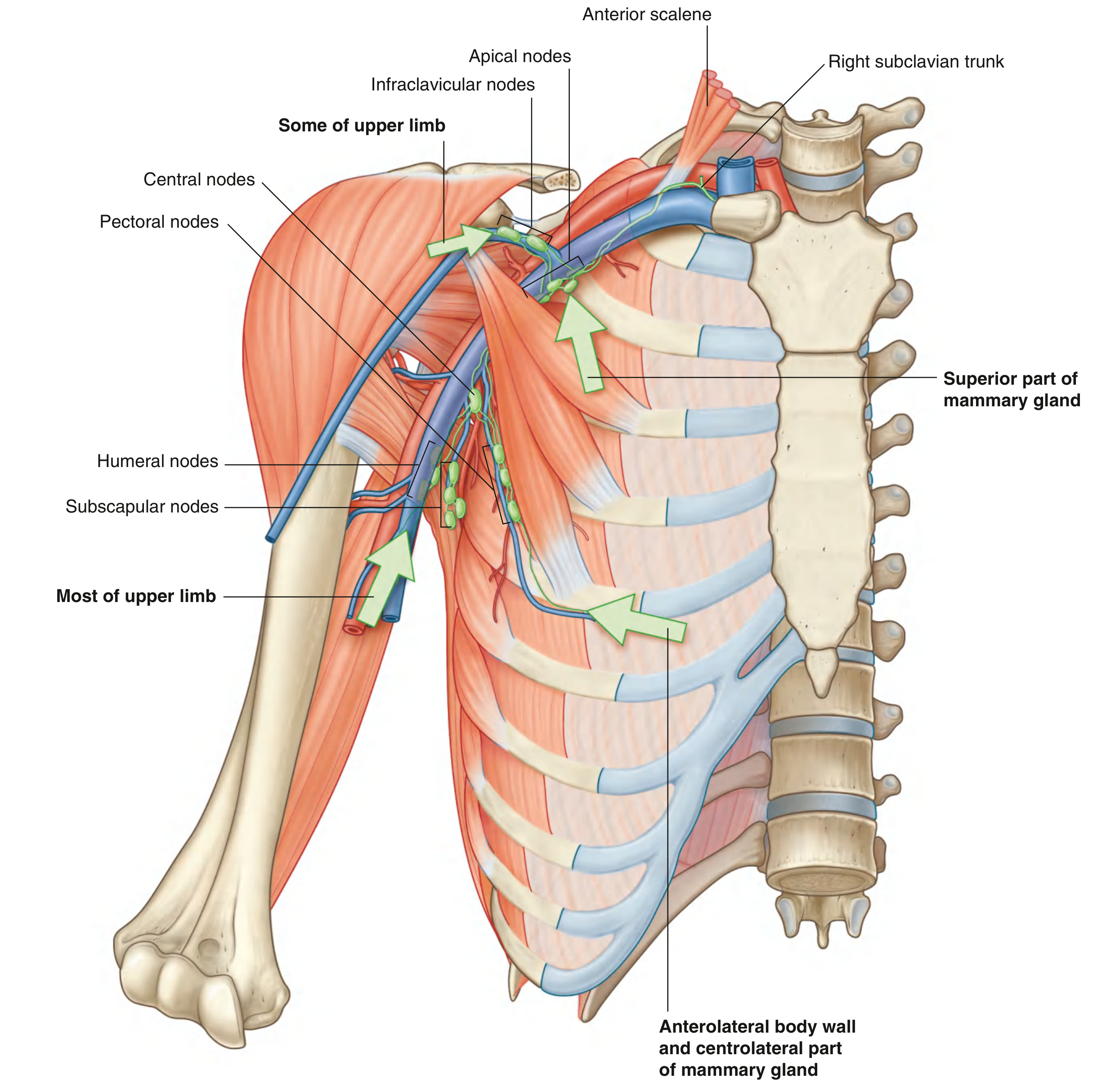
There are 30-60 lymph nodes in the axilla, arranged in five named groups and three surgical levels (defined by their relationship to pectoralis minor):
| Group | Level | Location | Drains |
|---|---|---|---|
| Humeral (lateral) | I | Posteromedial to axillary vein | Most of the upper limb |
| Pectoral (anterior) | I | Along lateral thoracic vessels/inferior pec minor margin | Breast, chest wall, abdominal wall |
| Subscapular (posterior) | I | Posterior axillary wall with subscapular vessels | Back, shoulder, neck |
| Central | II | Embedded in axillary fat | Receives from humeral, subscapular, pectoral groups |
| Apical (infraclavicular) | III | Apex of axilla, medial to pec minor | All other groups + cephalic vein lymphatics + superior breast |
Efferent vessels from the apical nodes form the subclavian trunk, which drains to the right lymphatic duct (right side) or thoracic duct (left side).
Clinical significance: Breast cancer metastasises to axillary nodes along this hierarchical chain. Axillary lymph node dissection or radiotherapy can damage lymphatic drainage, causing lymphoedema of the arm.
Other Axillary Contents
- Biceps brachii (short head from coracoid apex; long head via intertubercular sulcus from supraglenoid tubercle) - both innervated by musculocutaneous nerve (C5, C6)
- Coracobrachialis (from coracoid apex; musculocutaneous nerve pierces it) - innervated by musculocutaneous nerve (C5, C6, C7)
- Axillary process of the mammary gland - the superolateral breast may extend around pectoralis major into the axilla; clinically can enlarge during lactation
Key Clinical Correlations
-
Rib I fracture: Can injure the 1st part of the axillary artery / distal subclavian artery; collateral flow via the scapular anastomosis usually preserves limb viability
-
Anterior shoulder dislocation: Can compress the axillary artery and damage the axillary nerve or brachial plexus
-
Axillary vein catheterisation: Should be punctured at or lateral to the midclavicular line to avoid the subclavius muscle, which can kink or fatigue the line
-
Axillary nerve injury: Results in loss of deltoid and teres minor function (weak shoulder abduction) plus numbness over the regimental badge area
-
Long thoracic nerve injury: Paralysis of serratus anterior causes winging of the scapula
-
Thoracodorsal nerve: At risk in axillary dissection; its preservation is important for latissimus dorsi flap reconstruction
-
Gray's Anatomy for Students, pp. 841-868
Axillary artery
Finding Sources
Reading File
Reading File
Reading File
Reading File
Reading File
Loading Image
Reading File
Loading Image
Loading Image
Now let me get the relations of each part and the scapular anastomosis section:
Finding Sources
Loading Image
Finding Sources
Finding Sources
Reading File
Finding Sources
Reading File
Loading Image
I now have all the content I need for a comprehensive, well-illustrated response.
Axillary Artery
Overview and Extent
The axillary artery is the direct continuation of the subclavian artery and is the principal blood supply to the upper limb, axilla walls, shoulder, and related regions.
- Begins: at the lateral margin of rib I (where subclavian artery ends)
- Ends: at the inferior border of teres major (where it continues as the brachial artery)
It passes through the axilla enclosed in the axillary sheath (a fascial sleeve continuous with the prevertebral fascia of the neck), alongside the brachial plexus cords and the axillary vein.
Three Parts (defined by pectoralis minor)
The pectoralis minor muscle crosses the anterior surface of the axillary artery and divides it into three parts. The number of branches from each part follows the 1-2-3 rule:
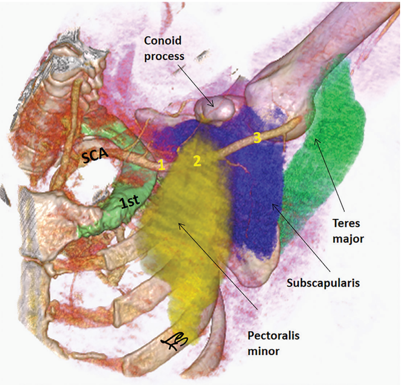
First Part
- Location: lateral border of rib I → medial border of pectoralis minor
- Branches (1): Superior thoracic artery
- Relations: the medial cord of the brachial plexus lies posteromedial; the axillary vein lies medial and anterior
Second Part
- Location: posterior to pectoralis minor
- Branches (2): Thoraco-acromial artery + Lateral thoracic artery
- Relations: lateral cord anterolateral, medial cord medial, posterior cord posterior - the three cords of the brachial plexus surround it on three sides
Third Part
- Location: lateral border of pectoralis minor → inferior border of teres major
- Branches (3): Subscapular artery + Anterior circumflex humeral artery + Posterior circumflex humeral artery
- Relations: the median nerve crosses anterior to this part; the ulnar nerve lies medially; this is the most accessible and most commonly injured part
Branches (6 total)
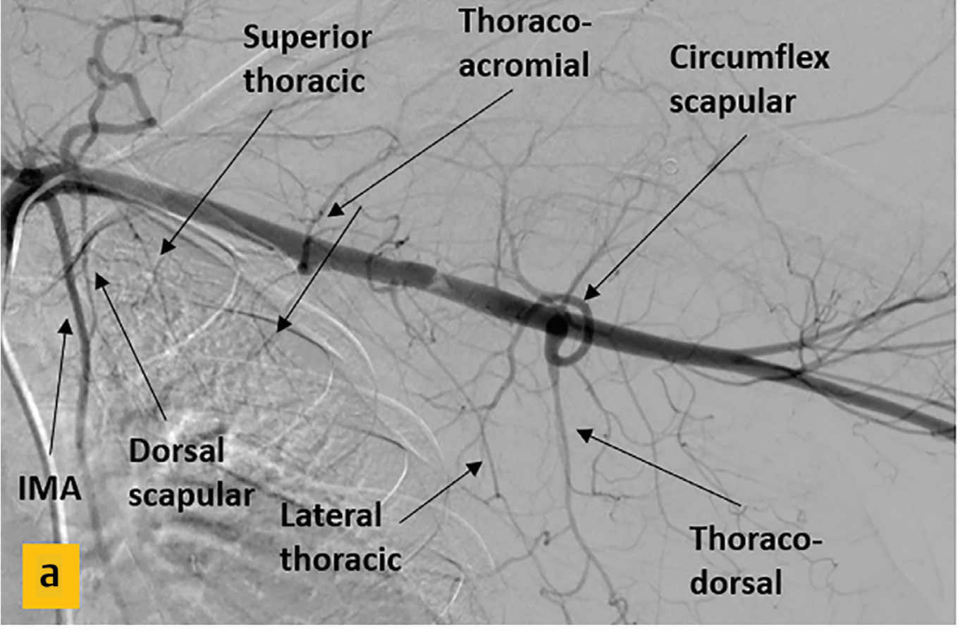
From the 1st Part
1. Superior Thoracic Artery
- Small; arises from the anterior surface
- Supplies the upper medial and anterior axillary walls (upper 2 intercostal spaces and adjacent muscles)
From the 2nd Part
2. Thoraco-acromial Artery
- Short trunk that curves around the superior margin of pectoralis minor and pierces the clavipectoral fascia
- Immediately divides into four branches:
- Pectoral branch - supplies pectoralis major and minor; also contributes to breast supply
- Deltoid branch - accompanies the cephalic vein in the deltopectoral groove; supplies deltoid and pectoralis major
- Clavicular branch - supplies subclavius and sternoclavicular joint
- Acromial branch - contributes to the acromial arterial network
3. Lateral Thoracic Artery
- Arises from the anterior surface, posterior to the lateral (inferior) margin of pectoralis minor
- Follows the inferolateral margin of pectoralis minor to the thoracic wall
- Supplies the medial and anterior axillary walls
- In females, branches emerge around pectoralis major to supply the breast (lateral mammary branches)
From the 3rd Part
4. Subscapular Artery (largest branch of the axillary artery)
- Arises from the posterior surface; runs along the inferior margin of subscapularis
- Divides into two terminal branches:
- Circumflex scapular artery - passes through the triangular space (bounded by subscapularis, teres major, and long head of triceps); reaches the infraspinous fossa and anastomoses with the suprascapular artery - forms the scapular arcade
- Thoracodorsal artery - descends along the lateral scapular border to supply latissimus dorsi; the thoracodorsal nerve travels alongside it
5. Anterior Circumflex Humeral Artery
- Small; arises from the lateral side of the 3rd part
- Passes anterior to the surgical neck of the humerus
- Supplies the glenohumeral joint capsule and the head of the humerus (contributes to avascular necrosis risk in surgical neck fractures)
- Anastomoses with the posterior circumflex humeral artery
6. Posterior Circumflex Humeral Artery
- Larger than the anterior; arises just posterior to ACHA
- Exits the axilla through the quadrangular space with the axillary nerve
- Winds around the surgical neck of the humerus
- Supplies the deltoid, teres minor, glenohumeral joint, and head of humerus
- Anastomoses with the ACHA, profunda brachii, suprascapular, and thoraco-acromial arteries
Relations Summary
| Part | Anterior | Posterior | Medial | Lateral |
|---|---|---|---|---|
| 1st | Clavipectoral fascia, pectoralis major | Medial cord, serratus anterior | Axillary vein | Lateral cord |
| 2nd | Pectoralis minor, pectoralis major | Posterior cord | Medial cord + axillary vein | Lateral cord |
| 3rd | Median nerve (crosses anterior) | Subscapularis, radial nerve | Ulnar nerve, axillary vein | Coracobrachialis, musculocutaneous nerve |
Throughout its course, the axillary vein lies medial and anterior to the artery, and the brachial plexus cords are arranged around it (lateral, medial, posterior - named for their positions relative to the 2nd part).
Scapular Anastomosis (Collateral Circulation)
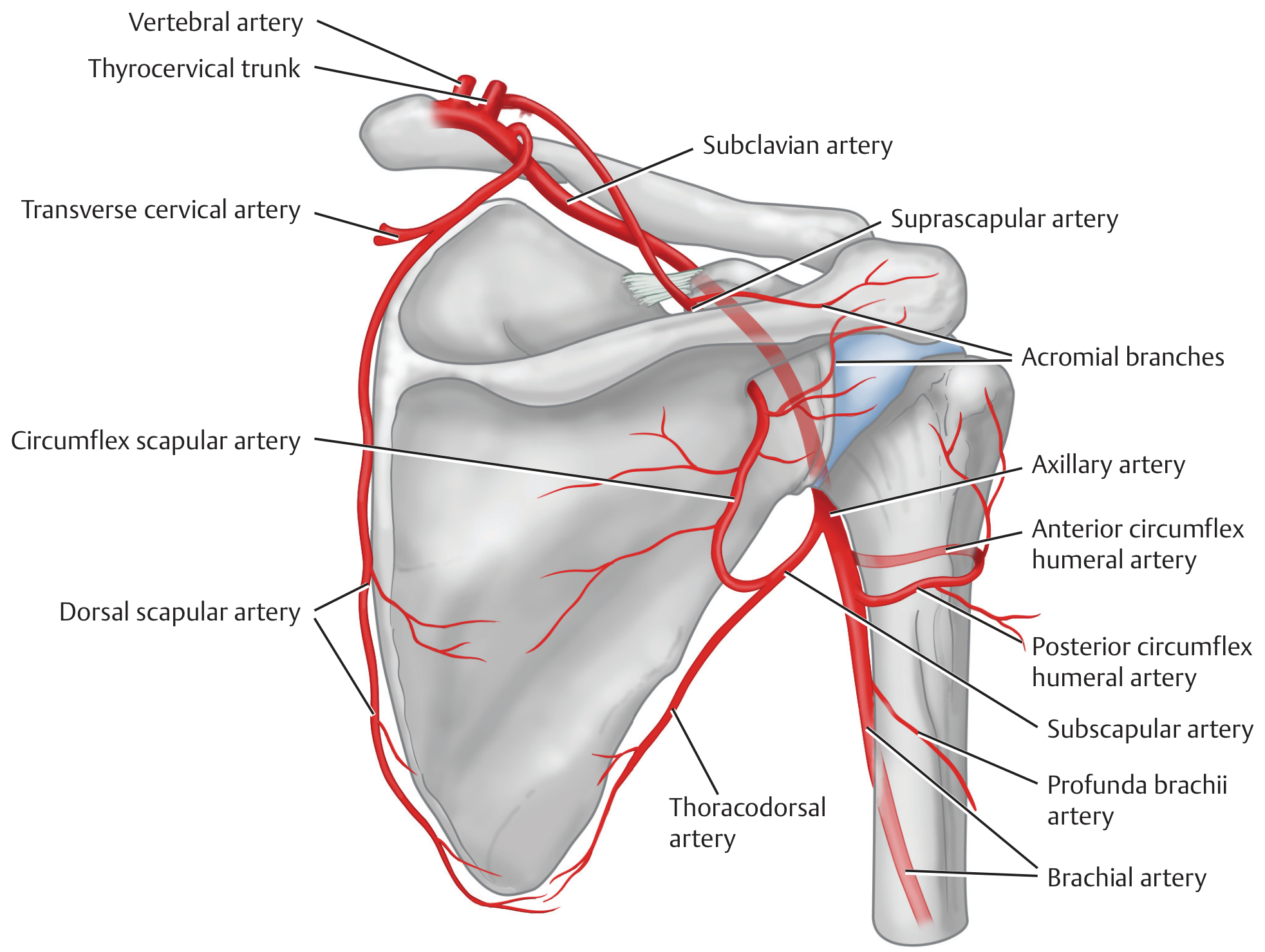
This is a clinically vital network that allows the limb to remain viable even if the axillary artery is occluded or ligated proximal to the subscapular artery:
| Subclavian side | Connects via | Axillary side |
|---|---|---|
| Suprascapular artery (thyrocervical trunk) | Scapular arcade | Circumflex scapular artery (from subscapular) |
| Transverse cervical / dorsal scapular artery | Scapular border | Subscapular branches |
The anastomosis between the suprascapular artery (subclavian territory) and the circumflex scapular artery (axillary territory) over the posterior scapula is the key collateral route. Ligation of the axillary artery proximal to the subscapular artery origin is usually tolerated without limb ischaemia because of this network.
Anatomical Variations
Branching patterns are quite variable - the classic 6-branch pattern occurs in only ~60% of people:
- High origin of the subscapular artery from the 2nd part (rather than 3rd): 29-36% of people
- Common origin of the subscapular and posterior circumflex humeral arteries: 12-42%
- Thoracodorsal artery arising directly from the axillary artery (rather than from subscapular): 3-10%
- Superior/lateral thoracic artery arising from the thoraco-acromial trunk
- Superficial brachial artery: early bifurcation of the axillary artery, with one branch taking a superficial course over the arm - Imaging Anatomy, Bones, Joints, Vessels and Nerves, p. 154
Clinical Correlations
| Scenario | Mechanism | Consequence |
|---|---|---|
| Rib I fracture | Fixes the subclavian/1st part of axillary artery; rapid deceleration injures vessel | Ischaemia (usually compensated by scapular anastomosis) |
| Anterior shoulder dislocation | Humeral head compresses the 3rd part | Axillary artery occlusion; co-injury to brachial plexus (especially axillary nerve) |
| Axillary artery aneurysm | Degenerative or traumatic (e.g. crutch use, effort thrombosis) | Thromboembolism to hand; compression of brachial plexus |
| Avascular necrosis of humeral head | Disruption of anterior/posterior circumflex humeral arteries in surgical neck fractures | Loss of blood supply to femoral head analogue |
| Thoracodorsal artery | At risk in axillary lymph node dissection | Denervates latissimus dorsi flap if not preserved - important for reconstructive surgery |
| Axillary-to-axillary vein bypass graft | Subclavian steal syndrome or proximal occlusion | Utilises 3rd part of axillary artery as inflow |
- Gray's Anatomy for Students, pp. 853-856
- Imaging Anatomy - Bones, Joints, Vessels and Nerves, pp. 154-156
- Fischer's Mastery of Surgery 8e, p. 1558
This is a shared conversation. Sign in to Orris to start your own chat.