attachment
attachment

What can we suspect from this nasal lesion of this cat. Highest differentials

analyse_media
Reading File
Finding Sources
Finding Sources
Searching Images

feline nasal dermatitis alopecia erythema crusting nasal bridge cat
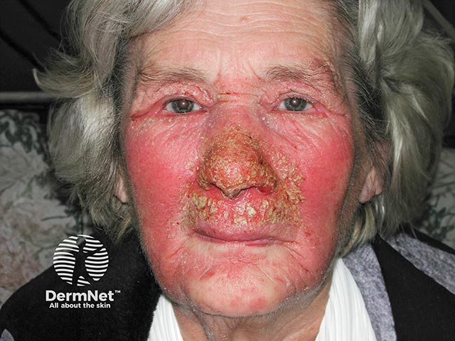
Clinical photograph illustrating allergic contact dermatitis of the face with prominent perinasal crusting and erythema following use of a nasal ointment containing chlorhexidine and neomycin. Modality is standard digital skin photography, frontal/anterior view, with close-up emphasis on the nasal bridge, alae, cheeks, and perinasal folds. The integumentary reaction is manifested by diffuse erythema, edema, excoriation, and bilateral crusted plaques around the nares and nasal tip, extending onto adjacent malar regions. The appearance is consistent with type IV hypersensitivity to topical antiseptics and antibiotic components, producing spongiotic dermatitis with inflammatory cell infiltrates in histology if biopsy were pursued. Clinically, the pattern suggests an allergic contact dermatitis rather than irritant dermatitis due to the localized, well-demarcated distribution and the presence of crusts rather than diffuse burning. Diagnostic significance includes the need to identify and discontinue the offending agent, implement patch testing for chlorhexidine and neomycin, and initiate topical corticosteroids with emollients to accelerate resolution while preventing secondary infection. Potential clinical use cases include dermatology teaching, allergology evaluation, patch-testing workflow, pharmacovigilance of topical agents, and documentation for medicolegal or clinical care pathways. This image is valuable for recognizing perinasal dermatitis and educating about contact allergen avoidance strategies.
Clinical photograph illustrating allergic contact dermatitis of the face with prominent perinasal crusting and erythema following use of a nasal ointment containing chlorhexidine and neomycin. Modality is standard digital skin photography, frontal/anterior view, with close-up emphasis on the nasal bridge, alae, cheeks, and perinasal folds. The integumentary reaction is manifested by diffuse erythema, edema, excoriation, and bilateral crusted plaques around the nares and nasal tip, extending onto adjacent malar regions. The appearance is consistent with type IV hypersensitivity to topical antiseptics and antibiotic components, producing spongiotic dermatitis with inflammatory cell infiltrates in histology if biopsy were pursued. Clinically, the pattern suggests an allergic contact dermatitis rather than irritant dermatitis due to the localized, well-demarcated distribution and the presence of crusts rather than diffuse burning. Diagnostic significance includes the need to identify and discontinue the offending agent, implement patch testing for chlorhexidine and neomycin, and initiate topical corticosteroids with emollients to accelerate resolution while preventing secondary infection. Potential clinical use cases include dermatology teaching, allergology evaluation, patch-testing workflow, pharmacovigilance of topical agents, and documentation for medicolegal or clinical care pathways. This image is valuable for recognizing perinasal dermatitis and educating about contact allergen avoidance strategies.

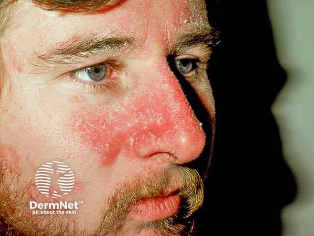
High-resolution color clinical photograph of an adult male’s central face focusing on the left cheek and nasal bridge. Modality: clinical photography; technique: close-up, color-accurate digital image captured under diffuse frontal lighting. The facial skin shows marked erythema centered on the malar region with diffuse redness extending toward the nasal ala and bridge. Subtle desquamation and fine scale may be present at the margins. The appearance is characteristic of seborrheic dermatitis, a common inflammatory dermatosis affecting sebaceous-rich areas of the face. The epidermis shows superficial inflammation without ulceration or vesiculation. The lesion margins are ill-defined and confluent, with variable textures from smooth erythema to flaky scales. There is no obvious crusting or infection. The surrounding skin appears unremarkable aside from mild erythema and oiliness. Clinically this image is valuable for dermatology education, differential diagnosis exercises (rosacea, contact dermatitis, atopic dermatitis), and monitoring therapeutic response to topical antifungal agents (ketoconazole) or anti-inflammatory regimens (selenium sulfide shampoo, zinc pyrithione, corticosteroids). In research, it supports studies of seborrheic dermatitis prevalence, severity grading, and cosmetic impact on quality of life. The image emphasizes visual cues used in bedside assessment, documentation, and tele-dermatology triage. Accurate annotation enhances searchability for educational archives and clinical databases for research.
High-resolution color clinical photograph of an adult male’s central face focusing on the left cheek and nasal bridge. Modality: clinical photography; technique: close-up, color-accurate digital image captured under diffuse frontal lighting. The facial skin shows marked erythema centered on the malar region with diffuse redness extending toward the nasal ala and bridge. Subtle desquamation and fine scale may be present at the margins. The appearance is characteristic of seborrheic dermatitis, a common inflammatory dermatosis affecting sebaceous-rich areas of the face. The epidermis shows superficial inflammation without ulceration or vesiculation. The lesion margins are ill-defined and confluent, with variable textures from smooth erythema to flaky scales. There is no obvious crusting or infection. The surrounding skin appears unremarkable aside from mild erythema and oiliness. Clinically this image is valuable for dermatology education, differential diagnosis exercises (rosacea, contact dermatitis, atopic dermatitis), and monitoring therapeutic response to topical antifungal agents (ketoconazole) or anti-inflammatory regimens (selenium sulfide shampoo, zinc pyrithione, corticosteroids). In research, it supports studies of seborrheic dermatitis prevalence, severity grading, and cosmetic impact on quality of life. The image emphasizes visual cues used in bedside assessment, documentation, and tele-dermatology triage. Accurate annotation enhances searchability for educational archives and clinical databases for research.

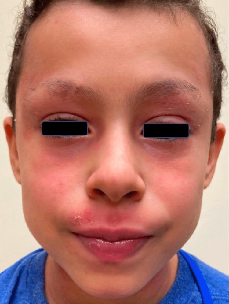
This clinical photograph shows the face of a pediatric patient exhibiting features of head and neck dermatitis (HND). The image displays diffuse erythema across the mid-face, including the cheeks and nasal bridge. Notably, there are erythematous plaques on the superior and inferior eyelids bilaterally, accompanied by mild edema and fine scaling. The perioral region demonstrates significant involvement, characterized by erythema and a localized cluster of pustules with honey-colored crusting lateral to the left oral commissure, suggestive of secondary impetiginization. The lips appear mildly inflamed and dry. This visual presentation is consistent with dupilumab-associated head and neck dermatitis, a known paradoxical reaction in patients treated for atopic dermatitis. The image serves as an educational example of regional dermatitis and secondary bacterial or fungal complications in the context of systemic immunomodulatory therapy. Clinical significance includes recognizing the distribution patterns that distinguish HND from primary flares of atopic dermatitis.
This clinical photograph shows the face of a pediatric patient exhibiting features of head and neck dermatitis (HND). The image displays diffuse erythema across the mid-face, including the cheeks and nasal bridge. Notably, there are erythematous plaques on the superior and inferior eyelids bilaterally, accompanied by mild edema and fine scaling. The perioral region demonstrates significant involvement, characterized by erythema and a localized cluster of pustules with honey-colored crusting lateral to the left oral commissure, suggestive of secondary impetiginization. The lips appear mildly inflamed and dry. This visual presentation is consistent with dupilumab-associated head and neck dermatitis, a known paradoxical reaction in patients treated for atopic dermatitis. The image serves as an educational example of regional dermatitis and secondary bacterial or fungal complications in the context of systemic immunomodulatory therapy. Clinical significance includes recognizing the distribution patterns that distinguish HND from primary flares of atopic dermatitis.

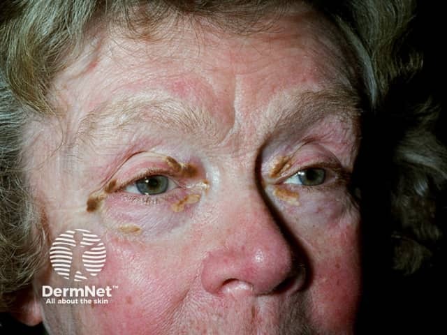
Imaging modality: Clinical photography. Frontal close-up view of the face focusing on the periorbital, malar and nasal regions. The patient is an elderly adult; skin shows diffuse facial erythema with pronounced redness across cheeks and nasal bridge. There are lichenified, crusted plaques along the upper and lower eyelids with yellow-brown crusts adherent to the lash margins. The periorbital skin exhibits mild edema and scale; surrounding skin is dry and sun-damaged. No obvious vesicles; no primary lesion on lips; the nose shows a slightly shiny, telangiectatic surface. The presentation is most consistent with inflammatory dermatitis involving the face, with periorbital blepharitis-like crusting. Differential diagnoses include seborrheic dermatitis (greasy scales, eyelid involvement), atopic dermatitis (lichenoid irritation), contact dermatitis to facial products, rosacea with centrofacial erythema, and psoriasis in the facial milieu. The image documents a chronic, possibly irritant-exposure driven dermatosis with eyelid involvement. Clinical significance includes the need to differentiate dermatitis from infectious etiologies and to guide topical therapy (emollients, mild topical steroids, calcineurin inhibitors) and eyelid hygiene. The image is valuable for education and research on facial dermatitis phenotypes, periorbital dermatitis, blepharitis-related crusting, and sun-damaged skin in elderly patients. High-resolution imaging supports telemedicine, documentation, and longitudinal assessment of treatment response outcomes.

Imaging modality: Clinical photography. Frontal close-up view of the face focusing on the periorbital, malar and nasal regions. The patient is an elderly adult; skin shows diffuse facial erythema with pronounced redness across cheeks and nasal bridge. There are lichenified, crusted plaques along the upper and lower eyelids with yellow-brown crusts adherent to the lash margins. The periorbital skin exhibits mild edema and scale; surrounding skin is dry and sun-damaged. No obvious vesicles; no primary lesion on lips; the nose shows a slightly shiny, telangiectatic surface. The presentation is most consistent with inflammatory dermatitis involving the face, with periorbital blepharitis-like crusting. Differential diagnoses include seborrheic dermatitis (greasy scales, eyelid involvement), atopic dermatitis (lichenoid irritation), contact dermatitis to facial products, rosacea with centrofacial erythema, and psoriasis in the facial milieu. The image documents a chronic, possibly irritant-exposure driven dermatosis with eyelid involvement. Clinical significance includes the need to differentiate dermatitis from infectious etiologies and to guide topical therapy (emollients, mild topical steroids, calcineurin inhibitors) and eyelid hygiene. The image is valuable for education and research on facial dermatitis phenotypes, periorbital dermatitis, blepharitis-related crusting, and sun-damaged skin in elderly patients. High-resolution imaging supports telemedicine, documentation, and longitudinal assessment of treatment response outcomes.

Searching the Web

feline nasal lesion alopecia erythema crusting nasal bridge differential diagnosis cat dermatology

Reading Web Page

https://academy.royalcanin.com/en/veterinary/diseases-of-the…

Here is a thorough clinical breakdown based on the image analysis and veterinary dermatology literature:

Feline Nasal Lesion - Top Differentials

What the lesion shows:

  • Focal alopecia (hair loss) on the nasal bridge/dorsum, extending toward the nasal planum junction
  • Erythema (pink-red discoloration) of the underlying skin
  • Mild dark crusting (small scabs/dried exudate) scattered across the area
  • Ill-defined, irregular borders fading into normal fur
  • No deep ulceration, no significant swelling, lesion is flat/superficial
  • White-furred cat (note: white/bicolor cats have known predispositions for certain conditions)

Top Differentials (Ranked by Likelihood)

1. Pemphigus Foliaceus - #1 suspect

The most common immune-mediated skin disease in cats affecting this exact location. Classic presentation is pustular dermatitis progressing to crusting, alopecia, and erythema on the bridge of the nose, nasal planum, and pinnae. Up to 50% of feline PF cases involve the nasal planum. Cytology showing acantholytic ("fried egg") keratinocytes surrounded by neutrophils is diagnostic. White and bicolor cats may be anecdotally predisposed. Key workup: skin cytology + punch biopsy.

2. Mosquito Bite Hypersensitivity

A very common and underdiagnosed cause of seasonal, recurring alopecia, crusting, and erosion specifically on the nasal bridge and planum. Strongly favors the dorsal nose and bridge, exactly as seen here. White and light-furred cats are anecdotally more predisposed. History of outdoor access is a key clue. Resolves or improves when the cat is kept indoors. No definitive test - diagnosis is clinical and by response to management.

3. Feline Herpesvirus-1 (FHV-1) Facial Dermatitis

FHV-1 can cause ulcerative/necrotizing facial dermatitis, often perinasal/periocular, that mimics pemphigus and mosquito bite hypersensitivity. Usually accompanied by a history of upper respiratory signs (sneezing, ocular discharge). Histopathology reveals epidermal necrosis with eosinophilic infiltrate; intranuclear inclusion bodies are diagnostic but not always present. PCR on biopsy tissue is the best confirmatory test.

4. Dermatophytosis (Ringworm - Microsporum canis)

A classic mimic of many facial skin conditions in cats. Produces alopecia, scaling, and mild erythema in irregular, poorly defined patches, often on the face and nasal area. Cats can be asymptomatic carriers. Fungal culture (toothbrush/MacKenzie technique or DTM culture) and Wood's lamp screening (positive in ~50% of M. canis) are essential first steps.

5. Solar/Actinic Dermatitis

White cats or those with white skin over the nasal bridge are highly predisposed to UV-induced damage. Presents as erythema, scaling, alopecia, and crusting on sun-exposed areas (nasal bridge, ear tips, periocular). This is a premalignant condition that can progress to squamous cell carcinoma if left untreated. Key history: outdoor exposure, light pigmentation.

6. Squamous Cell Carcinoma (SCC)

The primary concern when actinic dermatitis is present in older cats. SCC presents as erythematous, ulcerative, erosive, sometimes crusted lesions on the nasal planum and bridge. More common in older (7+ year) white/lightly pigmented cats with outdoor sun exposure. Biopsy is mandatory for diagnosis. A lesion that fails to respond to other treatments must be biopsied.

7. Eosinophilic Granuloma Complex

Though more common on the lips, chin, and oral cavity, eosinophilic plaques/granulomas can appear on the nasal region of allergic cats. Associated with underlying atopy, food allergy, or flea allergy. Presents as erythematous, sometimes ulcerated, pruritic plaques.

Recommended Diagnostic Workup

StepTestRules out / confirms
1Skin cytology (direct impression or tape prep)PF (acantholytic cells), bacteria, yeast
2Fungal culture / DTM + Wood's lampDermatophytosis
3Skin scrapingDemodex, other parasites
4Punch biopsy (essential if cytology unclear)PF, FHV-1, SCC, eosinophilic granuloma
5PCR on biopsy tissueFHV-1
6History (seasonality, outdoor access, vaccination status, other cats)Mosquito bite hypersensitivity, FHV-1

Clinical Bottom Line

Given the white-furred, bicolor cat with focal nasal bridge alopecia + erythema + mild crusting with ill-defined borders and no deep ulceration, the probability ranking is:
Pemphigus Foliaceus > Mosquito Bite Hypersensitivity > FHV-1 > Dermatophytosis > Solar/Actinic Dermatitis > SCC
Biopsy is ultimately required for a definitive answer in most of these cases. Cytology is a fast, cheap first step that may spare the need for biopsy if acantholytic keratinocytes or fungal elements are found.
This is a shared conversation. Sign in to Orris to start your own chat.