
Rifaximin
rifaximin
Rifaximin
Drug Class & Structure
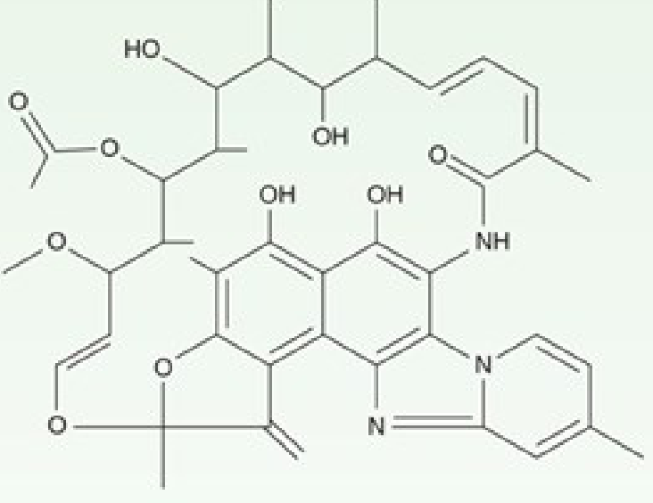
Mechanism of Action
Pharmacokinetics
| Parameter | Detail |
|---|---|
| Oral bioavailability | <0.5% (often stated as <1%) |
| Fecal concentrations | Extremely high - ~8,000 mcg/g stool after a 3-day traveler's diarrhea course |
| CYP450 interactions | None clinically significant (unlike other rifamycins, due to negligible absorption) |
| Route | Oral only |
Spectrum of Activity
- Gram-positive aerobes and anaerobes
- Gram-negative aerobes and anaerobes
FDA-Approved Indications & Dosing
1. Traveler's Diarrhea (non-invasive E. coli strains)
- Approved for patients >12 years
- Dose: 200 mg three times daily for 3 days (some sources cite 400 mg TID for 3 days)
- Not for diarrhea complicated by fever or bloody stools (invasive pathogens may not respond)
- Azithromycin is preferred in children and in regions with high fluoroquinolone resistance. - Sleisenger & Fordtran's, Red Book 2021
2. Hepatic Encephalopathy (HE) - Prophylaxis & Treatment
- Dose: 550 mg twice daily (for prophylaxis of recurrence)
- Acute treatment: 400 mg PO every 8 hours (Rosen's Emergency Medicine)
- Used in combination with lactulose (the combination achieves ~76% complete reversal vs. 50.8% with lactulose alone, with reduced mortality 23.1% vs. 49.1%) - Mulholland & Greenfield's Surgery, 7th Ed.
- Preferred over neomycin - fewer adverse effects (no ototoxicity or nephrotoxicity risk)
- The Washington Manual recommends it as second-line after lactulose or as add-on
- For grade I-II HE without complicating factors, outpatient management with lactulose + rifaximin is appropriate. - Rosen's Emergency Medicine
3. IBS with Diarrhea (IBS-D)
- Dose: 550 mg three times daily for 14 days
- Can be prescribed for up to 2 additional retreatment courses for recurrent IBS-D symptoms
- Meta-analysis (5 studies): rifaximin superior to placebo for global IBS symptom improvement (OR 1.57; 95% CI 1.22-2.01; NNT = 10.2) and bloating (OR 1.59; NNT = 10)
- In a network meta-analysis comparing IBS-D therapies (alosetron, ramosetron, rifaximin, eluxadoline), rifaximin had the fewest adverse events
- ACG and AGA grade evidence as moderate (2B) due to modest, transient effects
- Up to 64% of patients may relapse within 18 weeks - Yamada's Textbook of Gastroenterology, 7th Ed.; Harrison's Principles of Internal Medicine 22E
Off-Label / Investigational Uses
- Recurrent/refractory C. difficile infection - used as adjunct in adults (rifaximin is active against C. difficile, and resistance rates appear low per recent systematic review [PMID 41604218])
- Small intestinal bacterial overgrowth (SIBO) - rifaximin 400 mg TID x 10 days superior to placebo for global symptoms and bloating
- Diverticulitis recurrence prevention - cyclic rifaximin + fiber vs. fiber alone showed a trend to reduced recurrence (10.4% vs. 19.3%) but not statistically significant; AGA recommends against routine use for this indication - Yamada's, 7th Ed.
- SBP prophylaxis - theoretical basis; insufficient data to recommend for spontaneous bacterial peritonitis prophylaxis
Adverse Effects
- Nausea
- Abdominal pain
- Headache, upper respiratory infection, nasopharyngitis (rates similar to placebo in IBS trials)
- C. difficile colitis - rare; one case in retreatment trials
Drug Interactions
Resistance
- Resistance to rifaximin in gut pathogens has been a theoretical concern, but rates in C. difficile remain relatively low across ribotypes per a 2026 systematic review (PMID 41604218). Rifaximin appears to have minimal induction of clinically significant resistance during GI use.
Key Comparisons
| Feature | Rifaximin | Rifampin |
|---|---|---|
| Systemic absorption | <0.5% | High |
| CYP interactions | None significant | Many (inducer) |
| Primary site of action | GI lumen | Systemic |
| Use in TB | No | Yes |
| Hepatotoxicity risk | Very low | Yes |
Recent Evidence (2025-2026)
- A 2025 meta-analysis (PMID 39889173) confirms rifaximin efficacy and safety for prophylaxis of hepatic encephalopathy.
- A 2026 systematic review on IBS pharmacotherapy safety (PMID 40471839) further supports rifaximin's favorable safety profile among IBS agents.
GERD
"gastroesophageal reflux disease"[MeSH Terms] AND management
"gastroesophageal reflux" AND "proton pump inhibitor"
GERD gastroesophageal reflux disease pathophysiology lower esophageal sphincter diagram
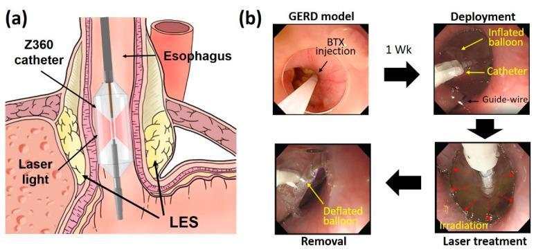
Educational figure illustrating Endoscopic Balloon-assisted Laser Treatment (EBLT) for Gastroesophageal Reflux Disease (GERD). (a) Anatomical diagram showing a Z360 balloon catheter positioned within the distal esophagus at the lower esophageal sphincter (LES). The catheter features a transparent inflated balloon and a central diffusing applicator that emits circumferential laser light to target the LES muscle layers. (b) A series of four endoscopic frames detailing the procedural stages in a porcine model: 1. GERD model induction via botulinum toxin (BTX) injection to weaken the LES; 2. Deployment stage showing the catheter and inflated balloon guided by a guide-wire; 3. Laser treatment stage showing active irradiation (indicated by red arrows); 4. Removal stage following balloon deflation. The educational focus is on the minimally invasive delivery of laser energy to the esophagogastric junction to induce tissue remodeling and increase LES pressure without mucosal injury.
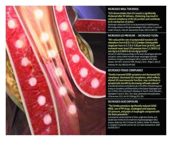
This medical illustration demonstrates the mechanism of action of the Stretta procedure, a minimally invasive treatment for gastroesophageal reflux disease (GERD). The visual depicts a cross-section of the esophageal wall near the gastroesophageal junction. Key components include multiple nickel-titanium needle electrodes delivering radiofrequency (RF) energy into the muscularis propria of the esophagus, represented by bright yellow circular glows within the red-textured tissue layers. The anatomical focus is on the lower esophageal sphincter (LES) region. Accompanying text panels summarize clinical outcomes and pathophysiology: increased wall thickness, increased LES basal pressure with decreased transient LES relaxations (TLESRs), decreased tissue compliance at the gastroesophageal junction, and significantly decreased esophageal acid exposure. The image highlights the device's ability to deliver thermal energy to induce tissue remodeling while protecting the overlying mucosa via irrigation. This illustration serves as an educational tool for gastroenterologists and surgical trainees to understand non-surgical antireflux interventions and their physiological impact on LES neuromuscular function.
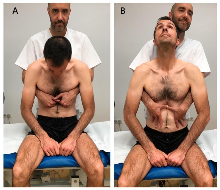
Two-panel clinical photograph demonstrating a visceral osteopathic manual technique targeting the lower esophageal sphincter, typically used for gastroesophageal reflux disease (GERD). Panel A (Initial Position) shows a male patient seated upright on a treatment table in spinal flexion with his neck flexed and head looking down. A practitioner stands behind him with hands placed horizontally in the epigastric region, specifically contacting the area just inferior to the costal margins. Panel B (Final Position) illustrates the completion of the maneuver: the patient has moved into spinal extension with his neck extended and gaze upward. Simultaneously, the practitioner has shifted hand placement to apply caudal (downward) pressure within the epigastrium. The visual demonstrates the coordination of patient respiratory/postural movement with manual tissue mobilization by the therapist. This procedural image is intended for medical students and physical therapy or osteopathic practitioners to understand the manual application and patient positioning for visceral abdominal therapy.
Gastroesophageal Reflux Disease (GERD)
Definition
Classification (Montreal)
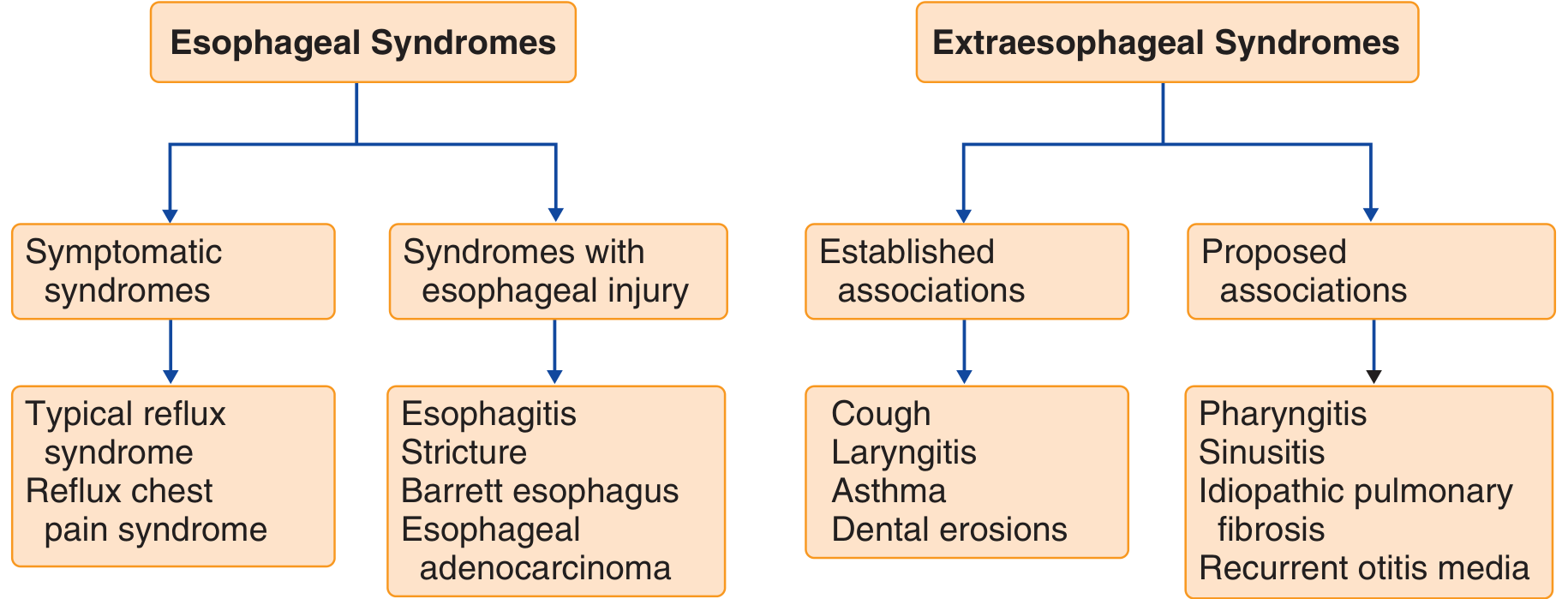
- Symptomatic: typical reflux syndrome, reflux chest pain syndrome
- With esophageal injury: reflux esophagitis, reflux stricture, Barrett esophagus, esophageal adenocarcinoma
- Reflux cough, reflux laryngitis, reflux asthma, reflux dental erosions
Pathophysiology
- Lower Esophageal Sphincter (LES) - the primary barrier; resting pressure normally 10-45 mmHg
- Crural diaphragm - acts as an external sphincter during inspiration and straining
- Esophagogastric junction anatomy - the intra-abdominal segment of esophagus
- Transient LES relaxations (TLESRs) - the dominant mechanism in most patients; reflex relaxation of the LES unrelated to swallowing, triggered by gastric distension
- Hypotensive LES - a persistently low basal LES pressure; associated with more severe reflux and erosive disease
- Hiatal hernia - disrupts the crural diaphragm contribution, impairs acid clearance, and creates a "trap" for acid
Clinical Features
Typical (Esophageal) Symptoms
- Heartburn - burning retrosternal sensation, worst postprandially, on bending, or lying supine
- Acid regurgitation - effortless return of acid/bitter contents into the mouth
- Dysphagia, odynophagia - suggest complications (stricture, severe esophagitis)
Atypical / Extraesophageal Symptoms
- Chronic cough
- Laryngitis / hoarseness
- Asthma (worsening or new)
- Dental erosions (palatal surfaces of maxillary teeth most common; enamel loss is irreversible)
- Chest pain (non-cardiac)
Asthma guidelines suggest treating GERD in asthmatics with heartburn or frequent nocturnal symptoms; however, PPI therapy alone does not reliably improve poorly controlled asthma without classic GERD symptoms. - Fishman's Pulmonary Diseases
Alarm Features (Require Urgent Endoscopy)
- Dysphagia or odynophagia
- Unintentional weight loss
- GI bleeding / iron deficiency anemia
- Persistent symptoms despite 4-8 weeks of therapy
- Age >60 with new-onset symptoms
Diagnosis
| Test | Role |
|---|---|
| Clinical diagnosis | Adequate for classic heartburn + regurgitation responding to PPIs - no workup needed initially |
| Upper endoscopy (EGD) | Alarm symptoms, PPI failure, screening for Barrett esophagus; detects erosive esophagitis, strictures, Barrett's |
| Ambulatory pH monitoring (24-hr or 48-96 hr Bravo capsule) | Gold standard for documenting abnormal acid exposure; use off-PPIs to diagnose, on-PPIs for refractory symptoms |
| Combined impedance-pH monitoring | Detects non-acid reflux; useful for breakthrough symptoms on PPIs |
| Esophageal manometry | Rule out achalasia; needed pre-operatively before antireflux surgery |
| Barium swallow | No role in evaluation of uncomplicated GERD |
- Goldman-Cecil Medicine; Sleisenger & Fordtran's
Treatment
Step-Up Strategy
1. Lifestyle Modifications
- Weight loss (if overweight/obese) - the most evidence-supported intervention
- Elevation of the head of the bed (wedge device) for nocturnal symptoms
- Avoid precipitants: alcohol, coffee, fatty/spicy foods, chocolate, late meals
- Avoid lying down within 2-3 hours after eating
- Smoking cessation
2. Pharmacological Management
- Rapid onset (minutes), short duration (1-2 hours)
- Role: on-demand relief of mild, infrequent symptoms
- Gaviscon (alginate): forms a viscous raft barrier above gastric contents
- Cimetidine 400 mg bid; Ranitidine 150 mg bid-qid; Famotidine 20-40 mg bid
- Duration of effect: 6-10 hours
- Better than antacids for infrequent heartburn (can be taken prophylactically before meals)
- Heal erosive esophagitis in <50% of patients - inferior to PPIs
- Useful if PPIs not tolerated; can be added as on-demand nocturnal dose
- Safe; adverse events <3%
| PPI | Standard Dose |
|---|---|
| Omeprazole | 20 mg once daily |
| Lansoprazole | 30 mg once daily |
| Pantoprazole | 40 mg once daily |
| Rabeprazole | 20 mg once daily |
| Esomeprazole | 40 mg once daily |
| Dexlansoprazole | 30-60 mg once daily |
- Take 30-60 minutes before the first meal (requires acid activation of the pump, which occurs with eating)
- No major differences in efficacy among available PPIs
- Twice-daily dosing produces faster endoscopic healing than once-daily (but not faster symptom relief)
- For infrequent heartburn (<3×/week): antacids or intermittent H2RAs
- For frequent heartburn or erosive esophagitis: PPIs preferred
- Dose should be titrated to the lowest effective dose for maintenance
- Vitamin B12 deficiency
- Clostridioides difficile infection
- Community-acquired pneumonia
- Hip fracture / metabolic bone disease
- Hypomagnesemia
- Small intestinal bacterial overgrowth
- Possible increased risk of diabetes mellitus
3. Surgical / Endoscopic Therapy
- Equivalent to continued PPI therapy for healing esophagitis and controlling chronic GERD in patients who respond initially to PPIs
- Indications: PPI intolerance, ongoing symptoms (especially regurgitation) despite PPIs, patient preference to avoid lifelong medication, large hiatal hernia
- Patients must have documented reflux and manometry pre-operatively to exclude achalasia
- Note: patients with persistent regurgitation despite PPIs may be better surgical candidates than those with heartburn alone - Goldman-Cecil Medicine; Sleisenger & Fordtran's
Complications
| Complication | Notes |
|---|---|
| Erosive esophagitis | ~50% of GERD patients; graded LA A-D; heals with PPI therapy |
| Peptic stricture | Fibrosis from chronic inflammation; presents with progressive dysphagia; treat with dilation + PPI |
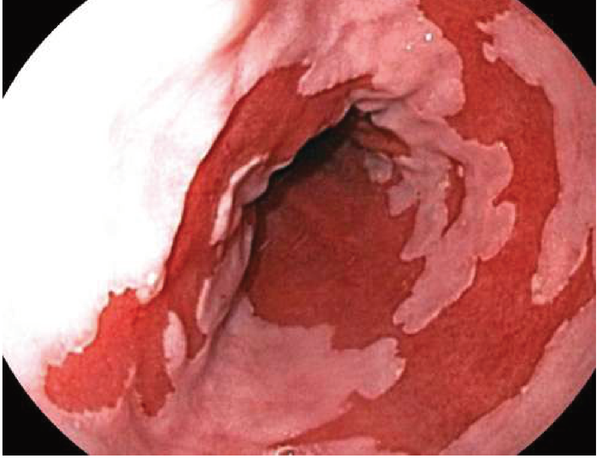
| Barrett esophagus | Columnar metaplasia (intestinal type) replacing squamous epithelium; pre-malignant; requires surveillance |
| Esophageal adenocarcinoma | Complication of long-standing Barrett's; obesity confers RR 4.8 (men) |
Endoscopic appearance of Barrett Esophagus
GERD in Special Populations
- Obesity: weight loss is therapeutic; gastric bypass resolves GERD in many patients
- Pediatric GERD: managed with positional measures and feeding modifications in infants; PPIs for severe/persistent disease
- Dental implications: enamel erosion on palatal surfaces; irreversible; PPIs most effective treatment in adults; reduce acidic food/beverage intake
- Asthma: treat co-existing GERD if classic symptoms present; PPI alone does not improve asthma control without concurrent GERD
Management Algorithm Summary
- Typical heartburn + regurgitation → empiric PPI trial (4-8 weeks) → if responds, continue at lowest effective dose
- No response or alarm features → Upper endoscopy
- Normal endoscopy + persistent symptoms → ambulatory pH/impedance monitoring off PPIs
- Confirmed GERD with esophagitis → PPI maintenance (long-term)
- Refractory to PPIs or patient preference → Antireflux surgery (after manometry + confirmed reflux)