
A 65 year old has pain back to leg of left side i think it's sciatica she has h/o hypertension give management
sciatica lumbar radiculopathy management
lumbar disc herniation sciatica dermatome diagram L4 L5 S1
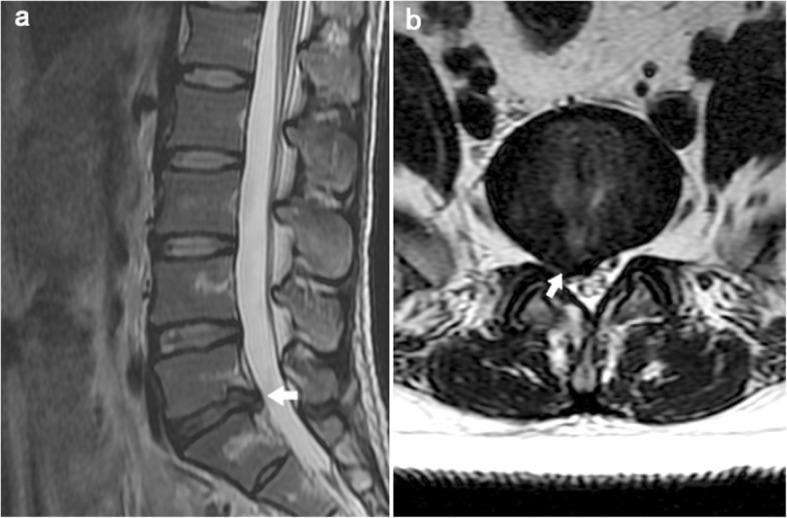
This diagnostic image consists of two T2-weighted magnetic resonance imaging (MRI) scans of the lumbar spine, demonstrating a lumbar disc herniation. Panel (a) is a sagittal view showing the lumbosacral spine. The intervertebral discs from L1 to L4 appear normal with high central signal intensity, while the L5-S1 disc exhibits decreased signal intensity (indicating dehydration/degeneration) and a posterior protrusion into the spinal canal (marked with a white arrow). Panel (b) provides an axial view at the L5-S1 level. It clearly illustrates a focal posterior disc herniation that is displacing and compressing the adjacent right-sided nerve root within the lateral recess. The thecal sac and posterior elements, including the lamina and facet joints, are visible. These findings are clinically relevant for diagnosing radiculopathy and sciatica. The image serves as an educational tool for identifying degenerative disc disease and neurovascular impingement in the lumbar region.
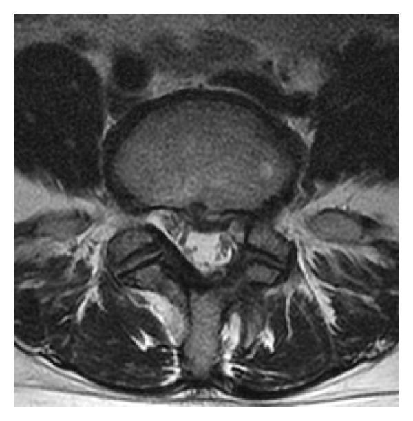
An axial-view T2-weighted magnetic resonance image (MRI) of the lumbar spine at the L4-L5 level. The image demonstrates a herniated nucleus pulposus (HNP) situated in the left lateral recess. The herniation presents as a soft-tissue protrusion extending from the intervertebral disc into the spinal canal, causing significant focal compression and displacement of the left L5 nerve root. Visible anatomical landmarks include the anterior vertebral body, the central spinal canal, the neural foramina, and the posterior elements including the lamina and spinous process. High-intensity signal suggests the acute or recurrent nature of the disc material. This diagnostic image illustrates common pathophysiology associated with lumbar radiculopathy and sciatica, specifically highlighting how localized disc displacement results in neurovascular compromise within the lateral recess.
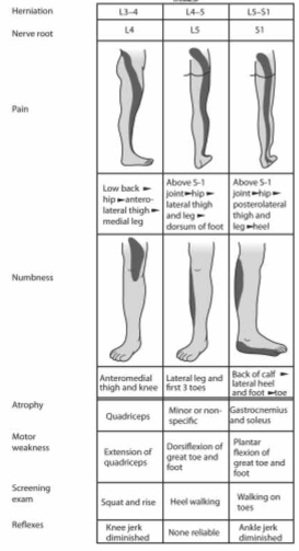
Summary : This figure is a comparative chart detailing the clinical features of lumbar disc herniation at three common levels (L3–4, L4–5, L5–S1), including affected nerve roots (L4, L5, S1), pain and numbness distribution, muscle atrophy, motor weakness, screening exams, and reflex changes. table/chart: # Herniation Levels and Nerve Roots : • L3–4 herniation affects L4 nerve root. • L4–5 herniation affects L5 nerve root. • L5–S1 herniation affects S1 nerve root. # Pain Distribution : • L3–4 (L4): Low back → hip → anterolateral thigh → medial leg. • L4–5 (L5): Above S-1 joint → hip → lateral thigh and leg → dorsum of foot. • L5–S1 (S1): Above S-1 joint → hip → posterolateral thigh and leg → heel. # Numbness Distribution : • L3–4 (L4): Anteromedial thigh and knee. • L4–5 (L5): Lateral leg and first 3 toes. • L5–S1 (S1): Back of calf, lateral heel, and foot/toe. # Muscle Atrophy : • L3–4 (L4): Quadriceps. • L4–5 (L5): Minor or nonspecific. • L5–S1 (S1): Gastrocnemius and soleus. # Motor Weakness : • L3–4 (L4): Extension of quadriceps. • L4–5 (L5): Dorsiflexion of great toe and foot. • L5–S1 (S1): Plantar flexion of great toe and foot. # Screening Exam : • L3–4 (L4): Squat and rise. • L4–5 (L5): Heel walking. • L5–S1 (S1): Walking on toes. # Reflex Changes : • L3–4 (L4): Knee jerk diminished. • L4–5 (L5): None reliable. • L5–S1 (S1): Ankle jerk diminished. # Visual Representation : • Each column includes a diagram showing the typical pain and numbness distribution for the affected nerve root. # Analysis : • The chart clearly distinguishes the clinical features of nerve root compression at different lumbar levels. • Pain and numbness patterns shift from anterolateral (L4) to lateral (L5) to posterolateral (S1) as the herniation level descends. • Reflex changes and motor weakness are specific to the affected root, aiding in clinical diagnosis. • Screening exams and muscle atrophy findings provide additional diagnostic clues for localization.
Management of Sciatica (Left Lumbar Radiculopathy) in a 65-Year-Old with Hypertension
First: Confirm and Localize
- Straight Leg Raise (SLR) test - positive if pain radiates below knee at 30-70°
- Neurological exam - check reflexes, motor strength, and sensation to localize the nerve root level:
- Red flag screen - rule out cauda equina syndrome (bowel/bladder dysfunction, saddle anesthesia, bilateral symptoms) - requires emergency MRI if present
- Imaging - MRI is the preferred modality; not needed acutely unless progressive neuro deficit or red flags are present
Step-by-Step Management Plan
1. Activity and Rest
- Do NOT prescribe bed rest - routine daily activity is as good as 2 weeks of bed rest for pain and functional outcomes
- Encourage gentle movement; use pain as the limiting factor
- Avoid prolonged sitting or positions that worsen radiculopathy
2. Analgesia - ⚠️ Special Caution in Hypertension
| Drug | Dose | Key Point |
|---|---|---|
| Paracetamol (acetaminophen) | 500-1000 mg TDS | First choice - safe in hypertension, no CV or renal risk |
| NSAIDs (ibuprofen/naproxen) | Ibuprofen 400-800 mg TDS or Naproxen 250-500 mg BD | ⚠️ Use with caution in hypertension - NSAIDs raise blood pressure, cause sodium retention, and can reduce antihypertensive drug efficacy. Use lowest dose, shortest duration. Monitor BP and renal function |
| Add PPI (omeprazole 20 mg OD) | - | If NSAIDs are used, add PPI to protect GI mucosa |
| Gabapentin | 300 mg OD titrating to 300 mg TDS | For neuropathic/radicular component; small study benefit shown for sciatica |
NSAIDs and hypertension: NSAIDs interfere with antihypertensives (especially ACE inhibitors and diuretics), can elevate BP, and may worsen renal function. Prefer paracetamol as first-line analgesia in this patient. If NSAIDs are needed, monitor BP and creatinine closely - Firestein & Kelley's Textbook of Rheumatology.
3. Neuropathic Pain Agents
- Gabapentin 300 mg once daily, uptitrate over 1-2 weeks to 300 mg three times daily (some evidence for sciatica)
- Pregabalin 75 mg twice daily (alternative; a trial showed benefit for acute and chronic sciatica)
- Note: These can cause dizziness and falls - use cautiously in a 65-year-old
4. Muscle Relaxants
- Methocarbamol 1000-1500 mg four times daily - can be used for associated muscle spasm
- Avoid benzodiazepines (addiction risk, greater side effects)
- Muscle relaxants + NSAIDs have no proven synergistic benefit
5. Opioids
- Only for moderate-severe pain, limited to 3 days, not first-line
- High caution in elderly (risk of falls, constipation, delirium)
6. Physiotherapy
- Refer to physiotherapy once acute pain settles
- Core strengthening, McKenzie exercises, nerve mobilization
- Spinal manipulative therapy gives small pain reduction up to 12 weeks
7. Epidural Steroid Injection
- Consider for moderate-to-severe persistent radiculopathy not responding to 4-6 weeks of conservative management
- Provides minor reduction in leg pain but does NOT reduce need for surgery
- Not an emergency procedure; arrange in follow-up
8. Antihypertensive Management
- Continue her current antihypertensive medications uninterrupted
- If on ACE inhibitor/ARB + diuretic, be extra vigilant when adding NSAIDs (risk of "triple whammy" AKI)
- Monitor BP at each visit; pain itself can transiently raise BP
When to Image (MRI)
- No emergent imaging unless red flags present
- Non-urgent MRI if: symptoms persist beyond 4-6 weeks despite conservative management, or if neurological deficit is present
When to Refer to Neurosurgery
- Definitive evidence of herniation on imaging
- Corresponding clinical picture and neurologic deficit
- Conservative treatment has failed over 6+ weeks
Emergency surgery is required only for acute cauda equina syndrome (bowel/bladder dysfunction + saddle anesthesia).
Summary for This Patient
| Priority | Action |
|---|---|
| Immediate | Paracetamol + activity modification; avoid prolonged bed rest |
| NSAID if needed | Short course with PPI; monitor BP and renal function closely |
| Neuropathic pain | Gabapentin 300 mg OD, uptitrate (watch for dizziness/falls) |
| Physiotherapy | Refer once acute phase settles |
| Follow-up in 4-6 weeks | If no improvement, order MRI and consider epidural steroid injection |
| Red flags | If bowel/bladder dysfunction or bilateral symptoms develop - emergency MRI |
I have pregabaline 150 mg
Using Pregabalin 150 mg for Sciatica in a 65-Year-Old with Hypertension
Dosing Protocol
| Phase | Dose | Schedule | Duration |
|---|---|---|---|
| Start low (elderly) | 75 mg/day | 75 mg at night (to reduce daytime dizziness) | First 3-5 days |
| Uptitrate | 150 mg/day | 75 mg morning + 75 mg night | Week 1-2 |
| Maintenance | 150-300 mg/day | In 2 divided doses (BD) | Ongoing as needed |
| Maximum | 600 mg/day | Only if tolerated and needed | - |
Since you have 150 mg tablets, you can start with half a tablet (75 mg) at night for the first few days if available, or if not splittable, start 150 mg once at night, then move to BD after 3-5 days.
Why Split the Dose?
- Pregabalin has a short half-life of 4.5-7 hours, so twice-daily dosing (BD) maintains better plasma levels than once daily
- Giving it at night first reduces daytime sedation and fall risk in an elderly patient
Key Pharmacology Points
- Acts on alpha-2-delta subunit of voltage-gated calcium channels - reduces neuropathic/radicular pain
- Bioavailability >90%, linear absorption - predictable response
- Eliminated unchanged in urine - dose reduction needed if renal impairment (check creatinine in this hypertensive patient)
- Takes several weeks to reach full effect - counsel the patient not to stop early
⚠️ Special Cautions for This Patient
| Risk | Detail |
|---|---|
| Falls | Dizziness and ataxia are common, especially in the elderly - warn patient, start at night |
| Somnolence | Common at initiation, usually improves with continued dosing |
| Peripheral edema | Pregabalin can cause fluid retention - monitor in a hypertensive patient as it may worsen BP control |
| Renal function | Pregabalin is renally cleared - if patient is on antihypertensives like ACE inhibitor/ARB, check renal function (creatinine/eGFR) and adjust dose if eGFR is reduced |
| Weight gain | Both pregabalin and gabapentin can cause weight gain - monitor |
| Do not stop abruptly | Taper slowly to avoid withdrawal - can cause seizures after prolonged use |
Practical Prescription for This Patient
Pregabalin 75 mg once at night x 5 days
→ Then 75 mg morning + 75 mg night (150 mg/day total) x 2 weeks
→ Review: if partial response, uptitrate to 150 mg BD (300 mg/day)
→ Maximum: 300-600 mg/day in divided doses

Combine with:
- Paracetamol 500-1000 mg TDS (safe in hypertension)
- Physiotherapy referral
- Avoid NSAIDs if possible (raises BP, worsens renal function)
Follow-up
- Review in 2 weeks - assess pain reduction, side effects, and blood pressure
- If no significant relief after 4-6 weeks at adequate dose, consider MRI lumbar spine and specialist referral
What about Gabapentin 300 ? Give prescription
Gabapentin 300 mg - Complete Prescription for Sciatica
How Gabapentin Works Here
📋 Prescription
━━━━━━━━━━━━━━━━━━━━━━━━━━━━━━━━━━━━━━
Patient: 65-year-old female
Diagnosis: Sciatica (Left lumbar radiculopathy)
H/O: Hypertension
━━━━━━━━━━━━━━━━━━━━━━━━━━━━━━━━━━━━━━

Rx 1:
Tab. GABAPENTIN 300 mg
- Day 1-3:   1 tab at NIGHT only (300 mg/day)
- Day 4-7:   1 tab MORNING + 1 tab NIGHT (600 mg/day)
- Week 2:    1 tab TDS - Morning, Afternoon, Night (900 mg/day)
- Week 3-4:  If partial relief → increase to 2 tabs at night
             + 1 tab BD = 1200 mg/day (max usual dose)
- Target maintenance: 900–1800 mg/day in 3 divided doses
- Maximum: 3600 mg/day (rarely needed)

Rx 2:
Tab. PARACETAMOL 500 mg
- 1 tab TDS after meals
- (Safe in hypertension - use as base analgesic)

Rx 3:
Tab. OMEPRAZOLE 20 mg
- 1 tab OD before breakfast
- (Gastroprotection, especially if any NSAID is co-prescribed)

━━━━━━━━━━━━━━━━━━━━━━━━━━━━━━━━━━━━━━
Duration: 4-6 weeks, then review
Dispense: 30 tablets Gabapentin 300 mg
━━━━━━━━━━━━━━━━━━━━━━━━━━━━━━━━━━━━━━
Why Slow Titration ("Start Low, Go Slow")?
| Reason | Detail |
|---|---|
| Elderly (65 yrs) | Higher sensitivity to CNS side effects - dizziness, falls |
| Bioavailability varies | Up to 50% of patients get side effects without benefit - titrate to response |
| Absorption is dose-limited | Gabapentin uses a saturable intestinal transporter - split doses absorb better than one large dose |
| Half-life is ~6 hours | Requires TDS dosing (not BD like pregabalin) to maintain steady levels |
⚠️ Important Cautions
| Issue | Action |
|---|---|
| Dizziness / falls | Start at night, warn patient not to drive initially |
| Somnolence | Usually resolves in 1-2 weeks with continued dosing |
| Renal clearance | Gabapentin is excreted unchanged in urine - check creatinine/eGFR in this hypertensive patient. Reduce dose if eGFR < 60 |
| Do NOT stop abruptly | Taper gradually if discontinuing - risk of withdrawal seizures |
| Avoid with opioids | Increased overdose/respiratory depression risk if combined |
| Monitor BP | Pain relief itself may help lower BP; reassess antihypertensives at follow-up |
Gabapentin vs Pregabalin - Which to Choose?
| Feature | Gabapentin 300 mg | Pregabalin 150 mg |
|---|---|---|
| Dosing | TDS (3 times/day) | BD (2 times/day) |
| Absorption | Variable, dose-limited | Linear, >90%, predictable |
| Titration | Slower, more steps | Faster |
| Cost | Much cheaper | More expensive |
| Efficacy for sciatica | Good (small study evidence) | Slightly better evidence |
| Verdict | ✅ Good choice, affordable | ✅ More convenient |
Follow-Up Plan
- 2 weeks: Check pain score, BP, renal function, tolerability
- 4-6 weeks: If inadequate relief at 900 mg/day, uptitrate to 1200-1800 mg/day
- 6 weeks: If no meaningful relief at adequate dose, order MRI lumbar spine and consider referral