How body feel pain and mechanism of Tramadol and contraindications of it and normal dose in ckd

Reading File
Finding Sources
Finding Sources
Finding Sources
Reading File
Reading File
Reading File
Reading File
Finding Sources
Finding Sources
Reading File
Finding Sources
Searching the Web

tramadol dose adjustment CKD chronic kidney disease renal impairment guidelines 2024

Reading File
Searching Images

pain pathway nociception spinal cord ascending diagram

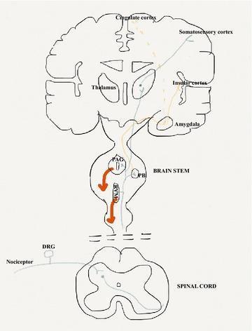
This medical anatomical diagram illustrates the neuroanatomical pathways of pain (nociception), including ascending sensory and descending modulatory systems. The diagram spans the spinal cord, brainstem, and cerebral cortex. The 'sensory-discriminative' pathway (blue line) originates from primary afferent nociceptors and the dorsal root ganglia (DRG), ascends the spinal cord to the thalamus, and terminates in the somatosensory cortex to convey stimulus location and nature. The 'affective-emotional' pathway (orange line) projects through the parabrachial nucleus (PB) in the brainstem and the amygdala, reaching the cingulate and insular cortices. Additionally, a descending inhibitory control pathway (red line/arrows) is shown originating in the periaqueductal grey (PAG) and rostroventral medulla (RVM) of the brainstem, projecting back to the spinal dorsal horn to modulate nociceptive output. This illustration serves as an educational tool for understanding the multi-dimensional processing of pain within the central nervous system.

This medical anatomical diagram illustrates the neuroanatomical pathways of pain (nociception), including ascending sensory and descending modulatory systems. The diagram spans the spinal cord, brainstem, and cerebral cortex. The 'sensory-discriminative' pathway (blue line) originates from primary afferent nociceptors and the dorsal root ganglia (DRG), ascends the spinal cord to the thalamus, and terminates in the somatosensory cortex to convey stimulus location and nature. The 'affective-emotional' pathway (orange line) projects through the parabrachial nucleus (PB) in the brainstem and the amygdala, reaching the cingulate and insular cortices. Additionally, a descending inhibitory control pathway (red line/arrows) is shown originating in the periaqueductal grey (PAG) and rostroventral medulla (RVM) of the brainstem, projecting back to the spinal dorsal horn to modulate nociceptive output. This illustration serves as an educational tool for understanding the multi-dimensional processing of pain within the central nervous system.

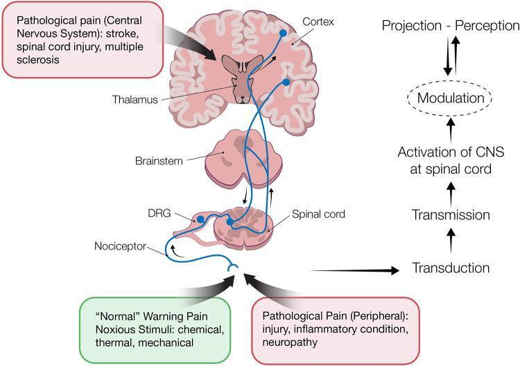
This pathophysiology diagram illustrates the ascending and descending pathways of pain processing in the human nervous system. The visual is divided into an anatomical illustration on the left and a functional flowchart on the right. Anatomical structures shown include peripheral nociceptors, the dorsal root ganglion (DRG), the spinal cord, brainstem, thalamus, and cerebral cortex. The diagram traces the sequence of nociception through four key stages: 1) Transduction (conversion of noxious stimuli like chemical, thermal, or mechanical signals into electrical impulses), 2) Transmission (conduction of action potentials via the DRG to the spinal dorsal horn), 3) Perception (projection of signals through the brainstem and thalamus to the cortex), and 4) Modulation (the enhancement or inhibition of pain signals via descending pathways, represented by a bidirectional arrow). Clinical context is provided through text boxes distinguishing between 'Normal' warning pain and 'Pathological Pain,' citing causes such as inflammation, neuropathy, stroke, spinal cord injury, and multiple sclerosis. This diagram is designed for medical students to understand the neuroanatomy of the pain pathway and the functional distinction between different pain types.

This pathophysiology diagram illustrates the ascending and descending pathways of pain processing in the human nervous system. The visual is divided into an anatomical illustration on the left and a functional flowchart on the right. Anatomical structures shown include peripheral nociceptors, the dorsal root ganglion (DRG), the spinal cord, brainstem, thalamus, and cerebral cortex. The diagram traces the sequence of nociception through four key stages: 1) Transduction (conversion of noxious stimuli like chemical, thermal, or mechanical signals into electrical impulses), 2) Transmission (conduction of action potentials via the DRG to the spinal dorsal horn), 3) Perception (projection of signals through the brainstem and thalamus to the cortex), and 4) Modulation (the enhancement or inhibition of pain signals via descending pathways, represented by a bidirectional arrow). Clinical context is provided through text boxes distinguishing between 'Normal' warning pain and 'Pathological Pain,' citing causes such as inflammation, neuropathy, stroke, spinal cord injury, and multiple sclerosis. This diagram is designed for medical students to understand the neuroanatomy of the pain pathway and the functional distinction between different pain types.

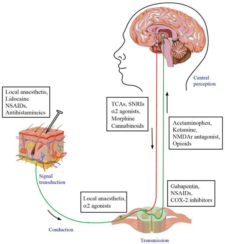
An educational pathophysiology diagram illustrating the ascending and descending pain pathways and sites of pharmacological intervention. The diagram begins at a skin cross-section where a nail stimulus triggers signal transduction. The signal travels via a green line representing conduction through a primary afferent neuron to the dorsal horn of the spinal cord (transmission). The signal then ascends to the brain's somatosensory cortex for central perception. A red line represents the descending modulatory pathway from the brain to the spinal cord. Pharmacological interventions are mapped at four levels: 1) Peripheral (skin): Local anesthetics (Lidocaine), NSAIDs, and antihistamines. 2) Spinal cord (dorsal horn): Gabapentin, NSAIDs, and COX-2 inhibitors. 3) Ascending pathway: Acetaminophen, Ketamine, NMDAr antagonists, and Opioids. 4) Descending/Central: TCAs, SNRIs, α2 agonists, Morphine, and Cannabinoids. The diagram demonstrates the multi-modal nature of pain management and the physiological stages of nociception: transduction, conduction, transmission, and perception.

An educational pathophysiology diagram illustrating the ascending and descending pain pathways and sites of pharmacological intervention. The diagram begins at a skin cross-section where a nail stimulus triggers signal transduction. The signal travels via a green line representing conduction through a primary afferent neuron to the dorsal horn of the spinal cord (transmission). The signal then ascends to the brain's somatosensory cortex for central perception. A red line represents the descending modulatory pathway from the brain to the spinal cord. Pharmacological interventions are mapped at four levels: 1) Peripheral (skin): Local anesthetics (Lidocaine), NSAIDs, and antihistamines. 2) Spinal cord (dorsal horn): Gabapentin, NSAIDs, and COX-2 inhibitors. 3) Ascending pathway: Acetaminophen, Ketamine, NMDAr antagonists, and Opioids. 4) Descending/Central: TCAs, SNRIs, α2 agonists, Morphine, and Cannabinoids. The diagram demonstrates the multi-modal nature of pain management and the physiological stages of nociception: transduction, conduction, transmission, and perception.

I now have all the information needed. Let me compile a thorough, well-structured answer.

Pain, Tramadol, and CKD Dosing

1. How the Body Feels Pain

Pain perception involves four sequential steps: transduction, transmission, modulation, and perception.
Pain pathway diagram showing ascending nociceptive and descending modulatory systems from spinal cord to cortex

Step 1 - Transduction (Peripheral)

Nociceptors are free nerve endings of Aδ and C fibers in skin, muscle, joints, and viscera. When tissue is damaged, chemicals are released locally:
  • Potassium (K+) from damaged cells depolarizes nociceptors directly
  • Prostaglandins (PGs) and bradykinin (BK) sensitize terminals (lower their threshold)
  • Low pH (H+) at the injury site activates TRPV1 channels
  • Histamine (from mast cells) and serotonin (5-HT) (from platelets) amplify the signal
This generates an action potential in the primary afferent nociceptor. The nociceptor also releases substance P and CGRP from its peripheral terminal, causing vasodilation and neurogenic edema - a process called nociceptor-induced inflammation (- Harrison's Principles of Internal Medicine 22E, p. 137).

Step 2 - Transmission (Spinal Cord)

Primary afferent axons enter the dorsal horn of the spinal cord via the dorsal root. They release:
  • Glutamate - fast, short-lasting excitation of second-order neurons
  • Substance P and CGRP - slower, longer-lasting excitation
The second-order neurons cross the midline and ascend via the spinothalamic tract (anterolateral system) to the thalamus. This pathway is critical - its interruption permanently abolishes pain and temperature sensation (- Harrison's, p. 138).

Step 3 - Perception (Brain)

From the thalamus, signals diverge to two key cortical areas:
  1. Somatosensory cortex - mediates location, intensity, and quality of pain (the "where and how bad")
  2. Anterior cingulate and insular cortex - mediates the emotional/affective dimension (the suffering, fear, and behavioral response)

Step 4 - Modulation (Descending Control)

The brain actively modulates pain. The periaqueductal grey (PAG) and rostroventral medulla (RVM) send descending inhibitory signals back to the dorsal horn. These pathways use endorphins, enkephalins (endogenous opioids), serotonin, and norepinephrine to suppress incoming nociceptive signals. This is the basis for placebo analgesia and stress-induced analgesia.
Diagram showing 4 stages: transduction, transmission, perception, modulation with pharmacological intervention sites

Types of Pain

TypeMechanism
NociceptiveDirect activation of nociceptors by tissue damage (acute, protective)
InflammatorySensitization by inflammatory mediators; lowered threshold (hyperalgesia, allodynia)
NeuropathicNerve injury causing ectopic firing, central sensitization, chronic burning pain
NociplasticAltered nociception without clear tissue/nerve damage (e.g., fibromyalgia)

2. Mechanism of Tramadol

Tramadol is a synthetic codeine analogue with a dual mechanism of action that distinguishes it from classical opioids (- Firestein & Kelley's Rheumatology, p. 4814).

Two Mechanisms Working Together

MechanismDetail
Weak μ-opioid receptor (MOR) agonismBinds MOR with ~1/6000th the affinity of morphine. The active metabolite M1 (O-desmethyltramadol) is 2-4x more potent than the parent compound and accounts for a significant part of analgesia
Monoamine reuptake inhibitionInhibits reuptake of serotonin and norepinephrine in the CNS, enhancing descending inhibitory pain pathways

Racemic Mixture Detail

Tramadol is supplied as a racemic mixture of two enantiomers, which is more effective than either alone:
  • (+)-enantiomer: binds MOR + inhibits serotonin reuptake
  • (-)-enantiomer: inhibits norepinephrine reuptake + stimulates α2-adrenergic receptors (- Firestein & Kelley's, p. 4826)

Pharmacokinetics

  • Oral bioavailability: 68% (single dose)
  • Protein binding: ~20%
  • Metabolism: Hepatic (CYP2D6 → M1 metabolite)
  • Excretion: Renal (90% excreted in urine)
  • Half-life: 6 hours (tramadol), 7.5 hours (M1 metabolite)
  • Onset: ~1 hour; peak effect: 2-3 hours; duration: ~6 hours
  • Maximum daily dose: 400 mg/day (normal renal function)

3. Contraindications of Tramadol

Absolute Contraindications

ContraindicationReason
MAO inhibitors (current or within 14 days)Risk of serotonin syndrome and hypertensive crisis
Triptans (e.g., sumatriptan)Serotonin syndrome risk
Uncontrolled epilepsy / seizure disorderTramadol lowers seizure threshold
Children under 12 yearsContraindicated (unpredictable metabolism)
Acute intoxication with alcohol, hypnotics, opioids, or psychotropicsCNS/respiratory depression risk

Serious Drug Interactions (use with extreme caution or avoid)

DrugRisk
SSRIs (e.g., fluoxetine, sertraline, paroxetine)Serotonin syndrome + seizures
SNRIs (venlafaxine, duloxetine)Serotonin syndrome + seizures
TCAs (amitriptyline)Serotonin syndrome + seizures
Neuroleptics/antipsychoticsSeizure threshold lowered
CarbamazepineReduces tramadol plasma levels; CYP3A4 induction
WarfarinExacerbated anticoagulant effect

Other Cautions

  • History of substance use disorder - abuse potential exists; avoid
  • Hepatic impairment - extended dosing interval needed
  • Head injury / raised ICP - opioid-related risks
  • Serotonin syndrome signs: hyperthermia, agitation, clonus, diarrhea - stop immediately if suspected
(- Firestein & Kelley's Rheumatology, p. 4840-4844; Katzung's Pharmacology 16e, p. 3099)

4. Tramadol Dosing in CKD

Key pharmacokinetic reason: Tramadol and its active metabolite M1 are ~90% renally excreted. In CKD, half-life can increase up to 2x, causing drug and metabolite accumulation, leading to CNS depression and seizures.

Dosing Table by CKD Stage

CKD StageeGFR (mL/min)Recommended Dose
CKD 1-2>60Normal: 50-100 mg every 6 hours (max 400 mg/day)
CKD 330-5950-100 mg every 6-8 hours; max 300 mg/day
CKD 415-2950 mg every 12 hours; max 200 mg/day
CKD 5 / eGFR <30<3050 mg every 12 hours; max 200 mg/day. Consider avoiding
eGFR <10 / ESRD<10AVOID if possible; if must use: 50 mg every 12 hours, max 100-200 mg/day

Hemodialysis

  • Tramadol is significantly removed by hemodialysis (~7% per session per some sources, more clinically significant by others)
  • Administer dose after each HD session to avoid sub-therapeutic levels
  • NOT removed by peritoneal dialysis

Special Populations in CKD

  • Elderly (>65 yrs) with CKD: Start at 25 mg every 12 hours
  • Elderly >75 yrs: Maximum 200 mg/day (same restriction as CKD)
  • Extended-release tramadol: AVOID in all stages of CKD
  • Patients on SSRIs/SNRIs + CKD: Double risk - seizure threshold lowered by drug accumulation. Avoid combination

Practical Clinical Summary for CKD

  • eGFR >30: Use with caution, reduce dose and extend interval
  • eGFR <30: Avoid if alternatives exist; if needed, max 50 mg every 12 hours, max 200 mg/day
  • Monitor for: Seizures, drowsiness, nausea, vomiting, respiratory depression
  • Prefer alternatives in advanced CKD: low-dose buprenorphine or hydromorphone with dose adjustment
Sources: Oxford University Hospitals renal pain guidelines | PMC - Safe Use of Opioids in CKD | PMC - Pain Management in CKD | Harrison's Principles of Internal Medicine 22E | Katzung's Basic and Clinical Pharmacology 16e | Firestein & Kelley's Textbook of Rheumatology

How body feel pain and mechanism of Tramadol indications and contraindications of it and normal dose in ckd in eat words

Here is everything explained in easy, simple words:

🧠 How the Body Feels Pain

Think of pain like a fire alarm system in your body.

Step 1 - Something hurts your body (Transduction)

When you get injured (cut, burn, hit), your body releases chemicals at that spot:
  • Bradykinin, prostaglandins, potassium pour out of damaged cells
  • These chemicals "wake up" special nerve endings called nociceptors (pain sensors)
  • The nociceptors send an electrical signal - like pressing an alarm button

Step 2 - Signal travels to the spinal cord (Transmission)

The electrical signal travels along two types of nerve fibers:
  • Aδ fibers (thin, myelinated) → carry sharp, fast pain (the first "ouch!")
  • C fibers (very thin, slow) → carry dull, burning, aching pain (the second, lingering pain)
The signal arrives at the dorsal horn of the spinal cord and releases chemicals (glutamate, substance P) onto the next nerve.

Step 3 - Signal goes up to the brain (Ascending Pathway)

From the spinal cord, the signal travels up the spinothalamic tract to:
  • Thalamus → the brain's relay station
  • Somatosensory cortex → tells you WHERE it hurts and HOW BAD
  • Emotional brain (cingulate cortex) → makes you SUFFER and fear the pain

Step 4 - Brain can reduce the pain (Descending Modulation)

Your brain is not helpless. It sends signals back down to the spinal cord to reduce pain using:
  • Endorphins (body's natural morphine)
  • Serotonin and norepinephrine (natural painkillers)
This is why athletes can play through pain, or soldiers don't feel injury during battle.

💊 Tramadol - Mechanism (How It Works)

Tramadol is a "dual action" painkiller - it works in two ways at the same time:

Way 1 - Weak Opioid Effect

  • Tramadol binds to mu-opioid (μ) receptors in the brain and spinal cord
  • This is like putting a "lock" on pain signals
  • Its metabolite M1 (formed in the liver) is even stronger - 2 to 4x more powerful than tramadol itself
  • Effect: reduces how strongly you feel pain

Way 2 - Blocks Reuptake of Serotonin and Norepinephrine

  • Tramadol increases serotonin and norepinephrine levels in the spinal cord
  • These chemicals boost the brain's own descending pain-relief system
  • Effect: brain suppresses pain signals coming up from the body
Simple summary: Tramadol blocks pain from below (opioid effect) AND activates your body's own pain-off switch from above (serotonin/norepinephrine effect).

✅ Indications (When to Use Tramadol)

ConditionNotes
Moderate to moderately severe acute painPost-surgery, injury, trauma
Chronic painOsteoarthritis, back pain, cancer pain (mild-moderate)
Neuropathic painDiabetic neuropathy, post-herpetic neuralgia
Musculoskeletal painFibromyalgia, joint pain
Premature ejaculationOff-label use (delays ejaculation via serotonin effect)
Tramadol is used when simple painkillers (paracetamol, ibuprofen) are not enough but strong opioids (morphine) are not yet needed.

🚫 Contraindications (When NOT to Use Tramadol)

Absolute - Never give these patients tramadol:

WhoWhy
Patients on MAO inhibitors (phenelzine, selegiline)Life-threatening serotonin syndrome - can be fatal
Patients with seizure disorder / epilepsyTramadol lowers the seizure threshold - can trigger fits
Children under 12Dangerous, unpredictable metabolism
Acute alcohol / drug intoxicationSevere respiratory depression (breathing stops)
Patients with allergy to tramadolObvious reason

Serious Caution - Use very carefully or avoid:

Drug/ConditionRisk
SSRIs (fluoxetine, sertraline) + TramadolSerotonin syndrome + seizures
SNRIs (venlafaxine, duloxetine) + TramadolSame risk
Tricyclic antidepressants (TCAs)Serotonin syndrome + seizures
Triptans (sumatriptan - used in migraine)Serotonin syndrome
WarfarinTramadol increases bleeding effect
CarbamazepineReduces tramadol levels (less pain relief)
History of addiction / substance abuseTramadol has abuse potential
Raised intracranial pressure / head injuryOpioid effect increases pressure further
Severe liver diseaseCannot metabolize tramadol properly

🫘 Tramadol Dose in CKD (Kidney Disease)

Why does CKD matter?

Tramadol and its active metabolite M1 are 90% removed by the kidneys. In CKD, the kidneys work slowly, so the drug builds up in the body → causes seizures, over-sedation, and breathing problems.

Dose Table - Easy Guide:

CKD StageeGFR (Kidney Function)Safe Dose
CKD 1-2 (Normal/Mild)>60 mL/minNormal: 50-100 mg every 6 hours, max 400 mg/day
CKD 3 (Moderate)30-59 mL/min50-100 mg every 6-8 hours, max 300 mg/day
CKD 4 (Severe)15-29 mL/min50 mg every 12 hours, max 200 mg/day
CKD 5 / Near dialysis<15 mL/minAvoid if possible. If must use: 50 mg every 12 hours, max 200 mg/day
On HemodialysisDialysisGive dose AFTER dialysis session. Dialysis removes tramadol from blood

Golden Rules for CKD Patients:

  • NEVER use extended-release tramadol in CKD - too dangerous
  • Start low - begin with 25-50 mg and see response
  • Watch for seizures, confusion, over-sedation
  • Prefer every 12 hours dosing instead of every 6 hours in CKD 3+
  • In elderly CKD patients: start with just 25 mg every 12 hours
  • If eGFR < 30: seriously consider other safer options (like low-dose buprenorphine)

Quick Memory Summary

PAIN: Tissue damage → nociceptors fire → 
      spinal cord → thalamus → brain → you feel it

TRAMADOL: Weak opioid (blocks pain receptor) 
        + Serotonin/Norepinephrine boost 
        = pain relief from two sides

CONTRAINDICATED: MAO inhibitors, SSRIs+SNRIs (seizure/serotonin), 
                 epilepsy, children <12, acute intoxication

CKD DOSE:
  Normal kidney  → 400 mg/day
  CKD 3          → 300 mg/day
  CKD 4 or worse → max 200 mg/day, every 12 hours
  eGFR < 30      → AVOID or use minimum dose
This is a shared conversation. Sign in to Orris to start your own chat.