
Enlargement of kidney
Reading File
Enlargement of the kidney (nephromegaly) can be unilateral or bilateral. Common causes include hydronephrosis, polycystic kidney disease, renal cell carcinoma, acute pyelonephritis, renal vein thrombosis, and compensatory hypertrophy. The approach depends on whether one or both kidneys are enlarged and associated symptoms.
Searching sources...
Finding Sources
Finding Sources
Searching PubMed
kidney enlargement nephromegaly causes
Good leads. Let me now pull the detailed content from the most relevant sources - S Das surgical section on enlarged kidney and the nephrology textbook.
Finding Sources
Reading File
Let me search directly for the enlarged kidney section in S Das and the nephrology textbook.
Finding Sources
Finding Sources
Reading File
Finding Sources
That block was a references section. Let me read the correct line range for the enlarged kidney entry.
Reading File
Finding Sources
Reading File
Finding Sources
Finding Sources
Finding Sources
Excellent! There's a flowchart image in the Comprehensive Clinical Nephrology textbook showing causes of abnormal kidney size. Let me fetch that image and read the surrounding text.
Loading Image
Reading File
I now have an excellent authoritative diagram plus detailed textbook content. Here is the full, well-organized answer:
Enlargement of the Kidney (Nephromegaly)
A kidney is considered enlarged when it measures >12 cm on imaging. The approach to the differential diagnosis is guided by two key features: unilateral vs. bilateral involvement, and smooth vs. irregular contour.
Classification by Laterality and Contour
The diagram below (from Comprehensive Clinical Nephrology, 7th ed.) summarizes this beautifully:
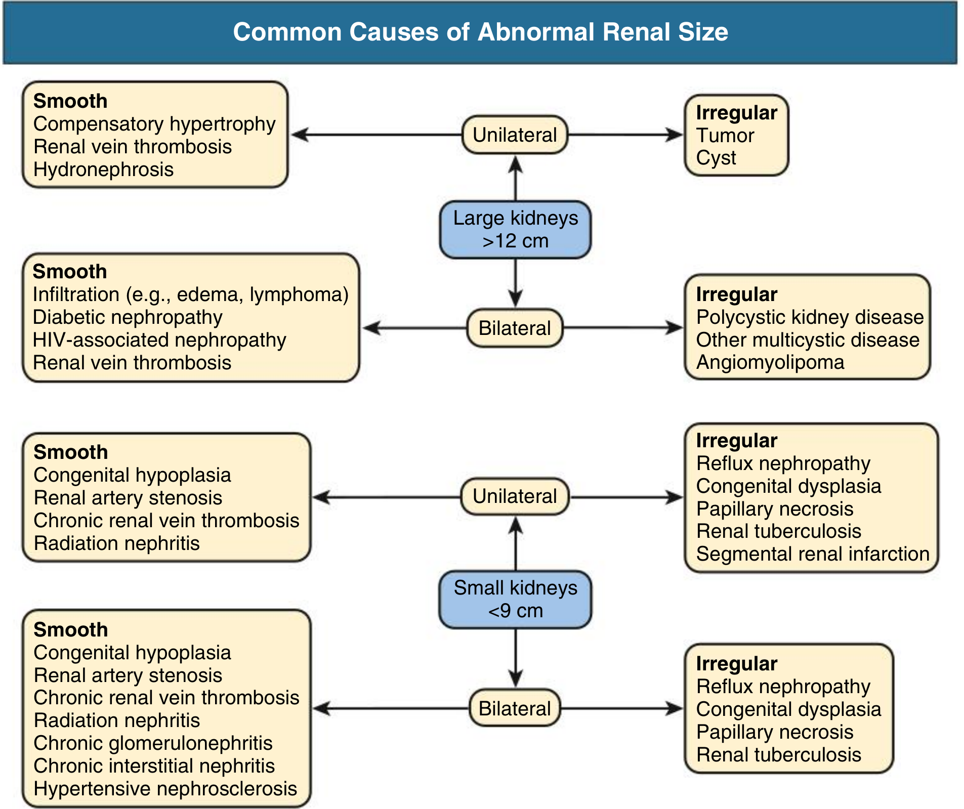
Large Kidneys (>12 cm)
Unilateral
| Contour | Causes |
|---|---|
| Smooth | Compensatory hypertrophy, Renal vein thrombosis, Hydronephrosis |
| Irregular | Tumor (Renal cell carcinoma, Wilms' tumor), Cyst |
Bilateral
| Contour | Causes |
|---|---|
| Smooth | Infiltration (edema, lymphoma), Diabetic nephropathy (early), HIV-associated nephropathy, Renal vein thrombosis |
| Irregular | Polycystic kidney disease (ADPKD/ARPKD), Other multicystic disease, Angiomyolipoma |
Key Individual Causes - Details
1. Hydronephrosis (Unilateral, smooth)
- Dilatation of the collecting system due to outflow obstruction (ureteric stone, stricture, pelviureteric junction obstruction, extrinsic compression)
- Diagnosed on ultrasound; CT urogram identifies the level/cause of obstruction
2. Compensatory Hypertrophy (Unilateral, smooth)
- Occurs in the contralateral kidney when one kidney is absent, non-functioning, or has been removed
- Physiological - not pathological
3. Renal Vein Thrombosis (Unilateral or Bilateral, smooth)
- Causes acute swelling and tenderness; may present with haematuria and nephrotic syndrome
- Risk factors: nephrotic syndrome (especially membranous nephropathy), dehydration, hypercoagulable states
4. Polycystic Kidney Disease (Bilateral, irregular)
- ADPKD (PKD1/PKD2 mutations): most common hereditary cause; bilateral, massively enlarged kidneys with multiple large cysts; associated with hypertension, haematuria, flank pain, and progressive renal failure. Kidney size increases at ~5.3% per year. "Kidney enlargement eventually occurs in 100% of patients with ADPKD." - Comprehensive Clinical Nephrology, 7th ed.
- ARPKD (PKHD1 mutation): presents in neonates/infants; bilateral echogenic enlarged kidneys; associated with hepatic fibrosis
5. Diabetic Nephropathy (Bilateral, smooth - early)
- Early diabetic nephropathy causes glomerular hyperfiltration and kidney enlargement
- Note: In advanced disease, kidneys become small and scarred
6. HIV-Associated Nephropathy (HIVAN) (Bilateral, smooth)
- HIVAN causes enlarged, echogenic kidneys on ultrasound (characteristically large despite impaired function)
- Predominantly affects patients of African ancestry
7. Renal Lymphoma / Infiltration (Bilateral, smooth)
- Kidneys enlarged without obstruction; rapid improvement with treatment supports the diagnosis
- Criteria supporting lymphomatous infiltration: (1) kidney enlargement without obstruction; (2) no other cause; (3) rapid improvement with therapy - National Kidney Foundation Primer on Kidney Diseases, 8th ed.
8. Renal Cell Carcinoma / Wilms' Tumour (Unilateral, irregular)
- A renal mass presenting as unilateral enlargement with an irregular contour
- Haematuria and flank pain are classical features
9. Acute Pyelonephritis / Renal Abscess
- Causes transient unilateral enlargement due to oedema and inflammation
- Associated with fever, loin pain, dysuria
10. Angiomyolipoma (Bilateral, irregular)
- Associated with tuberous sclerosis; bilateral in that setting; fat-containing lesions on CT
Normal Kidney Size (Reference)
- Normal adult kidney length: 9-12 cm (right slightly smaller than left)
- Measurement is typically done on ultrasound or CT in the longest axis
Approach to Investigation
- Ultrasound - first-line: detects hydronephrosis, cysts, solid masses, size asymmetry
- CT scan (contrast + non-contrast) - characterizes masses, identifies obstruction level, staging
- MRI - used in ADPKD to monitor total kidney volume; preferred in pregnancy
- Urinalysis and dipstick - haematuria, proteinuria
- Bloods - renal function (eGFR, creatinine), glucose, HIV serology, FBC (infiltration)
- Biopsy - when diagnosis uncertain (lymphoma, rare infiltrative diseases)
Sources: Comprehensive Clinical Nephrology, 7th Edition (Fig. 6.1); National Kidney Foundation Primer on Kidney Diseases, 8th ed.; S Das Manual on Clinical Surgery, 13th ed.
Cause of unilateral enlargement of kidney
The diagram from the previous answer already covers this directly. Here is a focused, complete answer:
Causes of Unilateral Kidney Enlargement
From the authoritative flowchart (Comprehensive Clinical Nephrology, 7th ed.), unilateral large kidney (>12 cm) is classified by contour:
Smooth Contour (Unilateral)
| Cause | Key Features |
|---|---|
| Compensatory hypertrophy | Contralateral kidney absent/non-functional; physiological, benign |
| Renal vein thrombosis | Acute painful swelling; haematuria, proteinuria; risk factors - nephrotic syndrome, hypercoagulable states |
| Hydronephrosis | Obstruction of outflow (stone, PUJ obstruction, stricture, external compression); diagnosed on USS |
Irregular Contour (Unilateral)
| Cause | Key Features |
|---|---|
| Tumour (Renal cell carcinoma / Wilms' tumour) | Haematuria, flank pain, palpable mass (classic triad); Wilms' in children |
| Cyst (simple or complex) | Simple cysts are common and benign; complex cysts (Bosniak III/IV) are potentially malignant |
Additional Causes (Not Shown in Chart but Recognized)
| Cause | Key Features |
|---|---|
| Acute pyelonephritis / Renal abscess | Transient enlargement due to inflammation/oedema; fever, loin pain, pyuria |
| Xanthogranulomatous pyelonephritis (XGP) | Chronic infection, usually associated with staghorn calculus; diffuse or segmental enlargement |
| Renal trauma / Subcapsular haematoma | History of trauma; smooth enlargement from blood collection under the capsule |
| Duplex kidney | Congenital duplication of the collecting system; kidney appears elongated/enlarged |
| Horseshoe kidney | Both kidneys fused at lower poles; each "half" may appear abnormally positioned/enlarged |
| Acute renal artery occlusion / infarction | Sudden flank pain, haematuria; kidney initially swells then shrinks |
| Angiomyolipoma (solitary) | Fat-containing benign tumour; can be large; associated with tuberous sclerosis |
Memory Aid - "CHART" for Smooth Unilateral Enlargement
C - Compensatory hypertrophy H - Hydronephrosis A - Acute pyelonephritis R - Renal vein thrombosis T - Trauma (haematoma)
Investigations
- Ultrasound - first line; differentiates hydronephrosis, cyst, solid mass
- CT (contrast) - characterises mass, staging, obstruction level
- Urinalysis - haematuria, pyuria, proteinuria
- Urine culture - if infection suspected
- Doppler USS / CT venogram - if renal vein thrombosis suspected
- IVP / CT urogram - for functional assessment and obstruction mapping
Source: Comprehensive Clinical Nephrology, 7th Edition, Fig. 6.1
This is a shared conversation. Sign in to Orris to start your own chat.